ORIGINAL RESEARCH ARTICLE
METHODS OF EVALUATION OF POSTURE IN ELDERLY: A BIBLIOMETRIC STUDY OF THE
SCIENTIFIC PRODUCTION OF THE BVS DATABASE
Caroline Fagundes, Diego da Silva Souza, Anna Regina Grings Barcelos, Victória Haas Masiero
Rita de Cássia Neumann and *Geraldine Alves dos Santos
University Feevale
ARTICLE INFO ABSTRACT
With the advancement of the years, it is observed the reduction of the tissues’ function that covers the joints, resulting in decreased range of motion and joint flexibility. These changes are perceptible in the curvatures of the spine, and as a consequence may limit the elderly. This study aims to identify the methods used to analyze the posture in elderly. To perform this analysis, the use of bibliometrics was used as a methodology, by inserting the keywords spine, curvatures and elderly in the Biblioteca Virtual da Saúde (BVS) database. With a total of 286 articles generated through the search, started the organization and categorization of the data. The articles were divided into two categories: 1) image exams, motion analysis systems, flexicurve and photogrammetry associated with softwares, and 2) x-ray and dual-emission x-ray absorptiometry. The results showed that radiography was the most used method to analyze vertebral curvatures in elderly (68%), followed by motion analysis systems (13%), flexicurva (11%) and photogrammetry associated with softwares (8%). Among the image exams it is possible to mention the radiography (96%) and dual-emission x-ray absorptiometry(4%). It is concluded the radiography is the most used method to evaluate the curvatures of the spine in elderly.
Copyright © 2018, Caroline Fagundes et al. This is an open access article distributed under the Creative Commons Attribution License, which permits unrestricted use, distribution, and reproduction in any medium, provided the original work is properly cited.
INTRODUCTION
According to data from the Brazilian Institute of Geography and Statistics (IBGE), 14.6% of the Brazilian population in 2017 was composed of individuals over 60 years old, surpassing the number of children from 0 to 9 years old. For years, the proportion of the elderly relative to the young was considerably lower due to the difficult conditions of survival. However, public investments in health and education have resulted in an increase in life expectancy of the population. According to the National Survey by Continuous Household Sample - Characteristics of Residents and Households the number of elderly people in Brazil has grow 18% in the last
five years (IBGE, 2018). According to Mendes et al. (2005),
aging is a natural and complex process characterized by biological, psychological and social changes that affect each individual in particular.
*Corresponding author: Geraldine Alves dos Santos
University Feevale
The biological modifications affect the organism as a whole and has as main characteristic the reduction of the
physiological functions, not necessarily resulting in
pathologies. From the age of 40, there is a loss of approximately 1% of the organic functions per year (Neumann, Schauren e Adami, 2016). Over the years, the tissues that covers the joints also undergo modifications that reduces their elasticity. Between 20 and 70 years of age, occurs declines of 20% to 50% in the range of certain movements, intervening in the independence of the elderly to perform their daily tasks. The reduction of flexibility of the joints, particularly the spine, hip and knees, is associated with difficulties in performing daily activities, and may be the main cause of discomfort, pain and functional disability in
individuals aged 60 years or over (Bandeira et al., 2010;
Gasparotto et al., 2012). Another alteration that can affect the
spine in aging is the accentuation of the curves. These misalignments can be perceived in the sagittal, frontal and / or transverse planes. It is worth mentioning that postural deviation in elderly can limit their mobility, interfering in body
ISSN: 2230-9926
International Journal of Development Research
Vol. 08, Issue, 10, pp.23303-23307, October, 2018
Article History:
Received 19th July, 2018
Received in revised form 09th August, 2018
Accepted 18th September, 2018
Published online 29th October, 2018
Key Words:
Curvatures, Elderly, Spine.
ORIGINAL RESEARCH ARTICLE
OPEN ACCESS
Citation: Caroline Fagundes, Diego da Silva Souza, Anna Regina Grings Barcelos, Victória Haas Masiero Rita de Cássia Neumann and Geraldine
Alves dos Santos. 2018. “Methods of evaluation of posture in elderly: a bibliometric study of the scientific production of the bvs database”, International
oscillations and making difficult the maintenance of the static balance and safe gait. Thus, the individuals 60 years or older are more predisposed to fall. Falling can affect the functional capacity in elderly, defined as the ability to maintain the physical and mental skills necessary for independent and
autonomous life. The functionally incapable elderly
compromises their social involvement and decreases their chances of enjoying the benefits of interaction social. These factors negatively influences the quality of life of this age group and are associated with an increased risk for mortality, morbidity, physical and cognitive disability, inactivity and depression. International and national studies identifies functional disability as one of the main predictive factors of mortality in elderly population (Aikawa, Braccialli e Padula,
2006; Veras, 2009; Burke et al., 2010; D’orsil, Xavier e
Ramos; 2011; Gasparotto et al., 2012; Porto et al., 2012; Pinto
e Neri; 2013). There are several ways of analyzing posture in elderly, and in view of the aforementioned elements, this article aims to identify the methods used to evaluate posture in individuals aged 60 years or older.
MATERIALS AND METHODS
In order to carry out the present study, the application of bibliometrics was adopted, which aims to map and present quantitative and statistical data of existing documentation according to the selected key words. This method is widely used to explain the panorama of a given field of knowledge, discipline or theme and, thus, allows the analysis of the characteristics of the object of study (Ribeiro, Costa e Ferreira, 2015). The present research examined only published articles referring to the terms spine, curvatures and elderly in the Biblioteca Virtual da Saúde (BVS) database, which was chosen for providing free access to technical and scientific productions in the health area. For this study to be carried out, the cited descriptors were inserted into the advanced search field of the BVS database on August 1, 2018. The search carried out with the mentioned terms generated a total of 286 articles published. During this period, these articles were read and categorized. To classify them, the following categories were adopted: image exams, motion analysis systems, flexicurve and photogrammetry associated with softwares. Subsequently, the articles were categorized in relation to the most frequently used image exams to evaluate posture in
elderly. They are: x-ray and dual-emission x-ray
absorptiometry.
RESULTS AND DISCUSSION
Two hundred and eighty six articles with the terms spine, curvatures and elderly were found. Of these productions, 37 were selected, because it was directly related to the posture analysis in elderly. The remaining articles were discarded because they dealt with surgeries in the spine, medical procedures, a sample not composed of individuals aged 60 years or more, among other subjects that do not fit into the characteristics proposed for the accomplishment of the present study. Among the thirty seven articles selected for analysis, we evaluated the methods adopted to evaluate the posture in elderly and how frequently they appear in the studied productions. In this sense, it is possible to point out 4 procedures, classified as: image exams, motion analysis systems, flexicurve and photogrammetry associated with softwares.
Graph 1. Evaluation method used for analysis of posture in elderly
According to Graph 1, the most used methods are the image exams and the least used is the photogrammetry associated with softwares. The others studies used motion analysis systems (electromagnetic motion tracking system, ultrasound-based motion analysis system and PEAK Motus 2D motion analysis system) and flexicurve. Among the image exams, 2 types of procedures were used: radiography, corresponding to 25 occurrences (96%) and dual-emission x-ray absorptiometry with 1 incidence (4%). The method considered gold standard to evaluate postural deviations is the Cobb angle, obtained through radiography, however, it exposes the subject to radiation. Therefore, this is not the most appropriateprocedure for frequent patient follow-up, as it will be harmful to the patient's health, including the risk of cancer, as well as being a high cost examination for underdeveloped and developing
countries (Bandeira et al., 2010; Legaye, 2012; Sedrez e
Candotti, 2013; Ribeiro et al., 2017). In this context, the
non-invasive techniques for the assessment of spinal curvatures are efficient because it does not expose the individual to radiation, their application is less complex and the cost is lower. Among these methods, it is possible to cite the flexicurve and photogrammetry associated with softwares, which can be
easily accessed and applied (Ferreira et al., 2011; Sedrez e
Candotti, 2013; Vacari et al., 2013).
The photogrammetry allows global assessment of posture in an objective and reliable way, through the registration of photographs in different planes, and later analysis in a specific software, such as the Postural Assessment Software (SAPO). This program allows the import and calibration of images, the marking of points in the photograph according to the protocol, and / or the free marking of points to determine linear measures, angular values, measures of distances and body angles. The SAPO is a free program that was developed by the University of São Paulo, and its reliability is well documented
(Ferreira et al., 2011; Pachioni et al., 2011; Preto et al., 2015;
Naves et al., 2017). The following materials and procedures
positions himself to the first photo. The verbal command is: "You will stand on this black cardboard carpet in a position that is familiar and comfortable, put on your feet in the way that is most comfortable for you." The digital camera will be positioned 3 meters away from the subject and at a height of
about half the height of the subject (Duarte et al., 2005; Preto
et al., 2015; Naves et al., 2017). The anatomical markers are located through the palpatory surface anatomy, in the orthostatic position of the subject. The points to be marked can follow the SAPO protocol or freely marked.This protocol was suggested by the initial team of the program development project and the choice of these, was based on clinical relevance, scientific basis, methodological feasibility and applicability. This protocol is used as a standard for postural evaluation and generates values for the posture analysis
database (Duarte et al., 2005; Pachioni et al., 2011; Preto et
al., 2015).
Pachioni et al. (2011) used the photogrammetry associated
with SAPO to evaluate the posture of thirty elderly people of both genders, divided into two groups. Group I was composed of 15 individuals with Chronic Obstructive Pulmonary Disease (COPD), and group II (control), by 15 people without respiratory disease. At the end of the study, it was possible to conclude that group I presented more postural alterations than group II. Among the modifications studied, the posterior pelvic deviation was 93.3%, and thoracic kyphosis was 73.3%. According to the authors, increased thoracic curvature occurs as a consequence of rectification and shortening of the
diaphragm. Similar findings were found by Oi et al. (2004), in
which the spinal curvatures of 71 elderly, 52 women and 25 men were evaluated using the photogrammetry method. The results showed that 36.6% of the elderly presented normal curvatures, 29.6% had thoracic hyperkyphosis and anterior head, 19.7% presented thoracic hyperkyphosis and lumbar hyperlordosis, 11.3% presented rectification of the thoracic and lumbar, and 2.8% hyperkyphosis thoracic and anterior head with the support of the hands on the thighs. Similar
results were demonstrated by Takahashi et al. (2005), where
46.2% of the participants had normal vertebral curvatures, whereas 19.9% presented thoracic hyperkyphosis, 17.4% inversion of the lumbar curvature, 11.9% thoracic and lumbar rectification, and 4,7% lumbar hyperlordosis. According to Oi
et al. (2004), the elderly with deformities in the spine, practiced less outdoor activities and lower scores regarding subjective health and satisfaction with life. The flexicurve consists of a flexible ruler capable of shaping the curvatures of the spine. This instrument, made of metal and coated in plastic, allows a quick analysis with information such as angular and linear measurements that represents the contours. It is worth mentioning that in relation to the angular measurement of the thoracic and lumbar region, flexicurve has already been
validated in relation to the gold standard (Ribeiro et al., 2017).
Bandeira et al. (2010) used the flexicurveto measure the
thoracic kyphosis of forty Brazilian elderly. Of these, 20 were sedentary and 20 were physical activity practitioners.
The method adopted showed an overall mean of 47.39° in this curvature, and it is possible to observe a difference between the studied groups. Those who exercised showed an average of 46.18°, while the others showed 48.60°. In another study, the flexible ruler was used to measure the thoracic and lumbar curvatures of 363 elderly women and related to organ prolapse. The results indicated that 25.3% showed abnormalities in the cited curvatures and 91% had organ prolapse. The most common alteration was the loss of lumbar lordosis (75%), suggesting that this deformation may be a factor for the
development of organ prolapse (Mattox et al., 2000). Finally,
Lojudice et al. (2006) evaluated the thoracic curvature of 72
elderly women using the flexicurve method. The results of the study indicated that 56.9% of the participants had a thoracic kyphosis index within normal range. The study considered the rates established by Takahashi and Atsumi (1955), who considers normal rates of 82.05 for individuals aged 60-69 years and 83.33 for those aged 70 years or over. Regarding to others methods used to evaluate spinal curvatures in elderly, it is possible to cite three types: electromagnetic movement tracking system PolhemusTM Patriot, ultrasound-based motion analysis system and PEAK Motus 2D motion analysis system. Below there is a description of each of them. 1) Electromagnetic movement tracking system PolhemusTM Patriot: It allows determining the position and the spatial orientation in three dimensions using electromagnetic sensors applied in certain anatomical points, and connected to a
computer and specific software in real time (Carneiro et al.,
2013). Singh, Bailey and Lee (2010) opted for this methodology to evaluate thoracic kyphosis and lumbar lordosis of 17 elderly people. In order to achieve the objective of the study, the electromagnetic sensor was dragged along the skin of the elderly, from the first thoracic vertebra to the third sacral tubercle. It was asked to the participants to remained standing in their comfortable posture. The findings showed an average of 46.95° for thoracic kyphosis and 27.11° for lumbar lordosis, and when comparing with the radiographic method, different angular values were observed, with a mean of 52.5° for thoracic kyphosis and 57, 4° for the curvature of the lumbar region. Regarding the thoracic region, similar data were found by Rahman, Singh and Lee (2017).
In their study, performed with the elderly and using the Polhemus TM Patriot system, the mean angulation of thoracic kyphosis was 46,30°. However, for the lumbar, different results were found pointing average of 14.10° of curvature. 2)
Ultrasound-based motion analysis system: Li et al. (2014)
[image:3.595.48.537.75.137.2]developed a wheelchair backrest, with the objective to offer a better support for the spinal curvatures, and therefore greater comfort for the wheelchairs. To test the effects of this backrest on the spinal curvatures, an experiment with ultrasound was performed. Twenty elderly people with disorders in the lower limbs and who used wheelchairs were invited to participate in the study. To check the pelvic, lumbar and thoracic angles, belts were designed and applied on subjects. Each belt had 3 miniature ultrasonic transmitters. The first belt was located around the level of the posterior superior iliac spines and
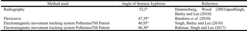
Table 1. Methods used to evaluate the angle of thoracic kyphosis
Method used Angle of thoracic kyphosis Reference
Radiography 52,5° Hammerberg, Wood (2003)apudSingh,
Bailey and Lee (2010)
Flexicurve 47,39° Bandeira et al. (2010)
anterior superior iliac spines. The second was placed at the level of the twelfth thoracic vertebra (T12) and the third level with the first thoracic vertebra (T1). The Win Data software was used to calculate the spinal curvatures. The findings of this study demonstrated that the developed backrest maintained a more neutral pelvic inclination and increased lumbar lordosis, helping to maintain proper seated posture and reducing the risk of back pain. PEAK Motus 2D Motion Analysis System: This system includes one or two video cameras with a 60 Hz or 180 Hz pickup frequency. These cameras must be located 5 meters from the individual, with minimum angulation between them (DINI; DAVID, 2009). In addition, markers are required, applied to the subject, and their location varies according to the purpose of each study. Then the recorded images are transferred to the PEAK Motus software and it is ready to be analyzed.
Kuo, Tully and Galea (2009) used this method to evaluate the spinal curvatures of 34 elderly subjects in standing and sitting position, pre and post Pilates intervention, twice a week for ten weeks. The only significant change was the reduction of 2.3 degrees in the thoracic curvature of the elderly participants of
the study. Regarding to the image exams, it is afford to
mention bone densitometry, obtained through dual energy X-ray absorptiometry (DXA), which aims to measure bone mineral density. This technique is considered the gold standard for diagnosing osteopenia and / or osteoporosis. In addition, it can also be used to evaluate body composition, being more reliable than Body Mass Index (BMI) (Silva, Damiani e
Cominato, 2013; Silva et al., 2015; Guimarães et al., 2017).
However, Maalouf et al. (2007) used X-ray absorptiometry to
measure the index of vertebral curvature irregularity, calculated by a specific equation that relates the anterior and posterior vertebral heights of one vertebra to that of the adjacent vertebra along the thoracolumbar spine (fourth thoracic vertebra (T4) - fourth lumbar vertebra (L4)). At the end of the study it was possible to observe that as higher the irregularity index of the vertebral curvature, the higher is the
number of fractures in the vertebral spine. At the end of the
present research, it is worth to cite that independently of the method used, the most of the studies demonstrates the increase of thoracic kyphosis as the main alteration related to the curvatures of the vertebral spine in elderly. This increase is also associated with osteoporosis, since this pathology modifies the shape and composition of bones, especially the vertebrae. Already the muscles are affected by the sarcopenia, resulting in the diminution of the capacity of generation of strength and flexibility. In addition, the elderly may present increased of body fat, mainly abdominal, and waist perimetry, leading to a reorganization of the body to ensure orthostatic
balance (Porto et al., 2012). As shown in Table 1, only 4
studies measured the angle of thoracic kyphosis. It can be observed that there is a consensus between the non-invasive methods (flexicurve and electromagnetic movement tracking system Polhemus TM Patriot); however, the results shows a divergence from the gold standard method, radiography, for angular measurement of the thoracic spine.
Conclusion
This study had as a general objective to present the scenario of the publications related to the thematic vertebral spine, curvatures and elderly in the BVS database. Postural changes in elderly can affect the amplitude of the movements, interfering in the balance and consequently, predisposing the
elderly to the fall, among other factors. Thus, it can be affirmed that the analysis of the posture in elderly is important and should be performed. There are several methods that can be applied and among them, stands out the radiography, as the most method used for this type of evaluation. It is considered as limitation and suggestion of the present research the use of different databases and keywords in order to present new information on the topic addressed in the present study.
REFERENCES
Aikawa, A.C., Braccialli, L.M.P., Padula, R.S. 2006. Efeitos das alterações posturais e de equilíbrio estático nas quedas
de idosos institucionalizados. Revista de Ciências Médicas,
15 (3): 189-196.
Bandeira, F.M., Delfino, F.C., Carvalho, G.A., Valduga, R.
2010. Comparação entre a cifose torácica de
idosossedentários e praticantes de atividade física
pelométodo flexicurva. Revista Brasileira de
Cineantropometria e Desempenho Humano 12(5): 381 – 386.
Burke, T.N., França, F.J.R., Meneses, S.R.F., Cardoso, V.I., Pereira, R.M.R., Danilevicius, C.F., Marques, A.P. 2010. Postural control among elderly women with and without
osteoporosis: is there a difference? São Paulo Medical
Journal, 128(4): 219-224.
Carneiro, J.A.O., Santos-Pontelli, T.E.G., Colafêmina, J.F., Carneiro, A.A.O., Ferrioli, E. 2013. Um estudo piloto na avaliação das estratégias posturais em jovens e idosos usando um sistema eletromagnético tridimensional.
Brazilian Journal of Otorhinolaryngology, 79(2): 219-225. D´orsil, E., Xavier, A.J., Ramos, L.R. 2011. Trabalho, suporte social e lazer protegem os idosos da perda funcional: estudo epidoso. Revista de Saúde Pública45(4): 685-92. Dini, P.D., David, A.C. 2009. Repetibilidade dos parâmetros
espaçotemporais da marcha: comparação entre crianças normais e com paralisia cerebral do tipo hemiplegia
espástica. Revista Brasileira de Fisioterapia, 13(3):
215-222.
Duarte, M., Ferreira, E.A., Maldonado, E.P., Freitas, A.Z.. 2005. Documentação sobre o SAPO - Softwares para avaliação postural. Retrieved in:<http://demotu.org/sapo/>. Ferreira, E.A., Duarte, M., Maldonado, E.P., Bersanetti, A.A., Marques, A.P. 2011. Quantitative assessment of postural alignment in young adults based on photographs of
anterior, posterior, and lateral views. Journal of
Manipulative and Physiological Therapeutics, 34(6):371-380.
Gasparotto, L.P.R., Reis, C.C.I., Ramos, L.R., Santos, J.F.Q. 2012. Autoavaliação da postura por idosos com e sem
hipercifose torácica. Ciência & Saúde Coletiva,
17(3):717-722.
Guimarães, M.F.B.R., Pinto, M.R.C., Raid, R.G.S.C., Andrade, M.V.M., Kakehasi, A.M. 2017. Qual o melhor ponto de corte de índice de massa corporal para diagnosticar a obesidade em mulheres com artrite reumatoide? Um estudo que usa a composic¸ão corporal pela absorciometria com raios X de dupla energia. Revista
Brasileira de Reumatologia., 57(4):279-285.
Kuo, Y., Tully, E.A., Galea, M.P. 2009. Sagittal spinal posture after pilates-based exercise in healthy older adults. Spine, 34(10):1046-1051.
Legaye, J. 2012. Follow-up of the sagittal spine by optical
technique. Annals of Physical and Rehabilitation Medicine,
55:76-92.
Li, C., Chen, Y., Chang, C., Tsai, K. 2014. The effects of backward adjustable thoracic support in wheelchair on spinal curvature and back muscle activation for elderly
people. Plos One, 9(11): 1-14.
Lojudice, D.C., Lavorato, C.S., Pereira, G.P., Cardoso, J.R. 2006. Avaliação da curvatura dorsal em mulheres idosas pelo índice da cifose torácica. Fisioterapia E Pesquisa, 13(3):32-37.
Maalouf, G., Maalouf, N.M., Schaaf, N., Zebaze, M., Nehme, A., TANNOUS, Z., WEHBE, J., Adib, G., Gannagé-Yared, M.H., Seeman, E. 2007. The spinal curvature irregularity
index independently identifies vertebral fractures.
Osteoporosis International., 18: 279–283.
Mattox, T.F., Lucente, V., Mcintyre, P., Miklos, J.R.,
Tomezsko, J. 2000. Abnormal spinal curvature and its
relationship to pelvic organ prolapse. American Journal of
Obstetrics and Gynecology 183(6): 1381-1384.
Mendes, M.R.S.S.B., Gusmão, J.L., Faro, A.C.M., Leite, R. C. B.O. 2005. A situação social do idoso no Brasil: uma breve consideração. Acta Paulista de Enfermagem18(4): 422-6. Naves, J.M., Soares, C., Svezzia, V.A., Cussolim, F.D.,
Mendonça, A.C. 2017. Correlação entre alinhamento
pélvico e fibroedema geloide. Fisioterapia e pesquisa
24(1):40-45.
Neumann, L., Schauren, B.C., Adami, F.S. 2016. Sensibilidade gustativa de adultos e idosos. Revista Brasileira de
Geriatria e Gerontologia., 19(5): 797-808.
Oi, N., Tobimatsu, Y., Iwaya, T., Okada, Y., Gushiken, S., Kusano, S., Yamamoto, M., Takakura, Y., Suyama, T. 2004. Reability and validity of classification of senile
postural deformity in mass examinations. The Tohoku
Journal Of Experimental Medicine, 202:105-112.
Pachioni, C.A.S., Ferrante, J.Á., Panissa, T.S.D., Ferreira, D.M.A., Ramos, D., Moreira, G.L., Ramos, E.M.C. 2011. Avaliação postural em pacientes com doença pulmonar obstrutiva crônica. Fisioterapia e Pesquisa18(4):341-345. Pinto, J.M., Neri, A.L. 2013. Doenças crônicas, capacidade
funcional, envolvimento social e satisfação em idosos
comunitários: Estudo Fibra. Ciência & Saúde Coletiva,
18(12):3449-3460.
Porto, F., Espinosa, G., Vivian, R.C., Itaborahy, A.S., Montenegro, R.A., Farinatti, P.T.V., Gurgel, J.L. 2012. O exercício físico influencia a postura corporal de idosas? Motriz, 18(3):487-494.
Preto, L.S.R., Santos, A.R.R., Rodrigues, V.M.C.P., Quitério, N.F.N., Pimentel, M.H., Manrique, G.A. 2015. Análise por Fotogrametria da Postura e Fatores de Risco Associados
em Crianças e Adolescentes Escolarizados. Revista de
Enfermagem Referência,4(7):31-40.
Rahman, N.N.A.A., Singh, D.V., Lee, R. 2017. Correlation between thoracolumbar curvatures and respiratory function
in older adults. Clinical Interventions in Aging, 12:523–
529.
Ribeiro, H.C.M., Costa, B.K., Ferreira, M.P. 2015. Governança corporativa nos esportes: análise dos últimos 23 anos de produção acadêmica em periódicos
internacionais. Revista de Administração e Contabilidade
da Unisinos., 12(2): 135-154.
Ribeiro, R.P., Marchetti, B.V., Oliveira, E.B., Candotti, C.T. 2017. Índice de cifose obtido em radiografia e com o
flexicurvena avaliação de crianças e jovens. Revista
Brasileira de Saúde Materno Infantil., 17(1):89-97. Sedrez, J.Á., Candotti, C.T. 2013. Métodos não invasivos de
avaliação postural da escoliose: Uma revisão sistemática.
Motricidade 9(4): 100-111.
Silva, A.C.V., Rosa, M.I., Fernandes, B., Lumertz, S., Diniz, R.M., Damiani, M.E.F.R. 2015. Fatores associados à osteopenia e osteoporose em mulheres submetidas à
densitometria óssea. Revista Brasileira de Reumatologia,
55(3):223-228.
Silva, M.M.X., Damiani, D., Cominato, L. 2013. Avaliação da densidade mineral óssea em adolescentes do sexo feminino
com transtorno alimentar. Arquivos Brasileiros de
Endocrinologia & Metabologia, 57(7):527-532.
Singh, D.V., Bailey, M., Lee, R. 2010. Biplanar Measurement of Thoracolumbar Curvature in Older Adults Using an Electromagnetic Tracking Device. Archives of Physical
Medicine andRehabilitation 91:137-142.
Takahashi, E., Atsumi, H. 1955. Age differences in thoracic
form as indicated by thoracic index. Human Biology.,
27(2): 65-74.
Takahashi, T., Ishida, K., Hirose, D., Nagano, Y., Okumiya, K., Nishinaga, M., Matsubayashi, K., Doi, Y., Tani, T., Yamamoto, H. 2005. Trunk deformity is associated with a reduction in outdoor activities of daily living and life satisfaction in community dwelling older people.
Osteoporosis International., 16:273–279.
Vacari, D.A., Ulbricht, L., Schneider, F.K., Neves, E.B. 2013. Principais métodos de diagnóstico postural da coluna lombar. Revista da Educação Física24(2):305-315.
Veras, R. 2009. Envelhecimento populacional contemporâneo:
demandas, desafios e inovações. Revista de Saúde Pública.,
43(3):548-554.