0095-1137/05/$08.00⫹0 doi:10.1128/JCM.43.11.5696–5704.2005
Copyright © 2005, American Society for Microbiology. All Rights Reserved.
Sensitive Phenotypic Detection of Minor Drug-Resistant Human
Immunodeficiency Virus Type 1 Reverse Transcriptase Variants
Dwight V. Nissley,
1,3* Elias K. Halvas,
2Nicole L. Hoppman,
3David J. Garfinkel,
3John W. Mellors,
2and Jeffrey N. Strathern
3Basic Research Program, SAIC-Frederick, Inc., NCI Frederick, Frederick, Maryland1; Division of Infectious Diseases,
University of Pittsburgh School of Medicine, Pittsburgh, Pennsylvania2; and Gene Regulation and
Chromosome Biology Laboratory, NCI Frederick, Frederick, Maryland3
Received 4 November 2004/Returned for modification 2 May 2005/Accepted 4 August 2005
Detection of drug-resistant variants is important for the clinical management of human immunodeficiency virus type 1 (HIV-1) infection and for studies on the evolution of drug resistance. Here we show that hybrid elements composed of theSaccharomyces cerevisiaeretrotransposon Ty1 and the reverse transcriptase (RT) of HIV-1 are useful tools for detecting, monitoring, and isolating drug-resistant reverse transcriptases. This sensitive phenotypic assay is able to detect nonnucleoside reverse transcriptase inhibitor-resistant RT domains derived from mixtures of infectious molecular clones of HIV-1 in plasma and from clinical samples when the variants comprise as little as 0.3 to 1% of the virus population. Our assay can characterize the activities and drug susceptibilities of both known and novel reverse transcriptase variants and should prove useful in studies of the evolution and clinical significance of minor drug-resistant viral variants.
Antiretroviral therapy has markedly reduced morbidity and mortality from human immunodeficiency virus (HIV) type 1 (HIV-1) infection in developed countries, but the selection of drug-resistant virus is frequent. Aside from rendering thera-pies ineffective, the evolution of resistance generates virus that is cross resistant to related inhibitors, thus limiting treatment options. Increasingly, the effective management of HIV infec-tion requires that the susceptibility of the replicating virus population to therapeutic options be known. Several expert panels (10, 19) now recommend drug resistance testing for determination of the best treatment regimen in all patients on a failing antiretroviral treatment regimen.
HIV-1 reverse transcriptase (RT) is a low-fidelity DNA poly-merase due to the absence of proofreading activity (24). The elevated error rate of HIV-1 RT coupled with high levels of replication and recombination result in extensive genetic di-versity and the production of innumerable variants, also termed the viral quasispecies. It has been estimated that every possible single nucleotide mutation is generated multiple times daily in an HIV-infected individual (5), suggesting that drug-resistant variants exist prior to antiretroviral therapy. The fre-quency of a given variant in the virus population is determined by its fitness (replication potential) relative to that of other viral variants exposed to the same environmental conditions. Drug-resistant variants that are present at low frequencies in the absence of drug selection can become dominant in the virus population with drug exposure. An accurate method for detection of drug-resistant variants and monitoring of their frequency over time would be helpful in monitoring the evo-lution of resistance and defining the role of minor variants in antiretroviral treatment failure.
Several approaches have been used to monitor HIV-1 drug resistance. Genotypic assays (33) infer drug resistance based on DNA sequence information and predicted amino acid pat-terns. Viral regions of interest are sequenced, detected by hybridization, or amplified in an allele-specific manner by PCR. While these approaches can be sensitive, their detection and/or interpretation is limited to known resistance mutations and may miss other relevant mutations or combinations of mutations. Phenotypic assays (9) assess drug susceptibility by determining the effects of inhibitors on the replication of viral isolates or recombinant vectors carrying patient-derived viral domains amplified from virus populations. These assays are useful for determining the average phenotype of the virus population but in routine practice are limited in their ability to detect minor drug-resistant variants.
We have developed a phenotypic assay based on hybrid elements derived from theSaccharomyces cerevisiaeTy1 retro-transposon in which reverse transcriptase is provided by HIV-1 RT (TyHRT) (27). TyHRT elements generate HIV-1 RT-mediated events at a high frequency, and the RT activity of HIV-1 RT variants can be differentiated and characterized over a 10,000-fold range. Since HIV-1 RT activity is inhibited by nonnucleoside reverse transcriptase inhibitors (NNRTIs) in yeast (26), the assay can be performed in the presence of these inhibitors to determine the drug resistance phenotype of individual RTs. Here we show that by constructing RT domain libraries in vivo, it is practical to characterize the basal RT activity and drug susceptibility of every RT isolates in libraries containing thousands of RT domains. This allows the phenotypic detection of drug-resistant viral variants present at frequencies of less than 1%. The TyHRT system detects known and novel drug-resistant RTs both in laboratory stocks of HIV-1 and in plasma samples from HIV-infected individuals.
* Corresponding author. Mailing address: BRP, SAIC-Frederick, NCI Frederick, P.O. Box B, Frederick, MD 21702. Phone: (301) 846-1181. Fax: (301) 846-6911. E-mail: [email protected].
5696
on May 15, 2020 by guest
http://jcm.asm.org/
MATERIALS AND METHODS
Yeast strains.Reverse transcription assays were carried out withS. cerevisiae
strain DG1251 (MAT␣ura3-167 trp1-hisG spt3-101 his3⌬200) (35).
Plasmids.Plasmid pH⌬RT1 was constructed by inserting the 489-bp BsrGI-PvuII fragment from pGFPuv (Clontech, Palo Alto, Calif.) into the BsrGI and PvuII sites in HIV-1 RT of pHART22 (27), resulting in the deletion of HIV-1 RT amino acids 37 to 250. HIV-1 RT variants cloned in pUC12 (pUC12HIVRT, pUC12HIVRTL100I, pUC12HIVRTY181C, pUC12HIVRTD185E, and pUC12HIVRTE478Q) were described previously (2, 20). The infectious plas-mids of HIV-1 used to generate viral stocks were pxxHIV-1LAI(36) and a
derivative (pxxHIV-1LAIK103N) produced by site-directed mutagenesis.
Generation of mixtures containing mutant and wild-type HIV-1.Stock viruses were generated by transfection of 293T cells with either the wild-type RT vector pxxHIV-1LAIor pxxHIV-1LAIK103N, which encodes the nonnucleoside reverse
transcriptase inhibitor (NNRTI) mutation 103N in RT. Each viral stock was characterized for infectivity (endpoint dilution assay with MT-2 cells), capsid level (p24 antigen), and HIV-1 RNA concentration (Roche AMPLICOR HIV Monitor assay, version 1.0) (E. Halvas, unpublished data). Viral stocks were mixed together based on the HIV-1 RNA concentrations to generate the 103N mutant at 0.4, 2, or 100%. In addition, a test panel of virus mixtures in HIV-1-seronegative human plasma was generated with the 103N mutant at the fol-lowing frequencies: 0.01, 0.1, 0.4, 1, 2, 5, 10, 25, 50, and 100%. Viral stocks and mixtures were stored at⫺80°C before use.
RNA extraction and cDNA synthesis from HIV-1 mixtures or plasmids.Viral RNA was isolated by using the QIA Amp Viral RNA extraction kit (QIAGEN Sciences, Germantown, Md.). RNA was extracted from 200l of each sam-ple and eluted with molecular biology-grade water (Quality Biological Inc., Gaithersburg, Md.). A total of 7.5l of viral RNA and 1l (10 pmol/l) of primer N1-L (5⬘-TTATCTGGTTGTGCTTGAAT-3⬘) were mixed, denatured at 70°C for 10 min, and then quenched on ice for 5 min. A total of 10.5l of the RT master mixture (1⫻1st Strand Superscript II buffer, 0.5 mM deoxynucleoside triphosphates [dNTPs], 0.01 M dithiothreitol, 40 U of RNase inhibitor, and 200 U of Superscript II; Gibco BRL) was added to each tube; and the samples were incubated at 45°C for 50 min, denatured at 70°C for 1 min, and quenched on ice for 5 min. Ten microliters of each reaction mixture containing cDNA was amplified with primers (40 pmol) N1-L and N1-U (5⬘-CTGTTGACTCAGATT GGTTG-3⬘), which span HIV-1 RT codons 10 to 496. Samples were amplified (1⫻Taqbuffer, 1 mM dNTPs, and 2.5TaqDNA polymerase) with the following cycling conditions: 94°C for 4 min (1 cycle); 94°C for 1 min, 56°C for 1 min, and 72°C for 3 min (35 cycles); and 72°C for 10 min (1 cycle). The PCR products were purified with a GFX PCR purification kit (Amersham Pharmacia, Piscataway, N.J.). Both xxLAI plasmid controls (2 pg of wild-type K103 or mutant 103N) were amplified with primers (40 pmol) xxLAI-L (5⬘-ACTATATTTACTTCT AGACCCGAAATCCTGCAAAGC-3⬘) and xxLAI-U (5⬘-GTAAAATTAAAG CCCGGGATGGATGGCCCAAAAGT-3⬘). The plasmids were amplified and the products were purified as described above.
RNA extraction and cDNA synthesis from HIV-1 mixtures in plasma.RNA was isolated from 500l of plasma following centrifugation at 16,000⫻gfor 1 h at 4°C. The pellet was resuspended in 50l RNase-free Tris-HCl (pH 8.0) with 10l of proteinase K (20 mg/ml) and incubated at 55°C for 30 min. Two hundred microliters of 5.8 M guanidinium isothiocyanate and 10l glycogen (20 mg/ml) were added, and the mixture was vortexed and incubated at room temperature for 5 min. RNA was precipitated by adding 270l of 100% isopropanol for 10 min at room temperature, followed by centrifugation at 16,000⫻gfor 10 min. The pellet was washed once with 70% ethanol and resuspended in 32l RNase-free Tris-HCl (pH 8.0). cDNA reactions were set up by adding 4l of 10 mM dNTPs and 4l of 50-ng/l random hexamers, denaturing at 65°C for 10 min, and cooling at 4°C for 2 min. Fifty microliters comprising the following compo-nents of the RT master mixture was added to the denatured RNA and annealed primer at the indicated final concentrations: 1⫻RT buffer, 5 mM MgCl2, 1 mM
dithiothreitol, 2 U of RNase-Out RNase inhibitor, and 2 U of Superscript II (Invitrogen, Carlsbad, Calif.). The reaction mixtures with cDNA were incubated at 25°C for 10 min and then at 42°C for 40 min. The reaction was stopped at 85°C for 10 min and stored at 4°C. One to 10l of the cDNA was amplified with primers 1849⫹(5⬘-GTAGACAGCATGTCAGGGAG-3⬘) and 3500⫺(5⬘-CTA TTAAGTATTTTGATGGGTCATAA-3⬘). The PCR amplifications were per-formed in 10-l reaction mixtures with the following components at the indicated final concentrations: 1⫻PCR buffer, 2 mM MgSO4, 0.2 mM dNTPs, 0.2M of
each primer, and 2.5 U ofTaqDNA polymerase (Invitrogen). The following cycling conditions were used: 94°C for 1 min, 60°C for 1 min, and 72°C for 2 min (35 cycles) and 72°C for 10 min (1 cycle). The PCR products were purified with a Qiaquick PCR purification kit (QIAGEN Sciences).
Yeast transformation and reverse transcription assay. BsrGI- and PvuII-linearized, gel-purified pH⌬RT1 (100 to 200 ng) and the PCR product contain-ing the HIV-1 RT domain (200 to 500 ng) were cotransformed into DG1251 by using a lithium acetate protocol (15). Transformants were selected on plates containing synthetic complete medium lacking uracil (SC-URA) with glucose (32). Transformants were arrayed in small (100/plate) or large (25/plate) patches on plates with SC-URA and glucose by using sterile toothpicks. Following incu-bation overnight at 30°C, the arrayed patches were replica plated with velveteen onto plates with SC-URA and galactose plates with or without inhibitor and were grown for 2 days at 30°C to induce expression of the hybrid retroelements, which are under the control of theGAL1promoter (26). The plates were then replica plated onto synthetic complete medium without histidine (SC-HIS) and with or without inhibitor and grown for 3 to 4 days at 30°C to select reverse transcription events. Elements carrying wild-type RT (H2BX) and the NNRTI resistance allele K103N were placed on each plate as controls. Efavirenz (EFV; kindly provided by Dupont Pharmaceuticals, Wilmington, Del.) and 8-Cl TIBO {(⫺)- (S)-8-chloro-4,5,6,7-tetrahydro-5-methyl-6-(3-methyl-2-butenyl)imdidazo[4,5,1-jk][1,4]benzodiazepine-2(1H)-thione monohydrochloride (R091767); kindly pro-vided by Janssen Research Foundation, Beerse, Belgium} were dissolved in dimethyl sulfoxide and used at 300 nM and 20M, respectively the final dimethyl sulfoxide concentration was 1%.
Sequencing of RT domains.HIV-1 RT domains in TyHRT elements were obtained by growing the isolates overnight in 10 ml SC-URA at 30°C and preparing DNA by a glass bead-phenol extraction method (21). DNA was sus-pended in 50l water, and 0.5 to 2.0l was used in a PCR with a 100-l reaction mixture to amplify the RT domain. Amplification was carried out for 30 cycles (94°C for 1 min, 60°C for 1 min, 72°C for 2 min) by using primers 44F (5⬘-GGA TGGATGGCCCAAAAGT-3⬘) and I1097 (5⬘-GCACTGCCTCTGTTAATTGT-3⬘), which span RT codons 15 to 356. The PCR products were purified with the GFX PCR purification kit (Amersham Pharmacia, Piscataway, N.J.). The purified PCR products were sequenced with internal primers B275 (5⬘-AGACTTCTGGGAAGTTCAAT-3⬘) and K489 (5⬘-AAGGCTCTAAGATTTTTGTCAT-3⬘).
Patient samples.Plasma samples were obtained at study entry from patients enrolled in AIDS Clinical Trials Group Protocol 398, which examined the effects of single versus dual protease inhibitor-containing regimens in 481 patients on a failing regimen containing a protease inhibitor (17). All patients had prior ex-perience with nucleoside analog reverse transcriptase inhibitors and one or more protease inhibitors; approximately half of the subjects had prior experience with an NNRTI other than efavirenz (e.g., delaviridine or nevirapine). Samples from NNRTI-experienced and NNRTI-naive subjects where standard genotyping (Celera HIV-1 ViroSeq, version 2.0) failed to identify NNRTI resistance muta-tions in isolates in plasma were selected for studies aimed at detecting minor resistant virus present at the baseline and correlating these variants with the predominant virus population at treatment failure (25). A subset of four samples from NNRTI-experienced, resistance genotype-negative subjects were used to determine whether the TyHRT assay detects minority NNRTI-resistant isolates in patient samples. Informed consent was obtained from all subjects. The guide-lines of the U.S. Department of Health and Human Services and the authors’ institutions were followed in the conduct of this research.
RESULTS
TyHRT assay.TyHRT retroelements are active inS. cerevi-siae, and HIV-1 RT activity can be monitored by a genetic assay (27). This assay is based on the reverse transcription indicator genehis3AI(7); it requires transcription, RNA splic-ing to remove an artificial intron, and reverse transcription to generate an intact cDNA copy of theHIS3gene. The produc-tion of yeast colonies that grow on medium lacking histidine is an indirect assay of reverse transcriptase activity. TyHRT ele-ments marked with his3AI (Fig. 1A) have the Ty1 reverse transcriptacoding region replaced by HIV-1 RT-coding se-quences.
The high efficiency of homologous recombination inS.
cer-evisiaemakes it possible to generate RT domain libraries in
TyHRT elements without conventional DNA cloning strate-gies. Cotransformation of linear DNA fragments (or PCR products) encoding HIV-1 RT with linearized vector-borne TyHRT elements from which part of RT is deleted results in
on May 15, 2020 by guest
http://jcm.asm.org/
the generation of intact TyHRT elements that carry the input RT domains (Fig. 1B). Assembly of novel TyHRT elements is selected genetically; only recircularized vectors give rise to colonies. Successful incorporation of an RT domain results in a clonal isolate of that particular RT domain. Isolates are easily manipulated, making it possible to construct and analyze RT domain libraries carrying several hundred to several thou-sand RT isolates. Isolates from these libraries are analyzed for RT activity and drug susceptibility (Fig. 1C).
HIV-1 RT activity and drug susceptibility are determined by use of a phenotypic readout. Previously, RT activity has been measured by growing cells containingGAL1 promoter-driven TyHRT elements on galactose to induce expression, followed by differential plating on rich medium to determine the cell number and on medium lacking histidine to select for reverse transcription (26). To achieve quick and simple drug susceptibility screening, we now use a semiquantitative visual approach to identify drug-resistant RTs. The activities
and drug susceptibilities of RT variants are determined by observing the ability of arrayed RT isolates to generate histidine-positive colonies. RT activity is qualitatively as-sessed in the absence of inhibitor to monitor the basal levels of activity. Drug susceptibility is assessed in the presence of inhibitor to identify resistant RT variants. With this visual screen it is possible to differentiate RTs that exhibit wild-type drug susceptibility, weak resistance, intermediate resis-tance, and strong resistance (Fig. 1C).
TyHRT libraries accurately represent input RTs.To accu-rately and sensitively monitor the presence of variants, a RT domain library must correctly reflect the frequency of the RT domains present in a viral pool. We assessed the accuracy of our approach by cotransformingS. cerevisiaewith known mix-tures of HIV-1 RT domains and a TyHRT vector from which part of the HIV-1 RT region was deleted. Wild-type, poly-merase-inactive (D185E), RNase H-inactive (E478Q), and NNRTI-resistant (encoding L100I or Y181C) RTs present in
FIG. 1. TyHRT elements. In TyHRT elements, Ty1 RT is replaced by HIV-1 RT. HIV-1 RT activity is monitored by a genetic assay in the budding yeast,Saccharomyces cerevisiae. (A) Expression and reverse transcription of TyHRT elements result in the conversion of a nonfunctional his3gene (his3AI) into a functionalHIS3gene. Each colony is the result of an independent reverse transcription event. RT activity and drug resistance are assessed qualitatively by visual inspection of RT isolates and quantitatively by counting the number of histidine-positive colonies. (B) TyHRT elements are used to assemble RT domain libraries to monitor RT activity and drug resistance. Efficient homologous recombination inS. cerevisiaemakes it possible to assemble libraries independent of conventional cloning. TyHRT elements from which part of the HIV-1 RT domain was deleted were cotransformed with RT-PCR products generated from viral RNA. Upon transformation, the RT domain DNA was incorporated into the RT region, generating novel TyHRT elements. Each isolate carries a unique RT domain, and the library is representative of the RT domains present in the original viral sample. (C) The TyHRT system monitors HIV-1 RT activity phenotypically. Reverse transcription is scored by placing cells on SC-HIS. The addition of efavirenz to the medium (SC-HIS⫹EFV) inhibits wild-type RT and selects for the reverse transcription events generated by drug-resistant variants. The number of RT events (the number of colonies) reflects the phenotype of a variant. Examples of the inactive, wild-type, weak resistance, intermediate resistance, and strong resistance phenotypes are shown.
on May 15, 2020 by guest
http://jcm.asm.org/
pUC vectors were mixed in equal amounts and amplified by PCR to generate full-length RT domain DNA. The resulting transformants were arrayed as patches, and reverse transcrip-tase activity was determined in the absence and presence of the NNRTI 8 Cl-TIBO.
An TyHRT RT domain library generated in this fashion (Table 1) is representative of the RT domains present in the original pool. Three of the five (60%) RT substrates used encode active RTs, and 56% of the isolates gave rise to histi-dine-positive colonies. Of the three active RTs, two (40% of the total) encoded the NNRTI-resistant variants L100I and Y181C; 36% of the isolates were resistant to 8-Cl TIBO. Wild-type RT was 20% of the substrate mix, and 20% of the isolates were sensitive to 8-Cl TIBO.
Since the linearized RT deletion plasmid may also circular-ize by nonhomologous end joining without regenerating a com-plete TyHRT element, the efficiency of homologous recombi-nation was tested by using an RT domain substrate generated by amplification of wild-type RT. Cotransformation of linear-ized TyH⌬RT vector plus HIV-1 RT DNA fragments resulted in yeast transformants in which 93% of the isolates had active HIV-1 RT domains, as determined by their ability to generate colonies on medium lacking histidine (Table 1). PCR and restriction enzyme analysis of the inactive TyHRT elements revealed that greater than 95% carried no RT insert (data not shown) and were the result of plasmid reclosure. The efficiency of recombination results in libraries where a high proportion of
the isolates have RT inserts. Almost all (⬎95%) of the inactive isolates lacked an insert, demonstrating that there is little in-activating mutagenesis of input RT domains (⬍0.5%) due to transformation and homologous recombination in yeast.
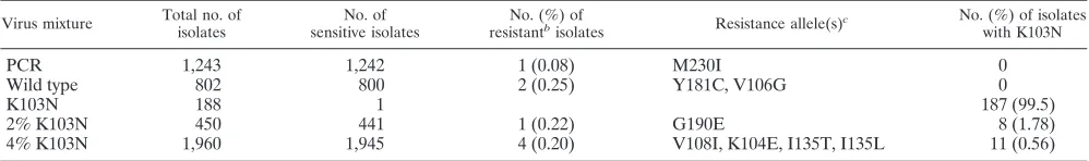
Accurate detection of drug-resistant RTs.Characterization of viral populations in many assays is facilitated by the ampli-fication of nucleic acids, such as by the use of RT-PCR for the amplification of HIV-1 RNA. Detection of minor drug-resis-tant RTs is complicated by the possibility that variants can also be generated during transcription of viral RNA, during in vitro reverse transcription of purified RNA, and during PCR ampli-fication. The sensitivity of any assay will be limited by the frequency of misincorporations during reverse transcription and amplification. To determine both the sensitivity and the background rate of resistance due to misincorporation of the TyHRT system, we generated wild-type and drug-resistant vi-ral stocks that were used alone or in mixtures for the assay (Table 2). The products generated by PCR amplification of the virus vectors were included as controls to determine the fre-quency of resistance due to PCR errors.
Amplification of the RT domain present in the wild-type HIV-1 plasmid clone (pxxHIV-1LAI) used to produce viral stocks generated EFV-resistant RT at a frequency of about 1 in 1,000 (Table 2). The one EFV-resistant variant had the codon change M230I. The extraction of HIV-1 RNA from wild-type virus (produced by transfection of 293T cells) and use of the RNA as a substrate to generate a wild-type RT domain library revealed that 99.8% of the isolates were active and EFV sensitive. Two isolates exhibited weak EFV resis-tance. As expected, 99.5% of isolates generated from the EFV-resistant virus stock encoding K103N exhibited strong EFV resistance and were indistinguishable from a cloned TyHRT element carrying K103N. One of the isolates from the K103N sample carried the codon change I180T and had less RT ac-tivity both in the presence and in the absence of EFV.
[image:4.585.42.284.81.164.2]To further define the accuracy of the TyHRT system, mix-tures containing low levels of drug-resistant virus were also analyzed (Table 2). RT isolates from a 2% mutant (K103N) and wild-type virus mixture were assayed, and 2% scored as EFV resistant by phenotyping. TyHRT DNA was purified from the EFV-resistant isolates, and DNA sequencing revealed that 1.8% of the isolates encoded K103N and 0.2% encoded a mutation elsewhere (G190E) that results in EFV resistance. RT isolates from a 0.4% mutant (K103N) and wild-type virus
TABLE 1. TyHRT system reproduces the phenotype of input RTsa
Mixture
No. (%) of isolates
Total HIS⫹histidine positive
8-Cl TIBO resistant
8-Cl TIBO sensitive
Vector 122 0
PCR mixture with wild-type virus only
200 186 (93)
PCR mixture 408 228 (56) 147 (36) 81 (20)
Expected 245 (60) 163 (40) 82 (20)
aWild-type RT plasmid and equal amounts of wild type, polymerase-inactive
(D185E), RNase H-inactive (E478Q), and two NNRTI-resistant (L100I and Y181C) RT plasmids were diluted and amplified by PCR to generate a PCR product containing the RT domain. DNA containing the RT domain was incor-porated into TyHRT elements by homologous recombination in vivo. Isolates were tested for RT activity and drug resistance. The number of isolates tested, the portion that were active, and the portion that were drug resistant are indi-cated. Also shown are the percentages expected based on the starting ratio of RT domains.
TABLE 2. Accurate detection of drug resistance with low backgrounda
Virus mixture Total no. of isolates
No. of sensitive isolates
No. (%) of resistantb
isolates Resistance allele(s)
c No. (%) of isolates
with K103N
PCR 1,243 1,242 1 (0.08) M230I 0
Wild type 802 800 2 (0.25) Y181C, V106G 0
K103N 188 1 187 (99.5)
2% K103N 450 441 1 (0.22) G190E 8 (1.78)
4% K103N 1,960 1,945 4 (0.20) V108I, K104E, I135T, I135L 11 (0.56)
aThe virus mixtures were analyzed to determine the sensitivity and background of the TyHRT system. Wild-type and drug-resistant virus (K103N) were cultured in
vitro and mixed as indicated in Materials and Methods. A control (PCR), where the RT substrate was generated by amplification of virus vector DNA, was included to determine the frequency of resistance due to PCR errors. The number of isolates analyzed for each sample, the proportion that were drug resistant or sensitive, and the codon changes present in the variants are shown.
bResistance (unexpected) due to an allele other than K103N. cOnly resistance alleles other than K103N are listed.
on May 15, 2020 by guest
http://jcm.asm.org/
[image:4.585.43.542.601.675.2]mixture were assayed, and 0.8% scored as EFV resistant. DNA sequencing revealed that 0.6% had K103N and 0.2% carried mutations elsewhere. Taken together, these results show that the TyHRT system is accurate and has a low background of resistance due to experimentally derived mutation.
Sensitive detection of drug-resistant RTs.The applicability and sensitivity of the TyHRT system for the detection of drug-resistant virus in HIV-1-seronegative plasma was tested by analyzing a panel of samples provided by the Virology Quality Assessment Program of the AIDS Clinical Trials Group. Mix-tures of wild-type and NNRTI-resistant (K103N) virus were assembled in HIV-1-seronegative human plasma and distrib-uted to 12 laboratories to test the samples for drug resistance by six different techniques in order to compare the sensitivities of the assays (16). Viral RNA from plasma samples was ex-tracted and subjected to RT-PCR as described above.
The TyHRT system detected EFV-resistant virus in human plasma (Table 3) when the resistant virus was present at a frequency as low as 0.4% in two of three experiments: 1 of 307 isolates (0.33%; assay I) and 1 of 247 isolates (0.40%; assay II) derived from the 0.4% mutant mixture. EFV-resistant virus was detected in one of three assays derived from the 0.1% mutant mixture: 1 of 291 isolates (0.34%). DNA sequencing confirmed the presence of K103N in these RTs. These results suggest that the sensitivity of the TyHRT system for the de-tection of drug resistance is below 1%.
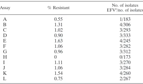
To test the reproducibility and variation of the TyHRT as-say, the assay with the 1% mutant mixture was replicated an additional 12 times (Table 4). RT domains were generated by independent PCR amplification of viral first-strand cDNA. By including the three 1% samples obtained previously (Table 3), the frequency of resistance ranged from 0 to 1.63%. The av-erage frequency of resistance for all 15 samples was 1.05%, with a standard deviation of 0.43%.
Detection of drug-resistant RTs in clinical samples. The sensitivity of the TyHRT system makes it possible to detect and monitor minor drug-resistant RTs among replicating HIV-1 populations. Since the RT domains present in infected indi-viduals are polymorphic, the various levels of basal RT activity
and drug resistance of the individual RTs result in complex phenotypic interpretations. A strong point of the TyHRT sys-tem is that it can be used to assess the phenotypes of individual RTs isolated from a heterogeneous population and to select a subset of RTs for further analysis. To simplify screening, RT isolates are divided into resistance phenotype groups based on the number of histidine-positive colonies (Fig. 1C; see above) in a patch of cells grown in the presence of EFV.
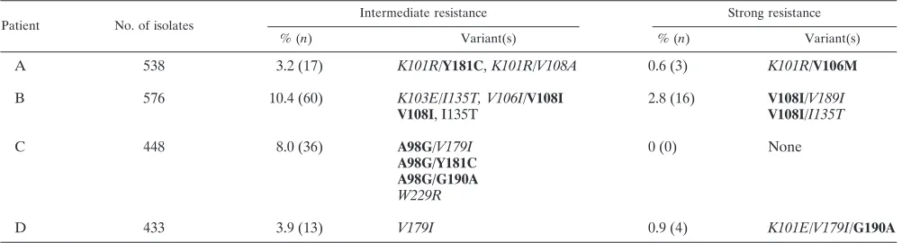
[image:5.585.45.542.80.231.2]Plasma samples from four individuals enrolled in AIDS Clinical Trials Group Protocol 398 (17) were screened for EFV resistance (Table 5). These samples were from NNRTI-expe-rienced individuals in whom NNRTI resistance was not de-tected by standard genotyping methods. Isolates with interme-diate resistance were detected at frequencies of 3 to 10%. Strong resistance was detected at frequencies of 0 to 2.8%. Resistant isolates were sequenced to determine whether these
TABLE 3. Sensitive detection of drug-resistant variants with various percentages of K103N cDNA in the samplesa
% K103N in sample
Assay I Assay II Assay III
% Resistant
No. of isolates EFVr/no. of
isolates HIS⫹
% Resistant
No. of isolates EFVr/no. of
isolates HIS⫹
% Resistant
No. of isolates EFVr/no. of
isolates HIS⫹
100 100 278/278 100 276/276 100 296/296
50 65 190/290 53 159/302 56 181/322
25 20 56/285 21 51/245 22 71/331
10 8.3 18/216 11 31/290 7.4 20/270
5 4.4 9/204 4.0 12/299 5.1 15/292
2 2.5 7/276 1.5 4/273 1.6 5/311
1 1.6 5/313 1.2 3/261 0.65 2/309
0.4 0.33 1/307 0.40 1/247 ⬍0.36 0/275
0.1 ⬍0.35 0/285 0.34 1/291 ⬍0.30 0/330
0.01 ⬍0.28 0/358 ⬍0.34 0/295 ⬍0.36 0/275
0 ⬍0.29 0/340 ⬍0.38 0/262 ⬍0.33 0/299
aSamples provided by the Virology Quality Assurance Program of the AIDS Clinical Trials Group were analyzed to determine the sensitivity of the assay. Mixtures
of wild-type and K103N mutant virus were tested as indicated in Materials and Methods. Three independent assays (assays I to III) of the K103N–wild-type virus mixture panel were done. The percent resistance for each mixture along with the number of histidine prototrophs (histidine positive [HIS⫹]) and efavirenz-resistant (EFVr) isolates is shown.
TABLE 4. Sensitive detection of drug-resistant variants with 1% K103N–wild-type cDNA mixturea
Assay % Resistant No. of isolates
EFVr/no. of isolates
A 0.55 1/183
B 1.31 4/306
C 1.02 3/293
D 0.90 3/333
E 1.63 4/245
F 1.06 3/282
G 0.96 3/312
H 0 0/173
I 1.11 3/270
J 1.06 3/284
K 1.54 4/260
L 0.75 2/267
aSamples provided by the Virology Quality Assurance Program of the AIDS
Clinical Trials Group were analyzed to determine the sensitivity of the assay. Mixtures of wild-type and K103N mutant virus were tested as indicated in Materials and Methods. cDNA from the 1% K103N–wild-type virus mixture was amplified by PCR 12 times (assays A to L) and tested as described in the text. The percent resistance along with the number of histidine prototrophs (histidine positive [HIS⫹]) and efavirenz-resistant (EFVr) isolates is shown for each
rep-licate.
on May 15, 2020 by guest
http://jcm.asm.org/
[image:5.585.302.540.520.660.2]RTs carried NNRTI resistance alleles. Indeed, efavirenz-resis-tant isolates from the samples encoded a variety of different NNRTI resistance mutations (Table 5). Clinically relevant NNRTI resistance alleles, such as V108I, Y181C, and G190A, were detected in these samples, demonstrating that the assay identifies and isolates NNRTI-resistant variants that are present at less than 1% in HIV-1-infected individuals. Non-standard and unexpected alleles (such as K103E and V108A) that may contribute to resistance were also readily identified.
DISCUSSION
Detection of minor drug-resistant variants may have impor-tant implications for the clinical management of HIV infection and for studies dissecting the mechanisms of antiretroviral treatment failure. Our experiments demonstrate that hybrid elements composed of the yeast retrotransposon Ty1 and the RT of HIV-1 are useful tools for the detection of drug-resis-tant RTs. This phenotypic assay is highly sensitive and able to detect drug-resistant RTs present in cultured virus and patient samples. This assay also monitors the activity and drug resis-tance of both known and novel RT variants.
Comparison to other assays.The genotypic assays used to monitor HIV drug resistance include bulk sequencing (6, 13, 23, 29, 34), oligonucleotide ligation assay (11, 14), line probe assay (37), microarray hybridization (38), heteroduplex forma-tion (1), allele-specific PCR (S. Palmer, 11th Conference on Retroviruses and Opportunistic Infections), and single-ge-nome sequencing (30). Other than allele-specific PCR, these assays are limited in their sensitivities for the detection of drug-resistant variants present at low frequencies. With the exception of sequencing approaches, these assays require a priori knowledge of the base substitutions that cause resistance so that detection of a specific genotype can be incorporated into the scheme. A drawback of genotypic assays is that they provide no phenotypic information, thus making it difficult to establish genotype-phenotype correlations for important phe-notypes such as replication capacity and drug resistance.
In contrast, the TyHRT system is highly sensitive and can be used to monitor the phenotype of any RT. The TyHRT system detects EFV-resistant viruses even when they comprise less
than 1% of the virus population (Tables 2 to 5). In a direct comparison to other genotypic assays (16), only allele-specific PCR is more sensitive than the TyHRT assay. The TyHRT assay also provides RT activity data for individual isolates so that basal RT activity as well as drug susceptibility can be established for each RT. The selective genetic readout of TyHRT is a powerful tool for the screening of viral populations to identify phenotypic variants of interest that can then be genotyped by DNA sequencing. This approach saves consider-able time, effort, and cost, especially when the variants of interest are present at a low frequency.
The phenotypic assays used to monitor HIV drug resistance entail recombinant viral vectors that are propagated in host cells (18, 22, 31, 36). Resistance testing by these assays pro-vides invaluable drug susceptibility data to researchers and clinicians for the management of HIV infection. These ap-proaches measure the cumulative phenotype of a viral popu-lation and may be used as predictors of the response to a particular treatment regimen. As routinely performed, this type of assay does not monitor the phenotype of individual virus genomes, and the phenotype of minor variants in the viral population may be obscured. Consequently, population-based phenotypic assays detect drug resistance only when resistant variants comprise a significant portion of the virus population. The TyHRT system individually assesses the phenotypes of all the RT domains present in a viral population, allowing the detection and characterization of minor variants. This ap-proach also makes it possible to determine the frequency of variants in a population and to detect drug-resistant variants before they become a significant portion of a population. Early detection of drug-resistant variants may be useful in the selec-tion of treatment opselec-tions for prevenselec-tion of the establishment of dominant resistant virus populations.
[image:6.585.43.544.81.216.2]The TyHRT system is limited in its ability to characterize other viral targets and widely used viral inhibitors. While the ability to look at RT phenotypes and NNRTI resistance inde-pendent of the effect(s) of variations elsewhere in the HIV-1 genome is advantageous for the analysis of RT activity, this assay does not provide the opportunity to assess drug resis-tance in protease, integrase, envelope, or other viral targets. It may be possible to modify TyHRT elements so that HIV-1
TABLE 5. Detection of drug-resistant virus in clinical samplesa
Patient No. of isolates
Intermediate resistance Strong resistance
% (n) Variant(s) % (n) Variant(s)
A 538 3.2 (17) K101R/Y181C,K101R/V108A 0.6 (3) K101R/V106M
B 576 10.4 (60) K103E/I135T, V106I/V108I 2.8 (16) V108I/V189I
V108I, I135T V108I/I135T
C 448 8.0 (36) A98G/V179I 0 (0) None
A98G/Y181C A98G/G190A W229R
D 433 3.9 (13) V179I 0.9 (4) K101E/V179I/G190A
aThe TyHRT system was used to analyze samples from NNRTI-experienced patients enrolled in AIDS Clinical Trials Group Protocol 398 where NNRTI-resistant
HIV-1 RT was not detected in plasma by standard genotyping methods. The number and percentage of strong and intermediate efavirenz-resistant isolates are shown for each sample. RT domain DNA from resistant isolates was isolated and sequenced to reveal the drug resistance mutations present. Established NNRTI resistance mutations are in boldface; potentially novel resistance alleles are italicized.
on May 15, 2020 by guest
http://jcm.asm.org/
protease activity is required for retrotransposition. The TyHRT assay is not restricted to NNRTI inhibitors; nonspe-cific RT inhibitors such as foscarnet and several RNase H inhibitors (4) block TyHRT activity (D. V. Nissley, unpub-lished data).S. cerevisiaestrains do not take up or phosphor-ylate nucleosides efficiently, which prevents the nucleoside re-verse transcriptase inhibitors (NRTIs) and NRTI-resistant variants from being analyzed by the TyHRT assay. However, the recent cloning and functional analysis of purine and py-rimidine transporters in S. cerevisiae (8, 39) should make it possible to construct strains where TyHRT activity is sensitive to NRTIs.
Sensitivity of TyHRT system.The TyHRT system is sensi-tive; NNRTI-resistant RT variants from cultured virus as well as from patient samples are detected when they are present at frequencies down to 0.4%. This level of sensitivity is a signif-icant improvement compared to that of most resistance assays currently used and is 25- to 50-fold more sensitive than stan-dard bulk genotype analyses, which detect resistant variants when they are present at frequencies of 10 to 20%. The sen-sitivity of any assay is limited by two factors: the number of isolates that it is practical to work with and the background signal generated by the experimental protocol. With the TyHRT system, the phenotypes of several hundred RT isolates per sample are easily monitored, allowing the detection of variants present at frequencies of 1% or less. In theory, the sensitivity of the TyHRT assay is also limited by the number of RT isolates assayed. For example, the failure to detect drug resistance in 1 of 15 samples derived from the 1% K103N-wild type mixture (Table 4, assay H) is most likely due to the fact that only 173 isolates were examined.
The sensitivity of the TyHRT assay is limited in practical terms by “background” drug resistance, which becomes an issue at frequencies below 0.5%. Amplification of the viral vectors used to generate viral stocks resulted in 1 EFV-tant variant of 1,243 (0.08%) isolates, suggesting that resis-tance due to PCR-induced errors occurs at a rate of about 0.1%. Although this background may be lowered by employing higher-fidelity polymerases, any resistance assay that uses PCR to amplify viral genomes will be limited by PCR-mediated error. The TyHRT system is unique in that it is sensitive enough to determine the contribution of PCR-induced errors to the frequency of resistance.
In examining the unexpected resistant isolates (i.e., those whose resistance was unexpected because it was due to an allele other than K103N), detected in the wild-type virus stock and the 0.4% and 2.0% K103N mixtures (Table 2), it is evident that the total background of the assay is about 0.2% (seven resistant RTs in 3,212 isolates [0.22%]). This background is likely the sum of the error rates from several sources. Follow-ing extraction, viral RNA undergoes an in vitro RT reaction. RT-PCR of viral RNA may result in a higher error rate than PCR of vector DNA. It is also important to note that viral RNA transcription in host cells potentially introduces errors during the generation of virus stocks. Therefore, the 0.2% resistance background seen in the viral mixtures is likely the result of the combined error rates in transcription, in vitro reverse transcription, and amplification by PCR.
NNRTI resistance. Many established EFV- and NNRTI-resistant variants (K103N, V108I, Y181C, G190A) were
iso-lated from virus cultured in vitro and from clinical samples. Not surprisingly, the TyHRT system also detected potentially novel NNRTI variants. Some of these variants, such as those with A98G, I135L, and I135T (3), have been recognized as contributing to NNRTI resistance. Other variants, such as those with V108A, K103E, V179I, W229R, and M230I, have changes at standard resistance-associated codons but not the known resistance allele. These variations are distinguished from neutral polymorphisms by correlation of the variations in isolates that are phenotypically resistant but that are not present in drug-susceptible isolates. Some of these RT variants may not efficiently replicate virus. The replicative capacities of these variant RTs are being tested by using host cell-based viral replication assays. The TyHRT activity phenotypes and repli-cation capacities in viruses are similar for the RTs tested (28), suggesting that the assay accurately measures HIV-1 RT ac-tivity.
Characterization of the NNRTI resistance mutations in Table 5 demonstrates the ability of the TyHRT assay to dis-tinguish NNRTI-specific phenotypic patterns. The primary re-sistance mutations Y181C and G190A are known to confer high-level resistance to nevirapine and lower-level resistance to efavirenz. As expected, the presence of Y181C in patient A and, likewise, the presence of Y181C and G190A in patient C confer intermediate levels of resistance to efavirenz. Both Y181C and G190A confer strong resistance to nevirapine in a TyHRT analysis of the drug resistance patterns that arise fol-lowing single-dose nevirapine therapy (12). It is notable that several mutations that confer intermediate levels of resistance to nevirapine alone (V108I and G190A) confer strong levels of resistance when they are paired with other mutations that are also expected to confer intermediate resistance (I135T, V189I, and K101E). This synergistic or additive effect of minor mul-tiple resistance mutations is most easily detected in a pheno-typic assay. Other primary NNRTI resistance mutations, such as K103N and M230L (strong resistance to both nevirapine and efavirenz), Y188C (intermediate resistance to efavirenz, strong resistance to nevirapine), and Y188H (intermediate re-sistance to efavirenz, weak rere-sistance to nevirapine), confer the expected phenotypic patterns in the TyHRT assay (12; unpub-lished data).
The detection of unexpected resistance variants demon-strates the capability of the TyHRT system to monitor the evolution of drug resistance and assess the status of a viral population at any point in time. The major resistance-associ-ated variations selected for by propagation of virus in the presence of NNRTIs and those present at therapy failure are well established. Less is known about the spectrum of RTs present in virus populations undergoing selective pressure prior to the emergence of the major resistance-associated vari-ations. The sensitivity and nonspecificity of the TyHRT system make it an ideal assay for monitoring the changes in a repli-cating viral population.
For the same reasons, the TyHRT system might be a useful diagnostic tool for the detection of drug resistance in HIV-infected individuals. In the initial studies presented here, NNRTI-resistant variants were detected at frequencies of less than 1% in reconstituted plasma and in samples from HIV-1-infected individuals. We are further testing this assay’s po-tential by examining the prevalence of EFV resistance in
on May 15, 2020 by guest
http://jcm.asm.org/
NNRTI-experienced and NNRTI-naive patients (J. Mellors, unpublished data).
Summary. The TyHRT system is a powerful yet simple method. The analysis of HIV-1 RT activity and drug resistance is accomplished in a model genetic organism; it is inexpensive and easily carried out. The assay requires no biocontainment facilities or facilities for the propagation of virus. The technical requirements for the assay are limited to the extraction and amplification of viral nucleic acid; the other steps require only basic microbiology techniques. The methods may be particu-larly useful for basic research and clinical diagnostics where early detection of resistance is important.
ACKNOWLEDGMENTS
We thank Sarah Palmer and Mary Kearney for assistance in prepar-ing viral cDNA. Thanks to Alison J. Rattray, Fransisco Malagon, and Sharon P. Moore for helpful comments.
This publication has been funded in part with federal funds from the National Cancer Institute, National Institutes of Health, under con-tract no. NO1-CO-12400.
The content of this publication does not necessarily reflect the views or policies of the Department of Health and Human Services, nor does mention of trade names, commercial products, or organizations imply endorsement by the U.S. Government.
REFERENCES
1.Barlow, K. L., J. Green, and J. P. Clewley.2000. Viral genome characteri-sation by the heteroduplex mobility and heteroduplex tracking assays. Rev. Med. Virol.10:321–335.
2.Boyer, P. L., A. L. Ferris, and S. H. Hughes.1992. Cassette mutagenesis of the reverse transcriptase of human immunodeficiency virus type 1. J. Virol.
66:1031–1039.
3.Brown, A. J., H. M. Precious, J. M. Whitcomb, J. K. Wong, M. Quigg, W. Huang, E. S. Daar, R. T. D’Aquila, P. H. Keiser, E. Connick, N. S. Hellmann, C. J. Petropoulos, D. D. Richman, and S. J. Little.2000. Reduced suscep-tibility of human immunodeficiency virus type 1 (HIV-1) from patients with primary HIV infection to nonnucleoside reverse transcriptase inhibitors is associated with variation at novel amino acid sites. J. Virol.74:10269–10273. 4.Budihas, S. R., I. Gorshkova, S. Gaidamakov, A. Wamiru, M. K. Bona, M. A. Parniak, R. J. Crouch, J. B. McMahon, J. A. Beutler, and S. F. Le Grice.
2005. Selective inhibition of HIV-1 reverse transcriptase-associated ribonucle-ase H activity by hydroxylated tropolones. Nucleic Acids Res.33:1249–1256. 5.Coffin, J. M.1995. HIV population dynamics in vivo: implications for genetic
variation, pathogenesis, and therapy. Science267:483–489.
6.Cunningham, S., B. Ank, D. Lewis, W. Lu, M. Wantman, J. A. Dileanis, J. B. Jackson, P. Palumbo, P. Krogstad, and S. H. Eshleman.2001. Performance of the applied biosystems ViroSeq human immunodeficiency virus type 1 (HIV-1) genotyping system for sequence-based analysis of HIV-1 in pediat-ric plasma samples. J. Clin. Microbiol.39:1254–1257.
7.Curcio, M. J., and D. J. Garfinkel.1991. Single-step selection for Ty1 ele-ment retrotransposition. Proc. Natl. Acad. Sci. USA88:936–940. 8.Damaraju, S., J. Zhang, F. Visser, T. Tackaberry, J. Dufour, K. M. Smith, M.
Slugoski, M. W. Ritzel, S. A. Baldwin, J. D. Young, and C. E. Cass.2005. Identification and functional characterization of variants in human concen-trative nucleoside transporter 3, hCNT3 (SLC28A3), arising from single nucleotide polymorphisms in coding regions of the hCNT3 gene. Pharma-cogenet. Genomics15:173–182.
9.Demeter, L., and R. Haubrich.2001. International perspectives on antiretroviral resistance. Phenotypic and genotypic resistance assays: methodology, reliability, and interpretations. J. Acquir. Immune Defic. Syndr.26(Suppl. 1):S3–S9. 10.Dybul, M., A. S. Fauci, J. G. Bartlett, J. E. Kaplan, and A. K. Pau.2002.
Guidelines for using antiretroviral agents among HIV-infected adults and adolescents. Ann. Intern. Med.137:381–433.
11.Edelstein, R. E., D. A. Nickerson, V. O. Tobe, L. A. Manns-Arcuino, and L. M. Frenkel.1998. Oligonucleotide ligation assay for detecting mutations in the human immunodeficiency virus type 1polgene that are associated with resistance to zidovudine, didanosine, and lamivudine. J. Clin. Microbiol.
36:569–572.
12.Flys, T., D. V. Nissley, C. W. Claasen, D. Jones, C. Shi, L. A. Guay, P. Musoke, F. Mmiro, J. N. Strathern, J. B. Jackson, J. R. Eshleman, and S. H. Eshleman.2005. Sensitive drug-resistance assays reveal long-term persis-tence of HIV-1 variants with the K103N nevirapine (NVP) resistance mu-tation in some women and infants after the administration of single-dose NVP: HIVNET 012. J. Infect. Dis.192:24–29.
13.Fontaine, E., C. Riva, M. Peeters, J. C. Schmit, E. Delaporte, K. Van Laethem, K.
Van Vaerenbergh, J. Snoeck, E. Van Wijngaerden, E. De Clercq, M. Van Ranst, and A. M. Vandamme.2001. Evaluation of two commercial kits for the detection of genotypic drug resistance on a panel of HIV type 1 subtypes A through J. J. Acquir. Immune Defic. Syndr.28:254–258.
14.Frenkel, L. M., L. E. Wagner II, S. M. Atwood, T. J. Cummins, and S. Dewhurst.1995. Specific, sensitive, and rapid assay for human immunode-ficiency virus type 1polmutations associated with resistance to zidovudine and didanosine. J. Clin. Microbiol.33:342–347.
15.Gietz, R. D., R. H. Schiestl, A. R. Willems, and R. A. Woods.1995. Studies on the transformation of intact yeast cells by the LiAc/SS-DNA/PEG pro-cedure. Yeast11:355–360.
16.Halvas, E., G. Adrovandi, J. P. Balfe, I. Beck, V. Boltz, L. Frenkel, J. Hazelwood, V. Johnson, M. Kearney, A. Kovacs, D. Kuritzkes, K. Metzner, D. Nissley, M. Nowicki, R. Ziermann, Y. Zhao, C. Jennings, J. Bremer, D. Brambilla, and J. Mellors.2003. Updated, blinded, multicentre comparison of the sensitivity of different technologies to detect and quantify a minor drug-resistant HIV-1 variant. Antivir. Ther.8:S102.
17.Hammer, S. M., F. Vaida, K. K. Bennett, M. K. Holohan, L. Sheiner, J. J. Eron, L. J. Wheat, R. T. Mitsuyasu, R. M. Gulick, F. T. Valentine, J. A. Aberg, M. D. Rogers, C. N. Karol, A. J. Saah, R. H. Lewis, L. J. Bessen, C. Brosgart, V. DeGruttola, and J. W. Mellors.2002. Dual vs single protease inhibitor therapy following antiretroviral treatment failure: a randomized trial. JAMA288:169–180.
18.Hertogs, K., M. P. de Bethune, V. Miller, T. Ivens, P. Schel, A. Van Cauwenberge, C. Van Den Eynde, V. Van Gerwen, H. Azijn, M. Van Houtte, F. Peeters, S. Staszewski, M. Conant, S. Bloor, S. Kemp, B. Larder, and R. Pauwels.1998. A rapid method for simultaneous detection of phenotypic resistance to inhibitors of protease and reverse transcriptase in recombinant human immunodeficiency virus type 1 isolates from patients treated with antiretroviral drugs. Antimicrob. Agents Chemother.42:269–276. 19.Hirsch, M. S., F. Brun-Vezinet, B. Clotet, B. Conway, D. R. Kuritzkes, R. T.
D’Aquila, L. M. Demeter, S. M. Hammer, V. A. Johnson, C. Loveday, J. W. Mellors, D. M. Jacobsen, and D. D. Richman. 2003. Antiretroviral drug resistance testing in adults infected with human immunodeficiency virus type 1: 2003 recommendations of an International AIDS Society—USA Panel. Clin. Infect. Dis.37:113–128.
20.Hizi, A., C. McGill, and S. H. Hughes.1988. Expression of soluble, enzy-matically active, human immunodeficiency virus reverse transcriptase in Escherichia coli and analysis of mutants. Proc. Natl. Acad. Sci. USA85:
1218–1222.
21.Hoffman, C. S., and F. Winston.1987. A ten-minute DNA preparation from yeast efficiently releases autonomous plasmids for transformation of Esche-richia coli. Gene57:267–272.
22.Jarmy, G., M. Heinkelein, B. Weissbrich, C. Jassoy, and A. Rethwilm.2001. Phenotypic analysis of the sensitivity of HIV-1 to inhibitors of the reverse transcriptase, protease, and integrase using a self-inactivating virus vector system. J. Med. Virol.64:223–231.
23.Lindstrom, A., and J. Albert.2003. A simple and sensitive ‘in-house’ method for determining genotypic drug resistance in HIV-1. J. Virol. Methods107:45–51. 24.Mansky, L. M., and H. M. Temin.1995. Lower in vivo mutation rate of human immunodeficiency virus type 1 than that predicted from the fidelity of purified reverse transcriptase. J. Virol.69:5087–5094.
25.Mellors, J., S. Palmer, D. V. Nissley, M. Kearney, E. Halvas, C. Bixby, L. Demeter, S. Eshleman, K. Bennett, S. Hart, F. Vaida, M. Wantman, J. Coffin, and S. Hammer.2003. Low frequency non-nucleoside reverse tran-scriptase inhibitor (NNRTI)-resistant variants contribute to failure of efa-virenz-containing regimens in NNRTI-experienced patients with negative standard genotypes for NNRTI mutations. Anitivir. Ther.8:S150. 26.Nissley, D. V., P. L. Boyer, D. J. Garfinkel, S. H. Hughes, and J. N. Strathern.1998.
Hybrid Ty1/HIV-1 elements used to detect inhibitors and monitor the ac-tivity of HIV-1 reverse transcriptase. Proc. Natl. Acad. Sci. USA 95:
13905–13910.
27.Nissley, D. V., D. J. Garfinkel, and J. N. Strathern.1996. HIV reverse transcription in yeast. Nature380:30.
28.Nissley, D. V., J. Julias, T. Flys, S. H. Hughes, J. Mellors, J. B. Jackson, J. N. Strathern, and S. H. Eshleman.2005. A sensitive phenotypic assay uncovers low frequency NNRTI-resistant HIV-1 RT variants in subtypes A, B, C and D from clinical samples. Antivir. Ther.10:S149.
29.O’Meara, D., K. Wilbe, T. Leitner, B. Hejdeman, J. Albert, and J. Lundeberg.2001. Monitoring resistance to human immunodeficiency virus type 1 protease inhibitors by pyrosequencing. J. Clin. Microbiol.39:464–473.
30.Palmer, S., M. Kearney, F. Maldarelli, E. K. Halvas, C. J. Bixby, H. Bazmi, D. Rock, J. Falloon, R. T. Davey, Jr., R. L. Dewar, J. A. Metcalf, S. Hammer, J. W. Mellors, and J. M. Coffin.2005. Multiple, linked human immunodeficiency virus type 1 drug resistance mutations in treatment-experienced patients are missed by standard genotype analysis. J. Clin. Microbiol.43:406–413.
31.Petropoulos, C. J., N. T. Parkin, K. L. Limoli, Y. S. Lie, T. Wrin, W. Huang, H. Tian, D. Smith, G. A. Winslow, D. J. Capon, and J. M. Whitcomb.2000. A novel phenotypic drug susceptibility assay for human immunodeficiency virus type 1. Antimicrob. Agents Chemother.44:920–928.
32.Rose, M. D., F. Winston, and P. Hieter.1990. Methods in yeast genetics: a laboratory manual. Cold Spring Harbor Laboratory Press, Plainview, N.Y.
on May 15, 2020 by guest
http://jcm.asm.org/
33.Shafer, R. W.2002. Genotypic testing for human immunodeficiency virus type 1 drug resistance. Clin. Microbiol. Rev.15:247–277.
34.Shafer, R. W., K. Hertogs, A. R. Zolopa, A. Warford, S. Bloor, B. J. Betts, T. C. Merigan, R. Harrigan, and B. A. Larder.2001. High degree of inter-laboratory reproducibility of human immunodeficiency virus type 1 protease and reverse transcriptase sequencing of plasma samples from heavily treated patients. J. Clin. Microbiol.39:1522–1529.
35.Sharon, G., T. J. Burkett, and D. J. Garfinkel.1994. Efficient homologous recombination of Ty1 element cDNA when integration is blocked. Mol. Cell. Biol.14:6540–6551.
36.Shi, C., and J. W. Mellors.1997. A recombinant retroviral system for rapid in vivo analysis of human immunodeficiency virus type 1 susceptibility to reverse transcriptase inhibitors. Antimicrob. Agents Chemother.41:2781–2785.
37.Stuyver, L., A. Wyseur, A. Rombout, J. Louwagie, T. Scarcez, C. Verhofstede, D. Rimland, R. F. Schinazi, and R. Rossau.1997. Line probe assay for rapid detection of drug-selected mutations in the human immunodeficiency virus type 1 reverse transcriptase gene. Antimicrob. Agents Chemother. 41:
284–291.
38.Vahey, M., M. E. Nau, S. Barrick, J. D. Cooley, R. Sawyer, A. A. Sleeker, P. Vickerman, S. Bloor, B. Larder, N. L. Michael, and S. A. Wegner.1999. Per-formance of the Affymetrix GeneChip HIV PRT 440 platform for antiretroviral drug resistance genotyping of human immunodeficiency virus type 1 clades and viral isolates with length polymorphisms. J. Clin. Microbiol.37:2533–2537. 39.Wormit, A., M. Traub, M. Florchinger, H. E. Neuhaus, and T. Mohlmann.
2004. Characterization of three novel members of the Arabidopsis thaliana equilibrative nucleoside transporter (ENT) family. Biochem. J.383:19–26.