JOURNAL OFCLINICALMICROBIOLOGY, Apr. 2005, p. 1694–1698 Vol. 43, No. 4 0095-1137/05/$08.00⫹0 doi:10.1128/JCM.43.4.1694–1698.2005
Copyright © 2005, American Society for Microbiology. All Rights Reserved.
Analysis of the Genetic Structure of Nontypeable Pneumococcal
Strains Isolated from Conjunctiva
Sonsoles Berro
´n,* Asuncio
´n Fenoll, Montserrat Ortega, Noemı´ Arellano, and Julio Casal
National Centre for Microbiology—National Institute of Health Carlos III, 28220 Majadahonda (Madrid), Spain
Received 23 April 2004/Returned for modification 17 August 2004/Accepted 7 October 2004
More than 50% of the nontypeable (NT) pneumococcal strains received in our laboratory for reference purposes are isolated in sporadic cases of conjunctivitis. To determine the genetic structure of the population of these NT conjunctival strains, we analyzed 75 pneumococci (40 NT and 35 typeable) isolated from conjunc-tivas and 30 (15 NT and 15 typeable) isolated from other sources. The NT and typeable conjunctival strains grouped in separate clusters, whereas NT and typeable pneumococci isolated from other sources were similarly distributed. NT conjunctival strains belonged to two well-differentiated clonal lineages. The first, represented by three newly described sequence types, featured fully antibiotic susceptible strains and appeared to be characteristic of conjunctival tissue; the second, represented by the previously described ST344, had a pattern of multiresistance to penicillin, tetracycline, and erythromycin and shared a genetic background with some NT strains isolated from other sources.
Streptococcus pneumoniaeis an important pathogenic bacte-rium associated with pneumonia, septicemia, meningitis, and otitis. It is also a common cause of acute conjunctivitis, partic-ularly in children, but also in adults (11, 19). For reference purposes, our laboratory receives many pneumococcus sam-ples from all origins that have been isolated in Spanish hospi-tals (8). Around one-third of the strains isolated from children below the age of 6 months that were studied in our laboratory between 1990 and 1999 caused acute conjunctivitis (9).
Pneumococcal serotyping usually fails to detect a small num-ber of strains that do not react with antipneumococcal typing sera. Nontypeable (NT) strains are infrequently isolated from sterile clinical specimens (2.2%), in which case they are rarely implicated as causes of invasive disease (2, 12), since they are otherwise relatively common in nonsterile samples (10%). The identification of these NT pneumococci is dubious (14, 17), particularly in nonsterile specimens, and they may be confused with otherStreptococcusspecies. The association between the presence of NT isolates and the occurrence of conjunctivitis was first suggested in 1977 (10) in a retrospective study of the incidence of capsular types in a Boston hospital between 1935 and 1974.
Further studies associated NT strains with outbreak and sporadic cases of conjunctivitis (1, 6, 18, 20), and a recent report has confirmed NTS. pneumoniae-like strains isolated from an outbreak of epidemic conjunctivitis as beingS. pneu-moniae(3).
In the last 10 years, approximately 50% of the NT pneumo-coccal strains received in our laboratory have been isolated from cases of conjunctivitis, and the frequency of these non-capsular strains was five times that found in other pathologies (laboratory data).
In general, the NT strains have been characterized in cases
related to outbreaks, but as yet we have little information concerning those strains isolated from sporadic cases.
The purpose of this study was to characterize NT pneumo-coccal strains isolated from conjunctivas in sporadic cases of conjunctivitis in Spain between 1997 and 2002. The overall objective was to determine whether the population genetic structure of this group of strains was similar to that found in typeable strains isolated from conjunctivas. In addition, the genetic relatedness of NT isolates from conjunctivas and NT strains from other origins was also analyzed. Pulsed-field gel electrophoresis (PFGE) (15) and multilocus sequence typing (MLST) (5) molecular markers were used for these pneumo-coccal strains.
MATERIALS AND METHODS
Strains. (i) Identification and typing.A total of 14,650 pneumococcal isolates
were received in our laboratory between 1997 and 2002. Of these, 1,068 strains were isolated from conjunctivas.
All isolates were identified asS. pneumoniaeby their distinctive colony mor-phology on sheep blood agar and were confirmed by the optochin susceptibility and bile solubility tests.
Serotyping was initially performed with a dot blot assay as previously described (7), using 46 anticapsular sera provided by the Statens Serum Institut (Copen-hagen, Denmark). Strains that gave uncertain results with this technique were typed by the Quellung reaction. Strains that did not react at all with any sera were classified as NT.
All NT strains were confirmed asS. pneumoniaeby using the AccuProbeS. pneumoniaeidentification kit (Gene Probe, Inc., San Diego, Calif.) (4).
(ii) Strains analyzed by PFGE.Thirty-five NT and 25 typeable strains isolated
from conjunctivas of different geographical origins within Spain between 1997 and 2000 were chosen at random to be analyzed by PFGE.
(iii) Strains analyzed by MLST.Sixty pneumococcal strains were analyzed by
MLST. Fifteen of these (5 NT and 10 typeable strains) represented the most frequent profiles obtained by PFGE analysis. Another 15 (10 NT and 5 typeable strains) were selected from the conjunctival strains isolated in 2001 and 2002. Finally, 30 strains (15 NT and 15 typeable) were selected from isolates recovered from other sources during the same period.
PFGE.Genomic DNA was prepared in agarose blocks as previously described
(13) and digested with SmaI (MBI Fermentans, Quimigranel, Spain) in the recommended restriction buffer. PFGE was performed in 1% agarose MP (Roche Diagnostics Corporation, Indianapolis, Ind.) in 0.5⫻Tris-borate-EDTA buffer at 12°C and 6 V/cm in a CHEF-DR II (Bio-Rad Laboratories, Hercules, Calif.) for 22 h with a switching time of 0.1 to 40 s.
* Corresponding author. Mailing address: Laboratory for Pneumo-cocci, National Centre for Microbiology—National Institute of Health Carlos III, 28220 Majadahonda (Madrid), Spain. Phone: 34918223620. Fax: 34915097966. E-mail: [email protected].
1694
on May 16, 2020 by guest
http://jcm.asm.org/
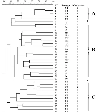
Band patterns were compared by using the Molecular Analyst Fingerprint (Bio-Rad Laboratories). A dendrogram was generated from the data by the unweighted-pair group method using average linkages, with the Dice coefficient and a tolerance of 1% (see Fig. 2).
MLST.Briefly, internal fragments of thearoE,gdh,gki,recP,spi,xpt, andddl
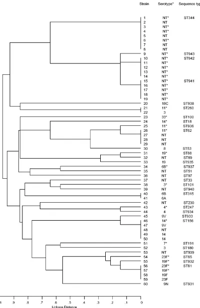
genes were amplified and then sequenced in each direction with primers de-scribed by Enright and Spratt (5). The sequences of each of the seven loci were compared with those of all the known alleles, by using the programs available on the MLST website (http://www.mlst.net/) to assign allele numbers. The same software was used to define the allelic profile and sequence type (ST) of each isolate. Data were analyzed, and the appropriate dendrogram was generated (see Fig. 3), with a sequence type analysis and recombinational test.
RESULTS
The conjunctival strains represented 7.2% of all pneumo-coccal isolates studied between 1997 and 2002. This percentage is similar to that found for otic strains (6.9%), while invasive isolates (i.e., isolates from blood or cerebrospinal fluid) recov-ered in the same period represented 45%.
NT strains were the most frequent (23.2%) of the conjunc-tival isolates. This NT percentage is higher than those found for strains isolated from other sources (Fig. 1). The most rep-resentative serotypes of the pneumococci causing conjunc-tivitis in Spain during the period 1997 to 2002 were 19A/F (14.1%), 6B (9.8%), 23F (8.8%), 14 (5.3%), 6A (4.6%), 23A/D (3.7%), and 11 (3.3%). This distribution does not differ signif-icantly from that of serotypes found among pneumococci iso-lated from cases of invasive disease.
PFGE analysis. Digestion of DNA with SmaI yielded a
total of 39 different band pattern profiles. Nineteen of the 35 (54.3%) NT strains from conjunctivas corresponded to only four closely related profiles, while the 25 typeable strains cor-responded to 22 profiles (Fig. 2).
In general, the isolates were distributed in clusters in accor-dance with their typeability. NT isolates appeared in two clus-ters, A and C (Fig. 2), and the typeable pneumococci were grouped in cluster B.
MLST typing analysis.The relationships based on the allelic
profiles of the 60 pneumococcal isolates analyzed by MLST are shown in Fig. 3. Thirty-three STs were found. Nevertheless, all
of the NT strains isolated from conjunctivas belonged to only four STs, three of which (ST941, ST942, and ST943) were closely related. The pneumococci grouped in these three re-lated STs belonged to PFGE cluster A (Fig. 2), and their allelic profiles shared six of the seven alleles analyzed. However, these strains shared only two or three of their alleles with the fourth profile (ST344) (Fig. 3).
The NT pneumococcal strains isolated from other sources were mainly distributed in other STs (Fig. 3) in the dendro-gram. It was particularly striking that the three related STs corresponded solely to NT strains isolated from conjunctivas, while ST344 grouped not only NT pneumococci isolated from conjunctivas but also other NT strains isolated from different sources. Likewise, typeable strains isolated from conjunctivas had no particular distribution pattern, appearing in different STs with typeable strains isolated from other sources.
DISCUSSION
Sporadic conjunctivitis has traditionally been associated with different microorganisms (11), including typeable pneumococ-cal strains (10). However, NT pneumococpneumococ-cal strains have been proposed to be an important cause of infection, particularly in outbreaks of conjunctivitis (18, 20). Nontypeability may result from a loss of capsular material due to specific but poorly understood mechanisms (20). In fact, to express little or no capsular polysaccharide can be advantageous in that it allows the colonization of several tissues, including the conjunctival surface (1, 21).
[image:2.585.120.466.68.268.2]In this study, a population of NT pneumococcal strains iso-lated from conjunctivas in sporadic cases was analyzed by com-paring it with other populations of strains (typeable and NT) isolated from conjunctivas and other sources. Between 20.5 and 28.7% of the conjunctival pneumococci studied in our laboratory were NT (Fig. 1), a frequency similar to that de-scribed by other authors (10, 20). This contrasts with the low percentage (2.2%) of NT strains isolated from other sources FIG. 1. Percentages of nontypeable pneumococcal strains isolated from different sources in Spain (1997 to 2002).
on May 16, 2020 by guest
http://jcm.asm.org/
(2) and with the infrequent appearance of NT strains in inva-sive diseases (12).
According to the data obtained by PFGE (Fig. 2), NT con-junctival strains were distributed in two separate clusters, A and C, which were well differentiated from that of the typeable isolates of the same origin. This suggests that NT strains iso-lated from conjunctivas may possess particular genetic charac-teristics suggesting a clonal nature.
On the basis of MLST, NT strains from conjunctivas again formed a group separate not only from the typeable strains isolated from conjunctivas but also from typeable strains of other origins, thereby yielding two distinct and well-differenti-ated clonal lineages, one made up of STs 941, 942, and 943, and one consisting of ST344 (Fig. 3). However, most of the patterns found in NT strains isolated from other sources were grouped with typeable isolates and had common or closely related STs. These may be considered pneumococci with sim-ilar genetic backgrounds that express little or no capsular poly-saccharide.
The population structure of the typeable pneumococci iso-lated from eyes is similar to that found in all pneumococcal strains (13). Throughout the dendrogram, typeable strains
iso-lated from eyes and from other sources were grouped within the same clonal lineages. Some typeable pneumococcal strains are thus able to survive in conjunctivas and give rise to sporadic cases of conjunctivitis. NT strains of STs 941, 942, 943, and 344 might be better adapted to the colonization of eye tissue. However, nontypeability alone cannot explain the specific lo-cation of these pneumococcal strains in the eyes, since most of the NT pneumococci isolated from other origins did not belong to these two clonal lineages. The isolates belonging to ST344 (Fig. 3) showed a typical pattern of multiresistance to penicil-lin, tetracycline, and erythromycin, while the other NT isolates (ST941, -942, and -943), grouped in the other cluster, were all susceptible. ST344 falls within cluster C by PFGE. Two strains with this ST, isolated from blood, have been described in the MLST database previously: one had the multiresistant pattern, and the other was resistant to penicillin but not to erythromy-cin. Therefore, NT ST344 pneumococci might not represent a clonal lineage exclusive to the conjunctiva. In fact, in our study, some NT strains from other sources were found in this ST. To address this matter, a large number of strains of this specific ST need to be analyzed.
[image:3.585.129.460.69.443.2]By contrast, the other NT pneumococci isolated from the FIG. 2. Dendrogram derived from cluster analysis of PFGE pattern profiles of typeable and nontypeable pneumococcal strains isolated from conjunctivas. Clusters A, B, and C are indicated by braces.
1696 BERRO´ N ET AL. J. CLIN. MICROBIOL.
on May 16, 2020 by guest
http://jcm.asm.org/
FIG. 3. Dendrogram derived from cluster analysis of allelic profiles generated by MLST analysis of typeable and nontypeable pneumococcal strains isolated from conjunctivas (marked with asterisks) and other sources.
on May 16, 2020 by guest
http://jcm.asm.org/
eye, which grouped together in three closely related STs, cor-responded to cluster A and might represent a characteristic conjunctival-tissue cluster. However, other authors have not found specific clusters among NT strains isolated from spo-radic cases of conjunctivitis (1). In the previous study, BOX-PCR was used to characterize the isolates; this methodology might not reflect the genetic structure of the pneumococcal population analyzed as well as MLST does.
The NT strains belonging to the two clonal lineages de-scribed in this study might represent Spanish endemic pneu-mococcal clones involved in conjunctivitis cases over a long period. In fact, several other NT pneumococci isolated from conjunctivas in Spain many years before, in the 1980s, also belong to the same clonal lineages (laboratory data). Two previous studies have shown that closely related NT strains were responsible for conjunctivitis outbreaks in different geo-graphical areas over at least 20 years (6, 16).
Further studies including a larger number of NT pneumo-coccal strains isolated from conjunctivas and other tissues from different and widely separated countries are required to con-firm our findings. Otherwise, we cannot discount the possibility that these NT pneumococci represent a group genetically di-vergent from typical pneumococcal strains. Additional studies with these strains, including the analysis of the capsular operon but also some other genes, might better clarify their real phy-logenetic position.
ACKNOWLEDGMENTS
This work was supported by The Spanish Pneumococcal Infection Study Network, Red Tema´tica de Investigacio´n Cooperativa (G03/ 103), Ministerio de Sanidad.
We thank the laboratories that sent us the pneumococcal strains.
REFERENCES
1.Barker, J. H., D. M. Musher, R. Silberman, H. M. Phan, and D. A. Watson.
1999. Genetic relatedness among nontypeable pneumococci implicated in sporadic cases of conjunctivitis. J. Clin. Microbiol.37:4039–4041.
2.Broome, C. V., and R. R. Facklam.1981. Epidemiology of clinically
signifi-cant isolates ofStreptococcus pneumoniaein the United States. Rev. Infect. Dis.3:277–280.
3.Carvalho, M. G. S., A. G. Steigerwalt, T. Thompson, D. Jackson, and R. R.
Facklam.2003. Confirmation of nontypeableStreptococcus pneumoniae-like
organisms isolated from outbreaks of epidemic conjunctivitis as Streptococ-cus pneumoniae.J. Clin. Microbiol.41:4415–4417.
4.Denys, G. A., and R. B. Carey.1992. Identification ofStreptococcus
pneu-moniaewith a DNA probe. J. Clin. Microbiol.30:2725–2727.
5.Enright, M. C., and B. G. Spratt.1998. A multilocus sequence typing scheme
forStreptococcus pneumoniae: identification of clones associated with serious invasive disease. Microbiology144:3049–3060.
6.Ertugrul, N., M. C. Rodrı´guez-Barradas, D. M. Musher, M. A. K. Ryan, C. S.
Agin, S. J. Murphy, M. Shayegani, and D. A. Watson.1997.
BOX-poly-merase chain reaction based DNA analysis of nonserotypeableStreptococcus pneumoniaeimplicated in outbreaks of conjunctivitis. J. Infect. Dis.176:
1401–1405.
7.Fenoll, A., I. Jado, D. Vicioso, and J. Casal.1997. Dot blot assay for the
serotyping of pneumococci. J. Clin. Microbiol.35:764–766.
8.Fenoll, A., I. Jado, D. Vicioso, A. Perez, and J. Casal.1998. Evolution of
Streptococcus pneumoniaeserotypes and antibiotic resistance in Spain: up-date (1990 to 1996). J. Clin. Microbiol.36:3447–3454.
9.Fenoll, A., I. Jado, D. Vicioso, S. Berron, J. E. Yuste, and J. Casal.2000.
Streptococcus pneumoniaein children in Spain: 1990–1999. Acta Paediatr. Suppl.435:44–50.
10.Finland, M., and M. W. Barnes.1977. Changes in occurrence of capsular
serotypes ofStreptococcus pneumoniaeat Boston City Hospital during se-lected years between 1935 and 1974. J. Clin. Microbiol.5:154–166.
11.Gigliotti, F., W. T. Williams, F. G. Hayden, J. O. Hendley, J. Benjamin, M.
Dickens, R. Ford, C. Gleason, V. A. Perriello, and J. Wood.1981. Etiology of
acute conjunctivitis in children. J. Pediatr.98:531–536.
12.Gross, J., and O. Fulco.1985. Pneumococcal pneumonia and septicemia
resulting from a nontypable strain ofPneumococcus. J. Am. Geriatr. Soc.
33:153.
13.Hall, L. M.1998. Application of molecular typing to the epidemiology of
Streptococcus pneumoniae.J. Clin. Pathol.51:270–274.
14.Kaijalainen, T., S. Rintama¨ki, E. Herva, and M. Leinonen.2002. Evaluation
of gene-technological and conventional methods in the identification of
Streptococcus pneumoniae.J. Microbiol. Methods51:111–118.
15.Lefevre, J. C., G. Faucon, A. M. Sicard, and A. M. Gasc. 1993. DNA
fingerprinting ofStreptococcus pneumoniaestrains by pulsed-field gel elec-trophoresis. J. Clin. Microbiol.31:2724–2728.
16.Martin, M., J. H. Turco, M. E. Zegans, R. R. Facklam, S. Sodha, J. A. Elliott,
J. H. Pryor, B. Beall, D. D. Erdman, Y. Y. Baumgartner, P. A. Sanchez, J. D.
Schwartzman, J. Montero, A. Schuchat, and C. G. Whitney.2003. An
out-break of conjunctivitis due to atypicalStreptococcus pneumoniae.N. Engl. J. Med.348:1112–1121.
17.Mundy, L., E. Janoff, K. Schwebke, C. Shanholtzer, and K. Willard.1998.
Ambiguity in the identification ofStreptococcus pneumoniae.Optochin, bile solubility, quellung, and the AccuProbe DNA probe tests. Am. J. Clin. Pathol.109:55–61.
18.Pease, A. A., C. W. Douglas, and R. C. Spencer.1986. Identifying
non-capsulate strains ofStreptococcus pneumoniaeisolated from eyes. J. Clin. Pathol.39:871–875.
19.Perkins, R. E., R. B. Kundsin, M. V. Pratt, I. Abrahamsen, and H. M.
Leibowitz.1975. Bacteriology of normal and infected conjunctiva. J. Clin.
Microbiol.1:147–149.
20.Shayegani, M. L., M. Parson, W. E. Gibbons, Jr., and D. Campbell.1982.
Characterization of nontypeableStreptococcus pneumoniae-like organisms isolated from outbreaks of conjunctivitis. J. Clin. Microbiol.16:8–14.
21.Weiser, J. N., R. Austrian, P. K. Sreenivasan, and H. R. Masure.1994. Phase
variation in pneumococcal opacity: relationship between colonial morphol-ogy and nasopharyngeal colonization. Infect. Immun.62:2582–2589.
1698 BERRO´ N ET AL. J. CLIN. MICROBIOL.