0095-1137/05/$08.00⫹0 doi:10.1128/JCM.43.2.733–739.2005
Copyright © 2005, American Society for Microbiology. All Rights Reserved.
Comparison of Hepatitis C Virus NS5b and 5
⬘
Noncoding Gene
Sequencing Methods in a Multicenter Study
Syria Laperche,
1Franc¸oise Lunel,
2Jacques Izopet,
3Sophie Alain,
4Paul De
´ny,
5Gilles Duverlie,
6Catherine Gaudy,
7Jean-Michel Pawlotsky,
8Jean-Christophe Plantier,
9Bruno Pozzetto,
10Vincent Thibault,
11Franc¸ois Tosetti,
12and Jean-Jacques Lefre
`re
13,14*
Centre National de Re´fe´rence pour les He´patites B et C en Transfusion, De´partement des Agents Transmissibles par le Sang, Institut
National de la Transfusion Sanguine,1Laboratoire de Virologie, Centre Hospitalo-Universitaire Pitie´-Salpeˆtrie`re,11and De´partement des
Agents Transmissibles par le Sang, Institut National de la Transfusion Sanguine,14Paris, Laboratoire de Virologie-Bacte´riologie, Centre
Hospitalo-Universitaire, Angers,2Laboratoire de Virologie, Centre Hospitalo-Universitaire Purpan, Toulouse,3Laboratoire de
Virologie-Bacte´riologie, Centre Hospitalo-Universitaire, Limoges,4Laboratoire de Virologie-Bacte´riologie Associe´ au CNR des
He´patites B et C, Centre Hospitalo-Universitaire Avicenne, Bobigny,5Laboratoire de Virologie, Centre
Hospitalo-Universitaire, Amiens,6Laboratoire de Virologie-Bacte´riologie, Centre Hospitalo-Universitaire Bretonneau,
Tours,7Laboratoire de Virologie-Bacte´riologie, Centre Hospitalo-Universitaire Henri-Mondor, Cre´teil,8
Laboratoire de Virologie-Bacte´riologie, Centre Hospitalo-Universitaire, Rouen,9Laboratoire
de Virologie-Bacte´riologie, Centre Hospitalo-Universitaire, Saint Etienne,10Laboratoire
de Virologie-Bacte´riologie, Centre Hospitalo-Universitaire, Grenoble,12and
Laboratoire d’He´matologie, Centre Hospitalo-Universitaire, Amiens,13France
Received 28 July 2004/Returned for modification 16 September 2004/Accepted 10 October 2004
A national evaluation study was performed in 11 specialized laboratories with the objective of assessing their capacities to genotype hepatitis C virus (HCV) and define the applicability of a given genotyping method. The panel consisted of 14 samples positive for HCV RNA of different genotypes (including 3 samples with two different artificially mixed genotypes) and 1 HCV-negative sample. Seventeen sets of data were gathered from the 11 participating laboratories. The sensitivities ranged from 64.3 to 100% and from 42.7 to 85.7% for the
methods that used sequencing of the NS5b region and the 5ⴕnoncoding (5ⴕNC) region, respectively. When the
data for the artificially mixed samples were excluded, NS5b genotyping gave correct results for 80% of the
samples, 1.7% of the samples were misclassified, and 18.3% of the samples had false-negative results. By 5ⴕ
NC-region genotyping methods, 58.3% of the results were correct, 29.7% were incomplete, 8.3% were misclas-sifications, 1.2% were false positive, and 2.4% were false negative. Only two procedures based on NS5b se-quencing correctly identified one of the three samples with mixtures of genotypes; the other methods identified the genotype corresponding to the strain with the highest viral load in the sample. Our results suggest that
HCV 5ⴕNC-region genotyping methods give sufficient information for clinical purposes, in which the
deter-mination of the subtype is not essential, and that NS5b genotyping methods are more reliable for subtype determination, which is required in epidemiological studies.
Hepatitis C virus (HCV) is responsible for chronic liver dis-ease, with a risk of evolution toward severe diseases such as cirrhosis and hepatocellular carcinoma (34). More efficient antiviral treatments have been developed in recent years (21), but their efficacies are largely influenced by several biological parameters, such as the virus genotype. For this reason, HCV genotyping is used to predict the response to antiviral therapy (12, 23, 30) and, in association with the determination of the viral load and related markers in different hosts, to optimize the duration of treatment (2, 31). Furthermore, HCV genotyping is an essential tool for epidemiological studies (3, 22, 29) and for tracing a source of contamination by HCV (1, 18–20, 27).
HCV isolates are characterized by a high degree of hetero-geneity: six main genotypes and more than 70 subtypes have been described (35). Many genotyping methods focused on the 5⬘ noncoding (5⬘ NC) region have been developed, and some of them are commercially available. However, the ability of the
sequence of this region to discriminate isolates at the subtype level is disputed (8), and alternative genomic regions have been proposed for use in genotyping (11, 26, 33).
Thus, before the initiation of large-scale epidemiological or therapeutic studies, the Action Coordonne´e 11 group of the Agence Nationale de Recherches pour le SIDA initiated an evaluation of the HCV genotyping methods used in 11 special-ized laboratories involved in multicenter clinical trials. The aim of this study was to assess the HCV genotyping capacities of these specialized laboratories and to define the best applica-bility of a given genotyping method.
MATERIALS AND METHODS
Panel.The panel was made up of 15 samples, including 10 undiluted samples (collected from HCV-infected blood donors and selected as a subset of HCV subtypes mainly encountered in clinical practice in Europe), 1 diluted HCV-positive sample (sample 6, which was a 1-in-200 dilution of sample 5), 3 samples with mixtures of two different genotypes (to mimic coinfections), and 1 HCV-negative sample. The characteristics of these samples are given in Table 1. Each HCV-positive sample was characterized by its viral load (Amplicor HCV Mon-itor, version 2.0; Roche, Meylan, France) and by its HCV genotype, determined by a method based on sequence analysis of the NS5b region (25). Briefly, after viral RNA extraction (QI Amp Viral RNA; Qiagen, Hilden, Germany) and * Corresponding author. Mailing address: Institut National de la
Transfusion Sanguine, 6 rue Alexandre-Cabanel, 75015 Paris, France. Phone: 01 44 49 30 51. Fax: 01 44 49 30 59. E-mail: [email protected].
733
on May 16, 2020 by guest
http://jcm.asm.org/
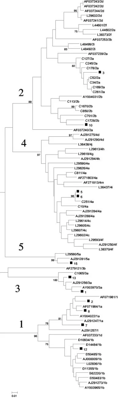
reverse transcription, performed by a random priming method (cDNA ramdom hexamers kit; Amersham Pharmacia Biotech, Orsay, France), cDNA was ampli-fied by a heminested PCR based on primers PR3 and PR4 in the first round, followed by PCR with primers PR3 and PR5 in the second round (Table 2). The DNA of each strain obtained from the purified PCR products (Quick Spin; Qiagen) was sequenced by PCR with primers PR3 and PR5. Cycle sequencing was undertaken by use of the fluorescent dye terminator technology (Big Dye terminator cycle sequencing; Applied Biosystems, Courtaboeuf, France) with AmpliTaq DNA polymerase, according to the instructions of the manufacturer. Electrophoresis and data collection were performed with an ABI 3100 genetic analyzer. The genotype of each sample was determined by comparison with those of HCV prototype strains from GenBank. Figure 1 shows the phylogenetic tree in which the samples of the panel are represented.
Among the three samples containing mixtures of genotypes, sample 11 had equivalent viral loads of two genotypes (genotypes 1a and 1b), whereas samples 9 and 14 had greater viral loads of genotypes 3a and 1b, respectively. All samples in the panel were prepared and aliquoted by an external investigator and were randomly coded and sent under appropriate transportation conditions to each participating laboratory for blind testing. Each laboratory was free to use any genotyping method of its choice.
Participating laboratories.The 11 participating laboratories were coded as laboratories A to K. Six of them used one genotyping method, four used two genotyping methods, and one used three genotyping methods, resulting in a total
of 17 results. The different genotyping methods used consisted of four in-house protocols of the NS5b sequencing assay (16, 20, 24, 33) in eight laboratories, a newly developed NS5b sequencing assay (Trugene HCV NS5b genotyping kit; Bayer Health Care Diagnostics, Puteaux, France) (28) in two laboratories, an in-house 5⬘NC region sequencing assay in two laboratories (13, 39), and two commercially available 5⬘NC region genotyping assays (Inno-LIPA [Innogenet-ics, Ghent, Belgium] and Trugene HCV genotyping kit [Bayer Health Care Diagnostics]) in five laboratories (32, 37). Table 2 includes the details for the PCR primers used only with the in-house methods.
Interpretation of results.For each HCV-positive sample, the result was con-sidered correct when both the correct genotype and the correct subtype were identified and, for samples with mixtures of genotypes, when both the genotype and the subtype of the strain with the highest viral load were identified. An incomplete result was defined as an exact genotype result with an unidentified subtype or with the absence of discrimination between two subtypes. A correct genotype in association with the incorrect subtype defined a misclassification. The sensitivity was defined as the percentage of correct results (correct genotype and correct subtype) among the 14 HCV-positive samples. The quality score was calculated by the percentage of correct results among all samples in the panel.
RESULTS
Performance of HCV genotyping. Overall, 17 sets of data
were generated by five different technical approaches (Table 3). In order to simplify the presentation, we considered each set of data as coming from an independent laboratory.
[image:2.585.44.283.79.246.2]The sensitivities ranged from 64.3 to 100% and from 42.7 to 85.7% for laboratories using the NS5b and 5⬘NC-region genotyping methods, respectively. Among the 10 NS5b region-based methods, incomplete results were observed for two (14.3%) samples, whereas the seven 5⬘NC-region-based meth-ods provided incomplete results for one to seven (0.7 to 50%) samples. Two of the NS5b-based analyses misclassified the ge-notype in one sample, while five of the 5⬘NC-region-based anal-yses misclassified the genotype in one or two samples. False-negative results were more commonly observed by NS5b-based analyses (9 of 10; 90%), which failed to identify the genotype in one to five samples. The virus in only one sample could not be genotyped by three of seven 5⬘NC-region-based methods. A false-positive result was observed for only one sample (by an in-house 5⬘NC-region-based assay). The quality scores ranged TABLE 1. Characteristics of the panel
Sample no.
Genotype (by NS5b sequencing)
HCV RNA load (log IU/ml)
1 3a 4.01
2 1a 4.93
3 2a 5.39
4 ⫺a 0
5 4a 5.62
6 4a 3.41
7 1a 3.33
8 1a 6.00
9 3a-1b 5.86/14.31
10 2b 5.44
11 1a-1b 4.93/14.31
12 1b 5.68
13 3a 5.86
14 1b-3a 5.68/4.01
15 5a 6.65
a
The sample was HCV negative.
TABLE 2. PCR primers used in NS5b-based in-house methods
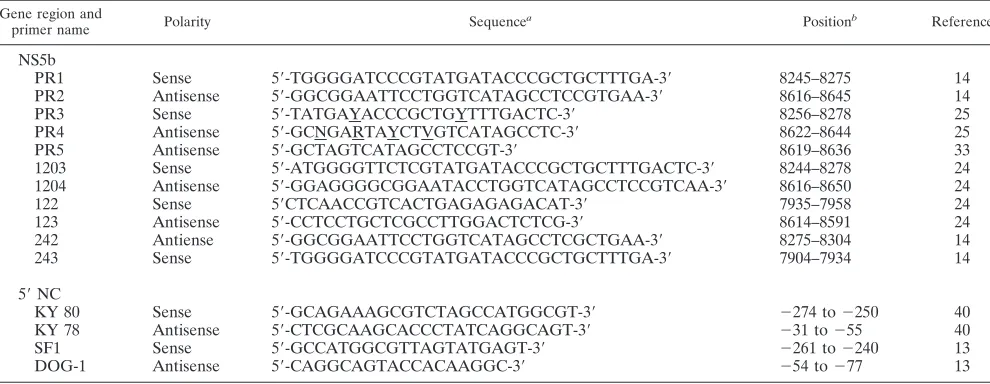
Gene region and
primer name Polarity Sequence
a Positionb Reference
NS5b
PR1 Sense 5⬘-TGGGGATCCCGTATGATACCCGCTGCTTTGA-3⬘ 8245–8275 14
PR2 Antisense 5⬘-GGCGGAATTCCTGGTCATAGCCTCCGTGAA-3⬘ 8616–8645 14
PR3 Sense 5⬘-TATGAYACCCGCTGYTTTGACTC-3⬘ 8256–8278 25
PR4 Antisense 5⬘-GCNGARTAYCTVGTCATAGCCTC-3⬘ 8622–8644 25
PR5 Antisense 5⬘-GCTAGTCATAGCCTCCGT-3⬘ 8619–8636 33
1203 Sense 5⬘-ATGGGGTTCTCGTATGATACCCGCTGCTTTGACTC-3⬘ 8244–8278 24
1204 Antisense 5⬘-GGAGGGGCGGAATACCTGGTCATAGCCTCCGTCAA-3⬘ 8616–8650 24
122 Sense 5⬘CTCAACCGTCACTGAGAGAGACAT-3⬘ 7935–7958 24
123 Antisense 5⬘-CCTCCTGCTCGCCTTGGACTCTCG-3⬘ 8614–8591 24
242 Antiense 5⬘-GGCGGAATTCCTGGTCATAGCCTCGCTGAA-3⬘ 8275–8304 14
243 Sense 5⬘-TGGGGATCCCGTATGATACCCGCTGCTTTGA-3⬘ 7904–7934 14
5⬘NC
KY 80 Sense 5⬘-GCAGAAAGCGTCTAGCCATGGCGT-3⬘ ⫺274 to⫺250 40
KY 78 Antisense 5⬘-CTCGCAAGCACCCTATCAGGCAGT-3⬘ ⫺31 to⫺55 40
SF1 Sense 5⬘-GCCATGGCGTTAGTATGAGT-3⬘ ⫺261 to⫺240 13
DOG-1 Antisense 5⬘-CAGGCAGTACCACAAGGC-3⬘ ⫺54 to⫺77 13
aY⫽C or T; R⫽A or G; V⫽A, C, or G; N⫽A, T, G, or G. bNumbering according to Choo et al. (10).
on May 16, 2020 by guest
http://jcm.asm.org/
[image:2.585.44.539.516.708.2]from 66.7 to 100% for the NS5b-based analyses and from 46.7 to 86.7% for the 5⬘NC-region-based analyses.
Comparison of NS5b- and 5ⴕNC-region-based genotyping
results by sample (excluding coinfected samples).A further
analysis that excluded the samples with artificially mixed ge-notypes was selectively performed (Table 4). By the 10 ap-proaches based on NS5b analysis, 120 results were expected. Among these, 96 (80%) results were correct. For each sample, the proportion of correct results ranged from 10 to 100%. Of the 24 incorrect results, 2 could be attributed to misclassifica-tions (in two samples) and 22 could be attributed to false-negative results (seven samples from which sequences could not be amplified by one to nine genotyping assays).
By the seven approaches based on 5⬘NC-region analysis, 84 results were expected. Among these, 49 (58.3%) results were correct. For each sample, the proportion of correct results ranged from 0 to 86%. Most (71%) of the incorrect results were due to incomplete genotype identification, while the ge-notypes in seven samples were misclassified. Two false-nega-tive results and one false-posifalse-nega-tive result were also noted.
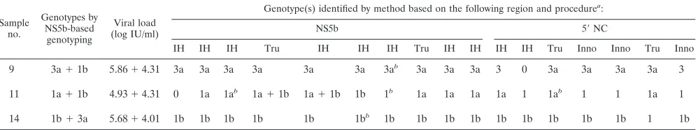
Comparison of NS5b and 5ⴕNC-region genotyping results
for coinfected samples.The results of the comparison of the
NS5b and 5⬘NC-region genotyping results for the coinfected samples are depicted in Table 5. Sample 9 (which contained genotypes 3a and 1b) was never identified as containing a mixed infection, and the genotype present in the sample was classified as 3a in 14 cases (all 10 NS5b-based analyses and 4 of the 7 5⬘NC-region-based analyses). One laboratory, using an NS5b-based method, identified the genotype 3a strain and mentioned the presence of a double population without giving the other subtype. All three incorrect results were obtained by 5⬘ NC-region-based methods: two because incomplete data were provided and one due to a lack of amplification.
Sample 11 (which contained genotypes 1a and 1b) was cor-rectly identified as containing a mixture of genotypes by two NS5b-based analyses; it was subtyped as genotype 1a in eight cases (5 of the 10 NS5b-based analyses and 3 of the 7 5⬘ NC-region-based analyses; a double population was mentioned in two cases. Among the seven remaining analyses, one provided a result of subtype 1b, five analyses could not completely char-acterize the genotype, and one gave a negative result.
Sample 14 (which contained genotypes 1b and 3a) was never identified as containing a mixture of genotypes by any of the assays; genotype 1b was detected in 16 analyses, and 1 analysis gave an incomplete result.
DISCUSSION
[image:3.585.66.259.73.735.2]The data obtained in this multicenter study have shown a wide heterogeneity of genotyping results, depending on the
FIG. 1. Phylogenetic analysis of the NS5b sequences of the samples containing HCV strains included in the panel (excluding those artifi-cially coinfected) compared with NS5b reference sequences from Gen-Bank and in-house NS5b sequences. HCV genotypes are designated by the numbers 1 to 5. Genomes from this study are indicated with black squares and with the sample number. The phylogenetic tree was con-structed by using the neighbor-joining method in the PHYLIP package (15). Bootstrap values are shown at each main branch.
on May 16, 2020 by guest
http://jcm.asm.org/
TABLE 3. Performance of the laboratories in HCV genotype determination Gene region and genotyping technique Labora- tory Primers used for PCR aor reference Primers used for sequencing or reference Results (no. of samples) for HCV RNA-positive samples ( n ⫽ 14) No. of samples with false-positive results ( n ⫽ 1) Quality score e ( n ⫽ 15) (no. [%] of samples) Correct results b Incomplete results c Misclassified samples d False-negative results NS5b In-house A PR3-PR4/PR3-PR5 PR3-PR5 10 (71.4) 0 0 4 0 11 (73.3) In-house B PR1-PR2/PR3-PR5 PR3-PR5 12 (85.7) 0 1 1 0 13 (86.7) In-house C1 PR3-PR4/PR3-PR5 PR3-PR5 12 (85.7) 0 0 2 0 13 (86.7) Trugene C2 28 13 (92.9) 0 0 1 0 14 (93.3) In-house D 1203-1204 123 9 (64.3) 0 0 5 0 10 (66.7) In-house E1 1203-1204 1203-1204 ( ⫺ 20 nt f at 3 ⬘ end) 11 (78.6) 1 0 2 0 12 (80) In-house F1 1203-1204/1203-123 1203-1204 11 (78.6) 1 0 2 0 12 (80) Trugene G1 28 12 (85.7) 0 1 1 0 13 (86.7) In-house H1 243-242/122-123 122-123 9 (64.3) 0 0 5 0 10 (66.7) In-house I PR3-PR4 PR3-PR4 14 (100) 0 0 0 0 15 (100) 5 ⬘ NC In-house E2 K80-K78 K80-K78 9 (64.3) 3 0 1 0 10 (66.7) In-house F2 K80-K78 DOG1-SF1 9 (64.3) 3 0 1 1 10 (60) Trugene G2 32 32 12 (85.7) 1 1 0 0 13 (86.7) InnoLipa G3 37 9 (64.3) 2 2 1 0 10 (66.7) InnoLipa H2 37 6 (42.7) 7 1 0 0 7 (46.7) Trugene J 32 32 6 (42.7) 7 1 0 0 7 (46.7) InnoLipa K 37 6 (42.7) 7 1 0 0 7 (46.7) aHeminested PCR or nested PCR was used when two pairs of primers are mentioned. bPercentage of correct results among the HCV RNA-positive samples. Percent sensitivity is presented in parentheses. cCorrect genotype with a subtype not identified. dCorrect genotype but incorrect subtype. eCorrect results among the 15 samples in the panel. fnt, nucleotides.
on May 16, 2020 by guest
http://jcm.asm.org/
[image:4.585.164.399.69.724.2]laboratory and the genotyping method used. Indeed, as the objective of the study was to compare the ability of expert laboratories to provide correct HCV genotyping results, no method was imposed. All laboratories used commercial RNA purification methods, and some of them used the same sets of primers; but all procedures were different. However, the per-formance variations observed allowed us to suggest two differ-ent strategies according to the HCV genotype determined.
The discrimination between major HCV genotypes, which is the strategy commonly adopted in clinical practice, was suc-cessfully performed independently of the method used by all laboratories for all samples except those containing artificially mixed genotypes. However, 5⬘NC-region genotyping-based meth-ods showed higher sensitivities. Indeed, in our study, while the assays based on analysis of the NS5b region missed 23 (16.4%) of the 140 expected positive results, 5⬘NC-region-based assays missed only 3 (3.1%) of the 98 expected positive results. Such false-negative results are probably due to the low levels if viremia in the samples tested, as described elsewhere (17, 26), as well as to the difficulty of amplifying products from samples infected with genotype 4 by NS5b-based methods (38). There-fore, unless the sensitivities of the present NS5b-based geno-typing methods are improved (especially in the choice of the primers used) or unless genotype C and E1 genotyping meth-ods are developed, as described elsewhere (4, 5, 11, 36), pro-cedures based on the 5⬘ NC-region gene (and, essentially, those that use commercial standardized assays) can be consid-ered the most adequate for genotyping in clinical practice, at least in countries where genotypes 1 to 5 are mainly encoun-tered. Indeed, it was shown that 5⬘NC-region-based genotyp-ing methods cannot distgenotyp-inguish certain isolates of genotype 6 from isolates of genotype 1 (9). This point must be empha-sized, especially for countries, such as Southeastern Asian countries, where genotype 6 is frequently found.
In the case of epidemiological studies requiring the precise determination of the HCV subtype, our results confirm that NS5b-based genotyping procedures are preferable to 5⬘ NC-region-based ones (6, 26, 33). Indeed, in our series, NS5b-based procedures were more accurate than 5⬘NC-region-based meth-ods, with the genotypes in only 2 samples misclassified by NS5b-based procedures, whereas 27 incomplete results or mis-classifications were obtained by 5⬘ NC-region-based genotyp-ing. One of these two misclassifications was for a sample in-fected with genotype 4a, which was misclassified as genotype 4c. The laboratory implicated in this misclassification observed after the study that this error was due to confusion over the nomenclature for genotype 4 in the sequence database. The database was subsequently modified to take this misclassifica-tion into account. Interestingly, we observed some previously described failures of 5⬘NC-region-based genotyping, such as misclassification of genotype 1a as genotype 1b (7, 38), the ab-sence of discrimination between subtypes 2a and 2c, and the lack of typing or subtyping of the strains in samples containing genotype 4 (38). For this reason, the use of NS5b-based geno-typing methods is preferable to the use of 5⬘NC-region-based genotyping methods for epidemiological investigations of HCV.
Although the results obtained with mixtures must be inter-preted cautiously due to their artificial constitution, we have observed that when mixtures of strains of two genotypes were present in the same sample, the identification of both
geno-TABLE 4. Results obtained for the 12 samples in the panel, infected only with single genotypes Sample no. HCV RNA load (log IU/ml) Genotype by NS5b-based genotyping No. of samples NS5b-based genotyping ( n ⫽ 10) 5 ⬘ NC-region-based genotyping ( n ⫽ 7) Correct results a Incomplete results Misclassified samples False-positive results False-negative results Correct results a Incomplete results Misclassified samples False-positive results False-negative results 1 4.01 3a 7 (70) 3 6 (86) 1 2 4.93 1a 6 (60) 4 6 (86) 1 3 5.39 2a 9 (90) 1 2 (29) 5 4 0 Negative 10 (100) 6 (86) 1 (1b) b 5 5.62 4a 7 (70) 1 (4c) b 2 2 (29) 4 1 (4c) b 6 3.41 4a 1 (10) 9 5 1 (4c) b 1 7 3.33 1a 7 (70) 1 (1b) b 2 1 5 (1b) b 1 8 6.00 1a 10 (100) 5 (71) 2 10 5.44 2b 10 (100) 5 (71) 2 12 5.68 1b 9 (90) 1 6 (86) 1 13 5.86 3a 10 (100) 5 (71) 2 15 6.65 5a 10 (100) 6 (86) 1 Total c 96/120 (80) 2/120 (1.7) 22/120 (18.3) 49/84 (58.3) 25/84 (29.7) 7/84 (8.3) 1/84 (1.2) 2/84 (2.4) a Values in parentheses in this column are in percent. b Designations in parentheses are the genotype identified. c Data represent the total number of samples with the indicated result/total number of results (percent).
on May 16, 2020 by guest
http://jcm.asm.org/
[image:5.585.317.517.73.724.2]types may be compromised if one of the two viral strains is present at a lower load. Although some participating labora-tories indicated the existence of a double population in such samples, none of them identified the genotype 1b strain in sample 9 or the genotype 3a virus in sample 14. Techniques based on direct sequencing, as well as commercial line probe assays, are not appropriate in all cases for the detection of mixtures of genotypes in the samples studied (only two NS5b-based techniques were able to discriminate genotypes 1a and 1b in sample 11).
A consensus HCV sequencing method would be useful. The divergences observed in our study could be linked to differ-ences in procedures (such as the RNA extraction methods, the primers, the types of enzymes, and the components in the mixture preparation used) and to the HCV sequence data-bases. However, the influence of each of these parameters is difficult to define in practice from the results of a multicenter study.
In conclusion, our results suggest, in agreement with previ-ous studies (6, 17, 33, 38), that HCV 5⬘NC-region-based geno-typing methods give sufficient information for clinical purposes and that NS5b-based genotyping methods are more reliable for the subtype determination required in epidemiological studies.
ACKNOWLEDGMENTS
This work was supported by a grant from the Agence Nationale de Recherches sur le SIDA et les Virus des He´patites.
We thank Franc¸oise Bouchardeau, Sandrine Castelain, Se´bastien Hantz, Nadine Le Marrec, Joe¨lle Lerable, Virginie Morel, Christo-pher Payan, Annie Razer, and Henia Saoudin for technical assistance and Camille Sureau for helpful reading of the manuscript.
REFERENCES
1.Ackerman, Z., E. Ackerman, and O. Paltiel.2000. Intrafamilial transmission of hepatitis C virus: a systematic review. J. Viral Hepat.7:93–103. 2.Anonymous.2002. Consensus conference. Treatment of hepatitis C virus.
Gastroenterol. Clin. Biol.26:B312–B320.
3.Bourliere, M., J. M. Barberin, M. Rotily, V. Guagliardo, I. Portal, L. Lecomte, S. Benali, C. Boustiere, H. Perrier, M. Jullien, G. Lambot, R. Loyer, O. LeBars, R. Daniel, H. Khiri, and P. Halfon.2002. Epidemiological changes in hepatitis C virus genotypes in France: evidence in intravenous drug users. J. Viral Hepat.9:62–70.
4.Bukh, J., R. H. Purcell, and R. H. Miller.1993. At least 12 genotypes of hepatitis C virus predicted by sequence analysis of the putative E1 gene of isolates collected worldwide. Proc. Natl. Acad. Sci. USA90:8234–8238. 5.Bukh, J., R. H. Purcell, and R. H. Miller.1994. Sequence analysis of the core
gene of 14 hepatitis C virus genotypes. Proc. Natl. Acad. Sci. USA91:
8239–8243.
6.Cantaloube, J., H. Venault, J. Zappitelli, P. Gallian, M. Touinssi, H. Attoui, P. Biagini, X. de Lamballerie, and P. de Micco.2000. Molecular analysis of HCV type 1 to 5 envelope gene: application to investigations of posttrans-fusion transmission of HCV. Transposttrans-fusion40:712–717.
7.Cantaloube, J. F., P. Gallian, H. Attoui, P. Biagini, P. De Micco, and X. De
Lamballerie.2001. Erroneous HCV genotype assignment by a hybridization typing assay in a case of posttransfusion HCV infection. Transfusion41:
429–430.
8.Chen, Z., and K. E. Weck.2002. Hepatitis C virus genotyping: interrogation of the 5⬘untranslated region cannot accurately distinguish genotypes 1a and 1b. J. Clin. Microbiol.40:3127–3134.
9.Chinchai, T., J. Labout, S. Noppornpanth, A. Theamboonlers, B. L. Haag-mans, A. D. Osterhaus, and Y. Poovorawan.2003. Comparative study of different methods to genotype hepatitis C virus type 6 variants. J. Virol. Methods109:195–201.
10.Choo, Q. L., K. H. Richman, J. H. Han, K. Berger, C. Lee, C. Dong, C. Gallegos, D. Coit, R. Medina-Selby, P. J. Barr, et al.1991. Genetic organi-zation and diversity of the hepatitis C virus. Proc. Natl. Acad. Sci. USA88:
2451–2455.
11.Corbet, S., J. Bukh, A. Heinsen, and A. Fomsgaard.2003. Hepatitis C virus subtyping by a core-envelope 1-based reverse transcriptase PCR assay with sequencing and its use in determining subtype distribution among Danish patients. J. Clin. Microbiol.41:1091–1100.
12.Davis, G. L., and J. Y. Lau.1997. Factors predictive of a beneficial response to therapy of hepatitis C. Hepatology26:122S–127S.
13.Doglio, A., C. Laffont, S. Thyss, and J. C. Lefebvre.1998. Rapid genotyping of hepatitis C virus by direct cycle sequencing of PCR-amplified cDNAs and capillary electrophoresis analysis. Res. Virol.149:219–227.
14.Enomoto, N., A. Takada, T. Nakao, and T. Date.1990. There are two major types of hepatitis C virus in Japan. Biochem. Biophys. Res. Commun.170:
1021–1025.
15.Felsenstein, J.1989. PHYLIP—phylogenic interference package (version 3.2). Cladistics5:164–166.
16.Gault, E., P. Soussan, Y. Morice, L. Sanders, A. Berrada, B. Rogers, and P. Deny.2003. Evaluation of a new serotyping assay for detection of anti-hepatitis C virus type-specific antibodies in serum samples. J. Clin. Micro-biol.41:2084–2087.
17.Germer, J. J., P. N. Rys, J. N. Thorvilson, and D. H. Persing.1999. Deter-mination of hepatitis C virus genotype by direct sequence analysis of prod-ucts generated with the Amplicor HCV test. J. Clin. Microbiol.37:2625– 2630.
18.Halfon, P., P. Trimoulet, M. Bourliere, H. Khiri, V. de Ledinghen, P. Couzi-gou, J. M. Feryn, P. Alcaraz, C. Renou, H. J. Fleury, and D. Ouzan.2001. Hepatitis C virus genotyping based on 5⬘noncoding sequence analysis (Tru-gene). J. Clin. Microbiol.39:1771–1773.
19.Izopet, J., C. Pasquier, K. Sandres, J. Puel, and L. Rostaing.1999. Molecular evidence for nosocomial transmission of hepatitis C virus in a French he-modialysis unit. J. Med. Virol.58:139–144.
20.Le Pogam, S., D. Le Chapois, R. Christen, F. Dubois, F. Barin, and A. Goudeau.1998. Hepatitis C in a hemodialysis unit: molecular evidence for nosocomial transmission. J. Clin. Microbiol.36:3040–3043.
21.Lerebours, E., P. Marcellin, and D. Dhumeaux.2002. Treatment of hepatitis C: advances and consensus. Gastroenterol. Clin. Biol.26(Spec. No. 2):B5– B6.
22.Martial, J., Y. Morice, S. Abel, A. Cabie, C. Rat, F. Lombard, A. Edouard, S. Pierre-Louis, P. Garsaud, O. Bera, R. Chout, E. Gordien, P. Deny, and R. Cesaire.2004. Hepatitis C virus (HCV) genotypes in the Caribbean island of Martinique: evidence for a large radiation of HCV-2 and for a recent intro-duction from Europe of HCV-4. J. Clin. Microbiol.42:784–791.
23.McHutchison, J. G., S. C. Gordon, E. R. Schiff, M. L. Shiffman, W. M. Lee, V. K. Rustgi, Z. D. Goodman, M. H. Ling, S. Cort, J. K. Albrecht, et al.1998. Interferon alfa-2b alone or in combination with ribavirin as initial treatment for chronic hepatitis C. N. Engl. J. Med.339:1485–1492.
24.Mellor, J., E. C. Holmes, L. M. Jarvis, P. L. Yap, P. Simmonds, et al.1995. Investigation of the pattern of hepatitis C virus sequence diversity in differ-ent geographical regions: implications for virus classification. J. Gen. Virol.
76:2493–2507.
[image:6.585.44.543.82.175.2]25.Morice, Y., D. Roulot, V. Grando, J. Stirnemann, E. Gault, V. Jeantils, M.
TABLE 5. Results obtained with the three samples containing mixtures of genotypes
Sample no.
Genotypes by NS5b-based
genotyping
Viral load (log IU/ml)
Genotype(s) identified by method based on the following region and procedurea:
NS5b 5⬘NC
IH IH IH Tru IH IH IH Tru IH IH IH IH Tru Inno Inno Tru Inno
9 3a⫹1b 5.86⫹4.31 3a 3a 3a 3a 3a 3a 3ab 3a 3a 3a 3 0 3a 3a 3a 3a 3
11 1a⫹1b 4.93⫹4.31 0 1a 1ab 1a⫹1b 1a⫹1b 1b 1b 1a 1a 1a 1a 1 1ab 1 1 1a 1
14 1b⫹3a 5.68⫹4.01 1b 1b 1b 1b 1b 1bb 1b 1b 1b 1b 1b 1b 1b 1b 1b 1 1b
a
IH, in-house genotyping method; Tru, Trugene procedure; Inno, InnoLipa line probe assay procedure. b
The presence of a double population was mentioned.
on May 16, 2020 by guest
http://jcm.asm.org/
Bentata, B. Jarrousse, O. Lortholary, C. Pallier, and P. Deny.2001. Phylo-genetic analyses confirm the high prevalence of hepatitis C virus (HCV) type 4 in the Seine-Saint-Denis district (France) and indicate seven different HCV-4 subtypes linked to two different epidemiological patterns. J. Gen. Virol.82:1001–1012.
26.Nolte, F. S., A. M. Green, K. R. Fiebelkorn, A. M. Caliendo, C. Sturchio, A. Grunwald, and M. Healy. 2003. Clinical evaluation of two methods for genotyping hepatitis C virus based on analysis of the 5⬘noncoding region. J. Clin. Microbiol.41:1558–1564.
27.Norder, H., A. Bergstrom, I. Uhnoo, J. Alden, L. Weiss, J. Czajkowski, and L. Magnius.1998. Confirmation of nosocomial transmission of hepatitis C virus by phylogenetic analysis of the NS5-B region. J. Clin. Microbiol.36:
3066–3069.
28.Othman, S. B., A. Trabelsi, A. Monnet, N. Bouzgarrou, F. Grattard, A. Beyou, T. Bourlet, and B. Pozzetto.2004. Evaluation of a prototype HCV NS5b assay for typing strains of hepatitis C virus isolated from Tunisian haemodialysis patients. J. Virol. Methods119:177–181.
29.Pawlotsky, J. M., L. Tsakiris, F. Roudot-Thoraval, C. Pellet, L. Stuyver, J. Duval, and D. Dhumeaux. 1995. Relationship between hepatitis C virus genotypes and sources of infection in patients with chronic hepatitis C. J. Infect. Dis.171:1607–1610.
30.Poynard, T., P. Marcellin, S. S. Lee, C. Niederau, G. S. Minuk, G. Ideo, V. Bain, J. Heathcote, S. Zeuzem, C. Trepo, J. Albrecht, et al.1998. Random-ised trial of interferon alpha2b plus ribavirin for 48 weeks or for 24 weeks versus interferon alpha2b plus placebo for 48 weeks for treatment of chronic infection with hepatitis C virus. Lancet352:1426–1432.
31.Poynard, T., J. McHutchison, Z. Goodman, M. H. Ling, J. Albrecht, et al.
2000. Is an “a la carte” combination interferon alfa-2b plus ribavirin regimen possible for the first line treatment in patients with chronic hepatitis C? Hepatology31:211–218.
32.Ross, R. S., S. O. Viazov, C. D. Holtzer, A. Beyou, A. Monnet, C. Mazure, and M. Roggendorf.2000. Genotyping of hepatitis C virus isolates using CLIP sequencing. J. Clin. Microbiol.38:3581–3584.
33.Sandres-Saune, K., P. Deny, C. Pasquier, V. Thibaut, G. Duverlie, and J. Izopet.2003. Determining hepatitis C genotype by analyzing the sequence of the NS5b region. J. Virol. Methods109:187–193.
34.Seeff, L. B.2002. Natural history of chronic hepatitis C. Hepatology36:
S35–S46.
35.Simmonds, P., A. Alberti, H. J. Alter, F. Bonino, D. W. Bradley, C. Brechot, J. T. Brouwer, S. W. Chan, K. Chayama, D. S. Chen, et al.1994. A proposed system for the nomenclature of hepatitis C viral genotypes. Hepatology19:
1321–1324.
36.Simmonds, P., J. Mellor, T. Sakuldamrongpanich, C. Nuchaprayoon, S. Tanprasert, E. C. Holmes, and D. B. Smith.1996. Evolutionary analysis of variants of hepatitis C virus found in South-East Asia: comparison with classifications based upon sequence similarity. J. Gen. Virol.77:3013–3024. 37.Stuyver, L., A. Wyseur, W. van Arnhem, F. Hernandez, and G. Maertens.
1996. Second-generation line probe assay for hepatitis C virus genotyping. J. Clin. Microbiol.34:2259–2266.
38.Tamalet, C., P. Colson, H. Tissot-Dupont, M. Henry, C. Tourres, N. Tivoli, D. Botta, I. Ravaux, I. Poizot-Martin, and N. Yahi.2003. Genomic and phylogenetic analysis of hepatitis C virus isolates: a survey of 535 strains circulating in southern France. J. Med. Virol.71:391–398.
39.Young, K. K., J. J. Archer, O. Yokosuka, M. Omata, and R. M. Resnick.
1995. Detection of hepatitis C virus RNA by a combined reverse transcrip-tion PCR assay: comparison with nested amplificatranscrip-tion and antibody testing. J. Clin. Microbiol.33:654–657.
40.Young, K. K., R. M. Resnick, and T. W. Myers.1993. Detection of hepatitis C virus RNA by a combined reverse transcription-polymerase chain reaction assay. J. Clin. Microbiol.31:882–886.