Journal compilation#2006 Blackwell Publishing Ltd doi: 10.1111/j.1600-0854.2006.00483.x Traffic 2006;7:1473–1481
Blackwell Munksgaard
Cargo Selectivity of the ERGIC-53/MCFD2 Transport
Receptor Complex
Beat Nyfeler1, Bin Zhang2, David Ginsburg2, Randal J. Kaufman2and Hans-Peter Hauri1,*
1
Biozentrum, University of Basel, CH-4056 Basel, Switzerland
2
Life Sciences Institute, the Departments of Biological Chemistry, Internal Medicine, Human Genetics Howard Hughes Medical Institute, University of Michigan, Ann Arbor, Mi 48109, USA
*Corresponding author: Hans-Peter Hauri, [email protected]
Exit of soluble secretory proteins from the endoplasmic reticulum (ER) can occur by receptor-mediated export as exemplified by blood coagulation factors V and VIII. Their efficient secretion requires the membrane lectin ER Golgi intermediate compartment protein-53 (ERGIC-53) and its soluble luminal interaction partner multiple coagulation factor deficiency protein 2 (MCFD2), which form a cargo receptor complex in the early secretory pathway. ERGIC-53 also interacts with the two lysosomal glycoproteins cathepsin Z and cathepsin C. Here, we tested the subunit interdependence and cargo selectivity of ERGIC-53 and MCFD2 by short interference RNA-based knockdown. In the absence of ERGIC-53, MCFD2 was secreted, whereas knocking down MCFD2 had no effect on the localization of ERGIC-53. Cargo binding properties of the ERGIC-53/ MCFD2 complex were analyzed in vivo using yellow fluorescent protein fragment complementation. We found that MCFD2 is dispensable for the binding of cathepsin Z and cathepsin C to ERGIC-53. The results indicate that ERGIC-53 can bind cargo glycoproteins in an MCFD2-independent fashion and suggest that MCFD2 is a recruit-ment factor for blood coagulation factors V and VIII.
Key words: cargo receptor, endoplasmic reticulum, ER– Golgi intermediate compartment, lectin, protein fragment complementation, protein retention, protein secretion
Received 14 June 2006, revised and accepted for publica-tion 4 August 2006
After folding, N-gylcosylation and oligomerization, newly synthesized secretory proteins leave the endoplasmic reticulum (ER) in coat protein II (COP II)-coated vesicles (1,2). Membrane proteins can be recruited into COP II-coated vesicles by interaction of their cytosolic tails with the Sec23/24 complex of the COP II coat (3–6). In contrast, soluble secretory proteins cannot rely on a direct interaction with the COP II coat for topological reasons. Their export is believed to occur by either bulk flow or receptor-mediated export (7). According to the bulk flow model, proteins enter COP II-coated vesicles by default due to their high
concen-tration in the ER (8). In the receptor-mediated export model, membrane receptors bind to soluble cargo proteins, thereby recruiting them into COP II-coated vesicles (9).
Interactions of cargo receptors with ER export signals of soluble secretory proteins have been characterized only recently. Two such cargo receptors have been studied in detail. The yeast membrane protein Erv29p binds a hydro-phobic ER export signal in its cargo protein glycosylated pro-alpha-factor (10,11). The mammalian membrane protein ER Golgi intermediate compartment protein-53 (ERGIC-53) recognizes an ER export signal in the cargo protein cathepsin Z that is composed of a combined oligosaccharide/peptide structure (12,13). ERGIC-53 is a 53-kDa type 1 membrane protein that operates as a mannose lectin cycling between the ER and the ERGIC (14–17). The cytosolic diphenylalanine motif in ERGIC-53 interacts with the COP II coat, thereby recruiting ERGIC-53 and its bound cargo to anterograde vesicles (6). A dilysine motif in the cytosolic tail of ERGIC-53 mediates retrieval back to the ER by interacting with the coat protein I (COP I) coat (18). ERGIC-53 acts as a cargo receptor for two lysosomal glycoproteins cathepsin Z and cathepsin C (12,19,20). Mutations in ERGIC-53 can lead to combined factor V and factor VIII deficiency in humans (OMIM #227300). Patients with loss of function mutations in ERGIC-53 show reduced levels of blood coagulation fac-tors V and VIII in their plasma (21). Biochemical studies established a role of ERGIC-53 as a cargo receptor required for efficient transport of factors V and VIII in cultured mammalian cells (22). Recently, the multiple coagulation factor deficiency 2 gene (MCFD2) was identified as a second locus responsible for blood coagulation factor V and VIII deficiency (23). The MCFD2 gene encodes a sol-uble 16-kDa protein in the lumen of the ER. The protein possesses two EF-hand domains and interacts with ERGIC-53 in a calcium-dependent manner. Chemical cross-linking showed an interaction of factor VIII with both MCFD2 and ERGIC-53, suggesting that ERGIC-53 and MCFD2 operate as a cargo receptor complex (23,24).
Results
Localization of endogenous MCFD2
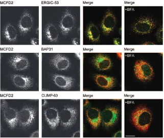
Overexpressed MCFD2 was previously shown to co-localize with ERGIC-53 to the ERGIC (23). To study the localization of endogenous MCFD2, immunofluorescence microscopy experiments were performed in HeLa cells. Figure 1 shows that endogenous MCFD2 co-localized with ERGIC-53 but only minimally with the two ER markers B-cell receptor associated protein 31 (BAP31) and cytoskeleton-linking membrane protein 63 (CLIMP-63). Next, we examined if MCFD2 cycles in the early secretory pathway. Cycling proteins are known to accumulate in ERGIC clusters in response to brefeldin A (BFA) (25). Brefeldin A treatment indeed led to the accumulation of MCFD2 in ERGIC-53 clusters, while the localization of the two ER resident proteins BAP31 and CLIMP-63 remained unchanged (Figure 1). These data suggest that endo-genous MCFD2 localizes to the ERGIC and cycles in the early secretory pathway.
ERGIC-53 retains MCFD2 in the early secretory pathway
Do MCFD2 and ERGIC-53 form a stable complex during their entire cycling journey? There are indications that ER and ERGIC may differ in their luminal pH and ionic properties (26). It is possible, therefore, that the calcium-dependent interaction of MCFD2 and ERGIC-53 does not persist throughout the entire cycling process. Co-immunoprecipitation of MCFD2 and ERGIC-53 from
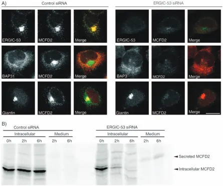
[image:2.595.53.374.428.697.2]BFA-treated cells (not shown) suggests that MCFD2 and ERGIC-53 interact in ERGIC clusters. Moreover, ERGIC-53-deficient lymphoblasts contain only trace amounts of intracellular MCFD2, indicating that MCFD2 requires ERGIC-53 either for stabilization or for intracellular retention (23). Experimental demonstration of a require-ment for intracellular retention of MCFD2 by ERGIC-53 would directly prove a physiologically relevant post-ER interaction of the two cycling proteins. To test this, we studied the effect of siRNA-based ERGIC-53 silencing on the localization of MCFD2. Three siRNA duplexes de-signed against different ERGIC-53 target sequences were tested for knockdown efficiencies. The most efficient siRNA duplex reduced ERGIC-53 levels in HeLa cells to less than 10% within 3 days. In ERGIC-53-depleted cells, only trace amounts of intracellular MCFD2 could be de-tected by immunoblotting. Immunofluorescence micro-scopy confirmed that MCFD2 was no longer detectable in these cells. The effect on MCFD2 is specific because the lo-calization of BAP31 and giantin was not affected (Figure 2A) and total secretion of35S-methionine-labeled proteins was unchanged (not shown). Although ERGIC-53 is a major protein of the ERGIC, its depletion does not seem to impair the morphology of the early secretory pathway as indi-cated by the normal localization of organelle markers of the early secretory pathway including numerous ER, ERGIC and Golgi proteins (not shown). An intact early secretory pathway after ERGIC-53 depletion is in line with previous observations showing that mistargeting of ERGIC-53 to the ER does not result in morphological changes of the early secretory pathway (19).
Figure 1: MCFD2 co-localizes with ERGIC-53. Localization of MCFD2 in HeLa cells visualized by double immuno-fluorescence microscopy using organelle marker antibodies for ERGIC (ERGIC-53, affinity-purified polyclonal antibody) and ER (BAP31 and CLIMP-63; mAbs). The cells were left untreated or incubated with brefeldin A (þBFA) for 90 min at a concentration of 10mg/mL. The MCFD2 is shown in green, while BAP31, CLIMP-63 and ERGIC-53 are shown in red. Bar¼10mm.
To investigate the disappearance of MCFD2 after ERGIC-53 knockdown, pulse–chase experiments with35S-methionine were performed. Cell lysate and conditioned medium of control and ERGIC-53 siRNA-transfected HeLa cells were probed for35S-methionine-labeled MCFD2 after 2 and 6-h chase periods. In ERGIC-53-depleted cells, intracellular MCFD2 disappeared and was detected as secreted protein in the conditioned medium (Figure 2B). Secreted MCFD2 showed a higher apparentMrthan the initially synthesized protein due to O-glycosylation (24). O-glycosylation may render MCFD2 less accessible to the antibody, accounting for the only partial recovery of MCFD2 in the conditioned medium. Alternatively, a fraction of MCFD2 may be degraded rather than secreted. Secreted,O-glycosylated MCFD2 can also be recovered from conditioned medium on overexpression of the protein (Figure 3). The results shown in Figure 2 show that ERGIC-53 is strictly required
for intracellular retention of MCFD2, indicating that MCFD2 and ERGIC-53 interact in post-ER compartments of unperturbed cells. In HeLa, COS, HepG2 and several glioblastoma cell lines, endogenous ERGIC-53 retains all MCFD2. No secreted MCFD2 could be detected in the conditioned medium (data not shown).
MCFD2 interacts with ERGIC-53 in the ER
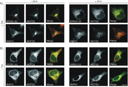
[image:3.595.80.525.48.420.2]Next, we examined if MCFD2 and ERGIC-53 can also interact in the ER. To this end, MCFD2 was localized in HeLa KKAA cells that express a dominant-negative form of ERGIC-53 that is retained in the ER by a C-terminal KKAA retention signal (19,27). In the presence of tetracycline, the expression of ERGIC-53-KKAA is repressed. Under these conditions, ERGIC-53 and MCFD2 co-localized to the ERGIC and were both BFA sensitive (Figure 4A) like in
wild-type HeLa cells. Removal of tetracycline, which induces the expression of ERGIC-53-KKAA, mislocalized MCFD2 to the ER (Figure 4B). ER localization of MCFD2 is indicated by co-localization with BAP31 as well as resistance to BFA. The mislocalization of MCFD2 by ER-retained ERGIC-53 shows that the two proteins can also interact in the ER.
ERGIC-53 binds certain cargo glycoproteins in an MCFD2-independent fashion
[image:4.595.52.400.49.193.2]Previous genetic and biochemical data showed that ERGIC-53 and MCFD2 form a cargo receptor complex, recognizing factors V and VIII (23,24). Factor VIII can be cross-linked to both ERGIC-53 and MCFD2 with similar efficiency, indicating the existence of a triple complex.
Figure 3: Overexpressed MCFD2 is secreted.HA-MCFD2-transfected and mock-transfected HeLa cells were labeled with 35S-methionine for 30 min and chased for the indi-cated times. MCFD2 was immuno-precipitated from cell lysates and conditioned medium using poly-clonal anti-MCFD2. Intracellular HA-MCFD2 disappears during the 3-h chase period and can be recovered from conditioned medium. Secreted HA-MCFD2 shows a shift inMrdue toO-glycosylation (24).
Figure 4: ERGIC-53-KKAA retains MCFD2 in the ER. MCFD2, BAP31 and ERGIC-53 were visualized by immunofluorescence microscopy in HeLa cells expressing ER-retained ERGIC-53-KKAA in a tetracycline-dependent inducible manner. Prior to fixation, the cells were treated with BFA (þBFA) (10mg/mL for 90 min) or left untreated (BFA). A) In the presence of tetracycline (þTet), the expression of ERGIC-53-KKAA is repressed and the cells express only endogenous ERGIC-53. MCFD2 co-localizes with ERGIC-53 and is BFA sensitive. B) Withdrawal of tetracycline (Tet) for 48 h induces ERGIC-53-KKAA that retains all ERGIC-53 in the ER due to a dominant-negative effect. Expression of ERGIC-53-KKAA relocalizes MCFD2 to the ER. Co-localization with BAP31 and BFA resistance show the ER retention of MCFD2. Bar¼10mm.
[image:4.595.75.511.322.619.2]ERGIC-53 also interacts with the two lysosomal glycopro-teins cathepsin Z and cathepsin C (12,19,20). To test if the MCFD2 subunit of the cargo receptor complex is required more generally for cargo binding, an siRNA-based knock-down approach was taken. Six siRNA duplexes, designed against different MCFD2 target sequences, were probed for knockdown efficiencies. The most efficient siRNA duplex reduced MCFD2 in HeLa cells to less than 10% within 3 days. Transfection efficiency of siRNA was very high as only very few cells stained positive for MCFD2 72 h after transfection (Figure 5B). Depletion of MCFD2 in HeLa cells affected neither the localization nor the protein level of ERGIC-53 (Figure 5A,C), which is consistent with the data from MCFD2-deficient lymphoblasts (23). Likewise, the localiza-tion of BAP31 and giantin was unaffected (Figure 5C).
[image:5.595.81.527.48.424.2]To analyze whether cargo binding of ERGIC-53 requires MCFD2, we combined siRNA-mediated MCFD2 depletion with a recently established yellow fluorescent protein (YFP)-based protein fragment complementation assay (PCA) (20). The basic concept of the YFP PCA relies on splitting YFP into two fragments (YFP1 and YFP2) that exhibit no fluorescence by themselves. When fused to two interacting proteins, the two YFP fragments can be brought into close proximity where they can complement to functional, fluorescent YFP by folding into an active 3D structure (28). Using YFP PCA, we have previously visual-ized the oligomerization of ERGIC-53, its interaction with MCFD2 and its lectin-mediated interactions with cathepsin Z and cathepsin C (20). Mutagenesis of the lectin domain of ERGIC-53 selectively abolished YFP complementation
with cathepsin Z and cathepsin C (20), demonstrating that YFP PCA is a powerful technique to study specific, carbohydrate-mediated interactions between ERGIC-53 and cargo proteinsin vivo.
Here, we applied the YFP PCA in MCFD2-depleted HeLa cells to investigate the role of MCFD2 on the oligomeriza-tion and glycoprotein-binding properties of ERGIC-53. Figure 6B shows that ERGIC-53 oligomerization and its interaction with cathepsin Z and cathepsin C are indepen-dent on the presence of MCFD2. Immunoblotting revealed equal expression of the YFP PCA constructs in control and MCFD2 siRNA-transfected cells, and silencing of MCFD2 was efficient (Figure 6C). As an internal control, the
[image:6.595.51.389.223.695.2]interaction between ERGIC-53 and MCFD2 was analyzed. After transfection of MCFD2-specific siRNA, no YFP complementation of ERGIC-53 and MCFD2 was observed because the expression of MCFD2-YFP2 was silenced. The results shown in Figure 6 clearly show that ERGIC-53 does not require MCFD2 to bind cathepsin Z and cathepsin C in vivo. Furthermore, MCFD2 is not required for ERGIC-53 oligomerization, which is consistent with data derived from MCFD2-deficient lymphoblasts (24). MCFD2-independent binding of cathepsin Z to ERGIC-53 is further supported by the fact that neither chemical cross-linking nor YFP PCA-based experiments showed a direct interaction of MCFD2 and cathepsin Z (not shown).
Figure 6: Yellow fluorescent protein PCA-based analysis of ERGIC-53-mediated protein in-teractions. A) The indicated pairs of YFP PCA constructs were ex-pressed in control and MCFD2 siRNA-transfected HeLa cells. B) YFP fragment complementation was detected by fluorometric analy-sis using microtiter plates with cell suspensions of transfected HeLa cells (20). Background fluorescence of mock-transfected cells was sub-tracted, and the relative fluores-cence units are shown. Data from three independent experiments were averaged and the error bars represent the standard deviation. MCFD2 siRNA affects neither the ERGIC-53 oligomerization nor the interactions between ERGIC-53 and cathepsin Z or cathepsin C. C) Lysates of fluorometrically analyzed cells were probed by Western blotting (WB) using mAbs against ERGIC-53, GFP, BAP31 and MCFD2. Equal expression of the YFP PCA constructs in control and MCFD2 siRNA-transfected cells is visualized by immunoblotting using ERGIC-53 and GFP anti-bodies. Equal protein amounts are present in all lanes as revealed by similar levels of endogenous ER-GIC-53 and BAP31. MCFD2 siRNA depletes endogenous MCFD2 and silences the expression of the MCFD2-YFP2 construct.
Discussion
Receptor-mediated export of secretory proteins from the ER is a universal feature of eukaryotic cells; yet, the diversity of this process is still largely unknown. Conceiv-ably, receptor-mediated ER export is required to increase transport efficiency of less abundant secretory proteins or for secretory cargo undergoing a second quality control step (2). Receptor-mediated ER export can also account for temporal and spatial co-ordination of the secretion of specific signaling molecules (29). The molecular basis underlying cargo receptor interactions is beginning to emerge although only few cases have been studied (11,13). ERGIC-53 and MCFD2 are particularly interesting because they are both required for the efficient secretion of blood coagulation factors V and VIII and constitute a cargo receptor complex composed of two subunits (23,24,30).
Here, we have described a detailed characterization of the ERGIC-53/MCFD2 receptor complex using a combination of immunofluorescence, gene silencing and PCA ap-proaches to provide insight into the interdependence and cargo selectivity of the two subunits. Endogenous MCFD2 co-localized with ERGIC-53 in untreated and BFA-treated HeLa cells and thus shows characteristic features of a pro-tein cycling between the ERGIC and the ER. Remarkably, both cycling and intracellular retention of MCFD2 are strictly dependent on ERGIC-53. ER-retained ERGIC-53-KKAA mis-localized MCFD2 to the ER, indicating that MCFD2 can bind to ERGIC-53 in the ER and requires ERGIC-53 for ante-rograde transport. Depletion of ERGIC-53 by siRNA re-sulted in the secretion of MCFD2; hence, MCFD2 requires ERGIC-53 also for retrograde transport back to the ER. We conclude that ERGIC-53 and MCFD2 form a stable com-plex and cycle together in the early secretory pathway due to the cytosolic diphenylalanine and dilysine motifs in ERGIC-53. In COS cells, endogenous ERGIC-53 and MCFD2 have similarly long half-lives (>26 h) (24), which is in line with a stable complex of the two proteins.
ERGIC-53-deficient patients have only trace amounts of intracellular MCFD2 (23). Our results provide now an explanation for this lack of MCFD2 by showng MCFD2 secretion on ERGIC-53 depletion in cell culture. Most soluble secretory proteins that localize to the early secre-tory pathway of mammalian cells carry a C-terminal KDEL tetrapeptide motif and are retained by binding to the KDEL receptor (31). This receptor captures the KDEL proteins in ERGIC andcisGolgi and recycles them back to the ER, providing a general mechanism for protein retention early in the secretory pathway (32). In contrast, MCFD2 is retained by a specific interaction with ERGIC-53, exempli-fying a KDEL-receptor-independent retention mechanism in the early secretory pathway.
Overexpression of MCFD2 also results in its secretion, suggesting that the protein is secreted as soon as the
retention capacity of ERGIC-53 is saturated. This finding may have physiological implications in view of a recent report proposing that rat MCFD2 (formerly termed stem cell-derived neuronal stem cell supporting factor (SDNSF)) can act as an autocrine or paracrine factor in maintaining stem cell potential and neurogenesis in the adult central nervous system (33). In HeLa, COS, HepG2 and several glioblastoma cell lines, however, we could not detect any secreted endogenous MCFD2, suggesting that endoge-nous ERGIC-53 is capable of retaining all MCFD2 in these cells, although secretion of minute amounts below the detection limit cannot be excluded. ERGIC-53 and MCFD2 levels are induced in response to cellular stress (34,35). This raises the intriguing possibility that abundant MCFD2 can be secreted and act in some signaling events under certain conditions.
Factor VIII can be cross-linked to both MCFD2 and ERGIC-53, arguing for a triple complex early in the secretory pathway (24). Interestingly, factor VIII can also be cross-linked to the MCFD2 D129E mutant that is unable to interact with ERGIC-53 (24). Thus, MCFD2 interacts with factor VIII in an ERGIC-53-independent manner. To deter-mine whether MCFD2 is a general cargo recruitment factor for ERGIC-53, we tested a putative interaction of MCFD2 with cathepsin Z or cathepsin C. Although serving as a cargo for ERGIC-53, cathepsin Z could not be directly cross-linked to MCFD2. Likewise, protein interaction stud-ies using YFP PCA could not reveal an interaction between MCFD2 and cathepsin Z or cathepsin C, arguing against a role of MCFD2 in the recruitment of these cargo proteins. To confirm these findings, the interaction between ERGIC-53 and cathepsin Z or cathepsin C was studied after MCFD2 knockdown. YFP PCA showed that the interaction between ERGIC-53 and cathepsin Z or cathepsin C in living cells is not affected by the depletion of MCFD2. Binding of cathepsin Z and cathepsin C to ERGIC-53 is unlikely to be mediated by residual MCFD2. First, siRNA-mediated knockdown is very efficient, and hardly any residual MCFD2 is detectable by immunoblot (Figures 5A and 6C). Second, immunofluorescence-based analysis of siRNA transfection efficiency (Figure 5B) shows that residual MCFD2 derives from only very few apparently nontrans-fected cells, while the big majority of cells is entirely depleted of MCFD2. Third, in the YFP PCA-based protein interaction analysis, endogenous ERGIC-53 and YFP1-ERGIC-53 compete for residual MCFD2, which am-plifies the MCFD2 depletion effect. In support of these in vivodata, ERGIC-53 can bind to immobilized mannose in vitrowhen purified without MCFD2 in the absence of calcium (26).
cathepsin Z or cathepsin C, in an MCFD2-independent fashion. Conversely, MCFD2 interacts with factors V and VIII in an ERGIC-53-independent manner. Binding of MCFD2 to 53 recruits factors V and VIII to ERGIC-53, thereby ensuring efficient ER export. In support of this model, unglycosylated factor VIII but not unglycosylated cathepsin Z can be cross-linked to the MCFD2/ERGIC-53 complex (12,24). Recruitment of specific cargo molecules by a luminal subunit of a receptor complex adds another layer of complexity to receptor-mediated ER export. It will be interesting in the future to elucidate how MCFD2 re-cruits factors V and VIII to ERGIC-53. This will require new methodology as we were unable to visualize this process by YFP PCA. Mechanistic insight into this recruitment process will be important to understand how soluble secretory proteins can be captured for ER export.
Materials and Methods
Antibodies
The following antibodies were used: mouse monoclonal antibody (mAb) G1/ 93 against human ERGIC-53 (14) (ALX-804-602; Alexis, Lausen, Switzerland), mouse mAb against human MCFD2 (23), goat polyclonal antibody (pAb) against human MCFD2 (R&D Systems, Minneapolis, MN, USA), rabbit pAb against human MCFD2 (23), mouse mAb A1/182 against BAP31 (16) 601; Alexis), mouse mAb G1/133 against giantin (36) (ALX-804-600; Alexis), mouse mAb against hemagglutinin (HA; Covance, Princeton, NJ, USA) and mouse mAb against green fluorescent protein (GFP; Roche Applied Science, Basel, Switzerland).
Cell culture
HeLa cells (CCL-2; ATCC, Manassas, VA, USA) were grown in DMEM, supplemented with 10% fetal bovine serum, 1nonessential amino acids and antibiotics. HeLa KKAA cells were cultured as described previously (19). For fluorometric analysis and metabolic labeling, cells were grown in six-well plates. For fluorescence microscopy cells, were grown on poly-L -lysine-coated glass slides.
siRNA transfection
siRNA oligos were purchased from Qiagen (Venlo, The Netherlands) and Eurogentec (Seraing, Belgium). Three and six siRNA oligos were designed against ERGIC-53 and MCFD2, respectively. The most efficient siRNA oligo was determined by immunoblotting and chosen for all further experiments. ERGIC-53 was knocked down using 50
-GGACAGAAUCGUAUUCAUCdTdT-30as sense and 50-GAUGAAUACGAUUCUGUCCdTdT-30as antisense oligo.
The MCFD2 was knocked down using 50
-AGAAGGUGUCAUCAA-CAAAdTdT-30 as sense and 50-UUUGUUGAUGACACCUUCUdAdG-30 as
antisense oligo. Nonsilencing control siRNA was purchased from Qiagen. A final siRNA concentration of 5 nM was used for transfection directly after cell plating using HiPerFect (Qiagen) according to the manufacturer’s instructions.
DNA transfection
Cloning of pcDNA3[YFP1-p53], pcDNA3[YFP2-p53], pcDNA3[MCFD2-YFP2], pcDNA3[YFP2-catZ] and pcDNA3[YFP2-catC] was described pre-viously (13,20). pcDNA3[MCFD2] was generated by inserting HA-MCFD2 without its signal sequence (generated by polymerase chain reaction amplification) into the pcDNA3 vector containing the artificial signal sequence of calreticulin (20). DNA constructs were transfected with FuGENE6 (Roche Applied Science) according to the manufacturer’s instruc-tions. For siRNA and DNA co-transfection, DNA constructs were
trans-fected 24 h after cell plating and siRNA transfection. Cells were analyzed 48 h after DNA transfection, which corresponds to 72 h after siRNA transfection.
Immunofluorescence microscopy
Brefeldin A (10mg/mL; Epicentre, Madison, WI, USA) was added to the cells 90 min prior to fixation. Cells were fixed in 3% para-formaldehyde and permeabilized for 5 min in PBS containing 3% BSA and 0.2% Triton-X-100. Primary antibodies were added for 30 min in PBS containing 3% BSA. After rinsing, the secondary antibodies conjugated with Alexa Fluor 488 or Alexa Fluor 568 (Molecular Probes, Leiden, The Netherlands) were added for 30 min. Cells were washed in PBS, embedded and analyzed by laser scanning confocal microscopy (TCS NT; Leica, Wetzlar, Germany). For indirect immunofluorescence using mouse mAb against human MCFD2, 0.5% SDS and 5%b-mercaptoethanol was added during permeabilization step (37). The use of mouse immunoglobulin G (IgG) 1 (for MCFD2) and mouse IgG2a (for BAP31 and CLIMP-63) specific secondary antibodies allowed double staining of MCFD2 with BAP31 or CLIMP-63.
Immunoblotting
Protein samples were prepared as described previously (20), separated by SDS–PAGE; transferred to nitrocellulose membranes; immunoblotted with anti-ERGIC-53, anti-BAP31, anti-MCFD2 and anti-GFP and visualized by enhanced chemiluminescence (Amersham Bioscience, Uppsala, Sweden).
Metabolic labeling
Cells were deprived ofL-methionine for 20 min, pulsed for 60 min with 100 mCi35
S-methionine (Perkin Elmer, Wellesley, MA, USA) and chased for the indicated times in HeLa culture medium containing 10 mML-methionine.
Cells were lysed in 1% Triton-X-100, 50 mM Tris–HCl (pH 7.5), 150 mM NaCl, 2 mM CaCl2and phenylmethylsulfonylfluoride (PMSF), and the lysate
was cleared by centrifugation at 100 000gfor 1 h. The chase medium was cleared from cell debris by centrifugation at 20 000gfor 5 min. Cleared samples were immunoprecipitated with anti-MCFD2. Immunoprecipitates were separated by SDS–PAGE, and radiolabeled bands were imaged using a phosphorimager (Molecular Dynamics, Sunnyvale, CA, USA).
Yellow fluorescent protein fluorometric analysis Fluorometric analysis was performed as described previously (20). Data from three independent experiments were averaged.
Acknowledgments
We thank K. Bucher for expert technical assistance. B. Z. is a recipient of a Career Development Award from the National Hemophilia Foundation, USA. D. G. and R. J. K. are Howard Hughes Medical Institute Investigators. This work was supported by the University of Basel, the Swiss National Science Foundation (H.-P. H) and the National Institutes of Health grants PO1 HL057346 (D. G.) and HL052173 (R. J. K.).
References
1. Lee MC, Miller EA, Goldberg J, Orci L, Schekman R. Bi-directional protein transport between the ER and Golgi. Annu Rev Cell Dev Biol 2004;20:87–123.
2. Ellgaard L, Molinari M, Helenius A. Setting the standards: quality control in the secretory pathway. Science 1999;286:1882–1888. 3. Miller EA, Beilharz TH, Malkus PN, Lee MC, Hamamoto S, Orci L,
Schekman R. Multiple cargo binding sites on the COPII subunit Sec24p ensure capture of diverse membrane proteins into transport vesicles. Cell 2003;114:497–509.
4. Mancias JD, Goldberg J. Exiting the endoplasmic reticulum. Traffic 2005;6:278–285.
5. Kappeler F, Klopfenstein DR, Foguet M, Paccaud JP, Hauri HP. The recycling of ERGIC-53 in the early secretory pathway. ERGIC-53 carries a cytosolic endoplasmic reticulum-exit determinant interacting with COPII. J Biol Chem 1997;272:31801–31808.
6. Nufer O, Guldbrandsen S, Degen M, Kappeler F, Paccaud JP, Tani K, Hauri HP. Role of cytoplasmic C-terminal amino acids of membrane proteins in ER export. J Cell Sci 2002;115:619–628.
7. Warren G, Mellman I. Bulk flow redux? Cell 1999;98:125–127. 8. Wieland FT, Gleason ML, Serafini TA, Rothman JE. The rate of bulk
flow from the endoplasmic reticulum to the cell surface. Cell 1987; 50:289–300.
9. Kuehn MJ, Herrmann JM, Schekman R. COPII-cargo interactions direct protein sorting into ER-derived transport vesicles. Nature 1998;391: 187–190.
10. Belden WJ, Barlowe C. Role of Erv29p in collecting soluble secretory proteins into ER-derived transport vesicles. Science 2001;294: 1528–1531.
11. Otte S, Barlowe C. Sorting signals can direct receptor-mediated export of soluble proteins into COPII vesicles. Nat Cell Biol 2004;6:1189–1194. 12. Appenzeller C, Andersson H, Kappeler F, Hauri HP. The lectin ERGIC-53 is a cargo transport receptor for glycoproteins. Nat Cell Biol 1999; 1:330–334.
13. Appenzeller-Herzog C, Nyfeler B, Burkhard P, Santamaria I, Lopez-Otin C, Hauri HP. Carbohydrate- and conformation-dependent cargo capture for ER-exit. Mol Biol Cell 2005;16:1258–67.
14. Schweizer A, Fransen JA, Bachi T, Ginsel L, Hauri HP. Identification, by a monoclonal antibody, of a 53-kD protein associated with a tubulo-vesicular compartment at the cis-side of the Golgi apparatus. J Cell Biol 1988;107:1643–1653.
15. Itin C, Roche AC, Monsigny M, Hauri HP. ERGIC-53 is a functional mannose-selective and calcium-dependent human homologue of legu-minous lectins. Mol Biol Cell 1996;7:483–493.
16. Klumperman J, Schweizer A, Clausen H, Tang BL, Hong W, Oorschot V, Hauri HP. The recycling pathway of protein ERGIC-53 and dynamics of the ER-Golgi intermediate compartment. J Cell Sci 1998;111: 3411–3425.
17. Ben-Tekaya H, Miura K, Pepperkok R, Hauri HP. Live imaging of bidirectional traffic from the ERGIC. J Cell Sci 2005;118:357–367. 18. Itin C, Schindler R, Hauri HP. Targeting of protein ERGIC-53 to the ER/
ERGIC/cis-Golgi recycling pathway. J Cell Biol 1995;131:57–67. 19. Vollenweider F, Kappeler F, Itin C, Hauri HP. Mistargeting of the lectin
ERGIC-53 to the endoplasmic reticulum of HeLa cells impairs the secretion of a lysosomal enzyme. J Cell Biol 1998;142:377–389. 20. Nyfeler B, Michnick SW, Hauri HP. Capturing protein interactions in the
secretory pathway of living cells. Proc Natl Acad Sci U S A 2005; 102:6350–6355.
21. Nichols WC, Seligsohn U, Zivelin A, Terry VH, Hertel CE, Wheatley MA, Moussalli MJ, Hauri HP, Ciavarella N, Kaufman RJ, Ginsburg D. Mutations in the ER-Golgi intermediate compartment protein ERGIC-53 cause combined deficiency of coagulation factors V and VIII. Cell 1998;93:61–70.
22. Moussalli M, Pipe SW, Hauri HP, Nichols WC, Ginsburg D, Kaufman RJ. Mannose-dependent endoplasmic reticulum (ER)-Golgi intermediate
compartment-53-mediated ER to Golgi trafficking of coagulation factors V and VIII. J Biol Chem 1999;274:32539–32542.
23. Zhang B, Cunningham MA, Nichols WC, Bernat JA, Seligsohn U, Pipe SW, McVey JH, Schulte-Overberg U, de Bosch NB, Ruiz-Saez A, White GC, Tuddenham EG, Kaufman RJ, Ginsburg D. Bleeding due to disruption of a cargo-specific ER-to-Golgi transport complex. Nat Genet 2003;34:220–225.
24. Zhang B, Kaufman RJ, Ginsburg D. LMAN1 and MCFD2 form a cargo receptor complex and interact with coagulation factor VIII in the early secretory pathway. J Biol Chem 2005;280:25881–25886.
25. Breuza L, Halbeisen R, Jeno P, Otte S, Barlowe C, Hong W, Hauri HP. Proteomics of endoplasmic reticulum-Golgi intermediate compartment (ERGIC) membranes from brefeldin A-treated HepG2 cells identifies ERGIC-32, a new cycling protein that interacts with human Erv46. J Biol Chem 2004;279:47242–47253.
26. Appenzeller-Herzog C, Roche AC, Nufer O, Hauri HP. pH-induced conversion of the transport lectin ERGIC-53 triggers glycoprotein release. J Biol Chem 2004;279:12943–12950.
27. Andersson H, Kappeler F, Hauri HP. Protein targeting to endoplasmic reticulum by dilysine signals involves direct retention in addition to retrieval. J Biol Chem 1999;274:15080–15084.
28. Kerppola TK. Visualization of molecular interactions by fluorescence complementation. Nat Rev Mol Cell Biol 2006;7:449–56.
29. Bokel C, Dass S, Wilsch-Brauninger M, Roth S. Drosophila Cornichon acts as cargo receptor for ER export of the TGFalpha-like growth factor Gurken. Development 2006;133:459–470.
30. Zhang B, McGee B, Yamaoka JS, Guglielmone H, Downes KA, Minoldo S, Jarchum G, Peyvandi F, de Bosch NB, Ruiz-Saez A, Chatelain B, Olpinski M, Bockenstedt P, Sperl W, Kaufman RJ et al. Combined deficiency of factor V and factor VIII is due to mutations in either LMAN1 or MCFD2. Blood 2006;107:1903–1907.
31. Lewis MJ, Pelham HR. A human homologue of the yeast HDEL receptor. Nature 1990;348:162–163.
32. Lewis MJ, Pelham HR. Ligand-induced redistribution of a human KDEL receptor from the Golgi complex to the endoplasmic reticulum. Cell 1992;68:353–364.
33. Toda H, Tsuji M, Nakano I, Kobuke K, Hayashi T, Kasahara H, Takahashi J, Mizoguchi A, Houtani T, Sugimoto T, Hashimoto N, Palmer TD, Honjo T, Tashiro K. Stem cell-derived neural stem/progenitor cell supporting factor is an autocrine/paracrine survival factor for adult neural stem/ progenitor cells. J Biol Chem 2003;278:35491–35500.
34. Nyfeler B, Nufer O, Matsui T, Mori K, Hauri HP. The cargo receptor ERGIC-53 is a target of the unfolded protein response. Biochem Biophys Res Commun 2003;304:599–604.
35. Spatuzza C, Renna M, Faraonio R, Cardinali G, Martire G, Bonatti S, Remondelli P. Heat shock induces preferential translation of ERGIC-53 and affects its recycling pathway. J Biol Chem 2004;279:42535–42544. 36. Linstedt AD, Hauri HP. Giantin, a novel conserved Golgi membrane protein containing a cytoplasmic domain of at least 350 kDa. Mol Biol Cell 1993;4:679–693.