The Unc-5 Receptor Is Directly Regulated by
Tinman in the Developing
Drosophila
Dorsal
Vessel
Jamshid Asadzadeh1,2¤, Niamh Neligan1, Judith J. Canabal-Alvear3, Amanda C. Daly1,2, Sunita Gupta Kramer3, Juan-Pablo Labrador1,2*
1Smurfit Institute of Genetics, Trinity College Dublin, Dublin, Ireland,2Institute of Neuroscience, Trinity College Dublin, Dublin, Ireland,3Department of Pathology and Laboratory Medicine, Robert Wood Johnson Medical School, Rutgers, The State University of New Jersey, Piscataway, New Jersey, United States of America
¤ Current address: Columbia University Medical Center, Department of Pathology and Cell Biology, New York, New York, United States of America
Abstract
During early heart morphogenesis cardiac cells migrate in two bilateral opposing rows, meet at the dorsal midline and fuse to form a hollow tube known as the primary heart field in vertebrates or dorsal vessel (DV) inDrosophila. Guidance receptors are thought to mediate this evolutionarily conserved process. A core of transcription factors from the NK2, GATA and T-box families are also believed to orchestrate this process in both vertebrates and invertebrates. Nevertheless, whether they accomplish their function, at least in part, through direct or indirect transcriptional regulation of guidance receptors is currently unknown. In our work, we demonstrate how Tinman (Tin), theDrosophilahomolog of the Nkx-2.5 tran-scription factor, regulates the Unc-5 receptor during DV tube morphogenesis. We use genetics, expression analysis with single cell mRNA resolution and enhancer-reporter assays in vitro or in vivo to demonstrate that Tin is required for Unc-5 receptor expression specifically in cardioblasts. We show that Tin can bind to evolutionary conserved sites within an Unc-5 DV enhancer and that these sites are required for Tin-dependent transactivation both in vitro and in vivo.
Introduction
Early stages of heart development, both in vertebrates and invertebrates, include the migration of bilaterally paired condensations of cardiac precursors and the formation of a linear tube. The tube is formed once these symmetrical groups of mesodermal cells meet, and attach to each other leaving a luminal space between them [1,2]. The coordinated migration of these mesodermal cells, bilateral interaction and the preservation of a lumen require complex inter-actions of multiple guidance receptors inDrosophiladuring DV morphogenesis [3–8]. Verte-brate homologs of the same ligand/receptor systems are expressed in the developing heart in OPEN ACCESS
Citation:Asadzadeh J, Neligan N, Canabal-Alvear JJ, Daly AC, Kramer SG, Labrador J-P (2015) The Unc-5 Receptor Is Directly Regulated by Tinman in the DevelopingDrosophilaDorsal Vessel. PLoS ONE 10(9): e0137688. doi:10.1371/journal.pone.0137688
Editor:Edward Giniger, National Institutes of Health (NIH), UNITED STATES
Received:July 4, 2015
Accepted:August 19, 2015
Published:September 10, 2015
Copyright:© 2015 Asadzadeh et al. This is an open access article distributed under the terms of the
Creative Commons Attribution License, which permits unrestricted use, distribution, and reproduction in any medium, provided the original author and source are credited.
Data Availability Statement:All relevant data are within the paper and its Supporting Information files.
many cases with strikingly similar patterns to the ones present inDrosophila[9,10]. Some, like the Robos and their Slit ligands [11,12] or plexins and semaphorins [13,14], have also been identified as key players at different stages of heart development. Nevertheless, how these guid-ance systems are regulated in place and time during heart morphogenesis is widely unknown.
Cardiogenesis in both vertebrates and invertebrates also requires the key regulatory actions of a core of evolutionarily conserved families of transcription factors (NK2, GATA, and T-box) [15]. They are required early in development during linear tube formation and function again at later stages of heart morphogenesis in vertebrates [16]. For example, Nkx2-5 members and itsDrosophilahomolog, Tinman (Tin), play an important role in early cardiogenesis starting with the specification of cardiac precursors to remodeling and functionality of the adult heart [1,17]. Given the role of guidance systems in heart morphogenesis, it is likely that they are direct or indirect targets of these families of transcriptional regulators.
To gain a better understanding if these transcription factors (TFs) control heart tubulogen-esis through the regulation of guidance receptors, we have studied Unc-5 receptor’s regulation in theDrosophiladorsal vessel (DV). The DV develops from mesodermal cardiac precursors. After precursor division, heart cells line up bilaterally into two rows where myocardial cells or cardioblasts (CBs) are positioned dorsally and pericardial cells (PCs) ventrolaterally. Finally, they migrate together towards the dorsal midline of the embryo where CBs fuse to form the tubular heart (Fig 1A). The CBs will constitute the pumping myocardium and PCs the pericar-dium. TheDrosophilaUnc-5 receptor is a repulsive receptor for Netrin A and B [18,19] and in the nervous system has been shown to be required for motoneuron guidance [20,21] and glial migration [22]. Unc-5 is expressed in both major cell types present in the DV and has recently been shown to be required in late dorsal vessel morphogenesis for lumen formation [7,8].tin is also expressed widely in PCs and most CBs (Fig 1A). Furthermore,tinandDoc, a T-box family TF, have been shown to regulate together an early cardiac mesodermUnc-5 enhancer [23]. In this work we show thattinis specifically required forUnc-5expression in CBs and is sufficient to induce its expression ectopically in the ectoderm. We identify a unique DV enhancer within theUnc-5regulatory region that fully recapitulates its expression at late stages of DV fusion. Cardioblast specific expression through this enhancer is strictly dependent ontinas is misexpression in the ectoderm. Additionally, Tin can induce transcription in vitro in a luciferase assay through theUnc-5DV enhancer but not from other knownUnc-5 enhanc-ers. Using ChIP analysis we identify three evolutionary conserved Tin-binding sites within this enhancer that are required in vitro for its activity. Finally, we demonstrate that these sites are the Tin-binding sites required in theUnc-5DV enhancer for its ectopic regulation in the ecto-derm and, more importantly, its specific expression in cardioblasts. Thus, Our work shows howtinregulates 5 receptor expression during late heart tube morphogenesis when Unc-5 is required for lumen formation. Our results provide a regulatory mechanism for a guidance receptor through a direct interaction with three conserved sites within its DV enhancer by one of the core transcription factors during tubulogenesis of the Drosophila DV.
Results
Tin is required for Unc-5 expression in the dorsal vessel
The Unc-5 receptor localizes preferentially at the luminal side in CBs at the onset of tubulogen-esis and it is required to preserve the luminal space between CBs [7,8]. However, how this receptor is regulated at this late stage is not known. Genome-wide chromatin immunoprecipita-tion screens to identify Tin target genes in cardiac mesoderm and cardiac precursors have iden-tifiedUnc-5as one of its targets [23,24]. Previous studies have established that early cardiac specification inDrosophilais dependent on the homeobox transcription factor Tin [2,25,26]. Competing Interests:The authors have declared
As a consequence,tinloss-of-function mutants lack a DV due to its early role in the mesoderm to specify cardiac progenitors [26,27]. Nevertheless, in cardiac specifictinmutant animals (tin-ABD;tin346/tin346), wheretinis re-expressed in a mutant background under the control of enhancer elements (ABD) recapitulating its entire endogenous expression pattern except in the dorsal vessel. Myocardial cells are specified and the DV forms in these mutants [17]. To deter-mine whethertinregulatesUnc-5, we analyzed its expression intinmutant DVs (Fig 1B and 1C).Unc-5mRNAs is significantly reduced when compared withtinheterozygous DVs (com-pare 1B with 1C). Thus,tinis required forUnc-5expression in the DV at the onset of
tubulogenesis.
Tinman is sufficient to induce Unc-5 expression ectopically
We further testedtin’s sufficiency to induceUnc-5transcriptionin vivoby misexpressingtinin ectodermal stripes with anengrailed-Gal4(en-Gal4) driver. We detectedUnc-5mRNA through in situ hybridization and we confirmed that, indeed, ectopictinexpression in the ectoderm is sufficient to induceUnc-5in the characteristicengrailedstripes (Fig 2B and 2B’) where neither
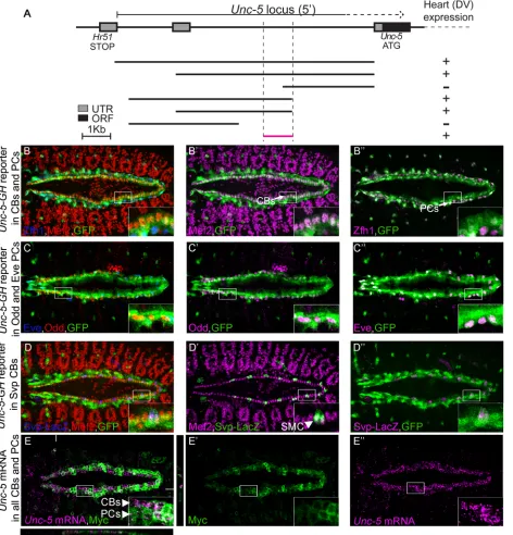
Fig 1.tinregulatesUnc-5expression in vivo.(A) Organization and cellular composition and development of the Drosophila dorsal vessel. Schematic represents Drosophila DV at embryonic stages 15 (migrating cells, top) and 17 (tubular heart already formed, bottom) with different cell types color-coded based on the marker TF expressed. Aortic portion is oriented anteriorly and the beating portion (heart) posteriorly. CBs are divided into Tin- (light green) or Svp-expressing SMCs (dark green) subtypes. The three most posterior pairs of Svp-expressing CBs will make the future ostial (inflow valve) cells. Pericardial cells (gray) surround the CBs on their ventrolateral side and fall into two major types; Tin-positive or Tin-negative PCs (not colored). Bottom is a schematic cross section of the heart lumen at stage 17 where CBs on opposite sides take a crescent-like shape after contact, leaving in between them a hollow luminal space. (B)Unc-5mRNA (magenta) is present in the DV in embryos heterozygous fortin346(B). IntinABD;tin346/tin346homozygous mutant embryos, however,Unc-5mRNA expression is significantly reduced (C). Anterior side of the embryo is to the left in panels B and C.
[image:3.612.204.563.79.358.2]tinnorUnc-5are normally not expressed (Fig 2A and 2A’). Thus,tinis not only required for Unc-5expression in the dorsal vessel but it is also sufficient to induce its expression in other tissues.
Identification of the Unc-5 cardiac enhancer
[image:4.612.201.559.78.268.2]In order to identify regulatory regions required forUnc-5expression in the DV, we dissected theD.melanogaster Unc-5genomic locus into overlapping fragments of varying length starting from the preceding gene (Hr51) to the 5th intron within theUnc-5locus [28] (Fig 3A). All the fragments were fused to a GFP ORF and inserted into the same locus to avoid any variability due to position effect. We identified a unique 1kb minimal fragment upstream of theUnc-5 ATG sufficient to drive GFP expression in the DV at late stages of PCs and CBs migration and during tube formation (stage 13 onwards,Fig 3A) largely overlapping with the early mesoderm enhancer previously described [23]. To characterize the expression pattern of this enhancer we co-stained embryos carrying the DV enhancer driving GFP (GH reporter) with anti-GFP and markers for PCs or CBs, Mef2. The reporter drives GFP expression in all Tin-expressing CBs and PCs (Fig 3B–3D”) including Eve- and Odd-positive PCs (Fig 3C–3C”) and the Tin-nega-tive Seven Up (Svp)-expressing myocardial cells (SMCs,Fig 3D–3D”). Finally, to confirm that the enhancer faithfully recapitulatesUnc-5endogenous expression in theDrosophilaDV we also performed double labeling of the cells expressing the reporter andUnc-5mRNA by in situ hybridization. Our data shows that theUnc-5mRNA expression pattern in the DV fully matches that of the enhancer (Fig 3E–3E”).
Fig 2. Ectopic expression of Tin inducesUnc-5mRNA expression in vivo.(A-A’) in situ hybridization for endogenous mRNA expression ofUnc-5does not show any striped ectodermal signal (magenta,
arrowheads with asterisks). (B–B’) ectopic expression oftinin ectodermal stripes (green) from aUAS-tin transgene usingengrailed-Gal4induces endogenousUnc-5mRNA in a striped pattern (compare A’with B’, arrowheads). Engrailed stripes are labelled green by co-expression of Tau-Myc from aUAS-tau-myc construct. All panels are lateral views with anterior to the left. A magnification of the regions delineated by insets is shown for each panel.Unc-5in situ fuorescence inengrailedstripes was quantified (0.33±0.06 s.e. m. or -0.03±0.01 s.e.m. intin-expressingen-stripes A, A’oren-stripes not expressingtinrespectively B, B’, p<0.005, n = 12). Confocal XZ sections are presented below each panel (location of the sections is indicated in the upper panel a white line) where Unc-5 specific signal can be detected colocalizing withen-stripes when tin is expressed (B, B’) but not in awild-typeembryo (A, A’).
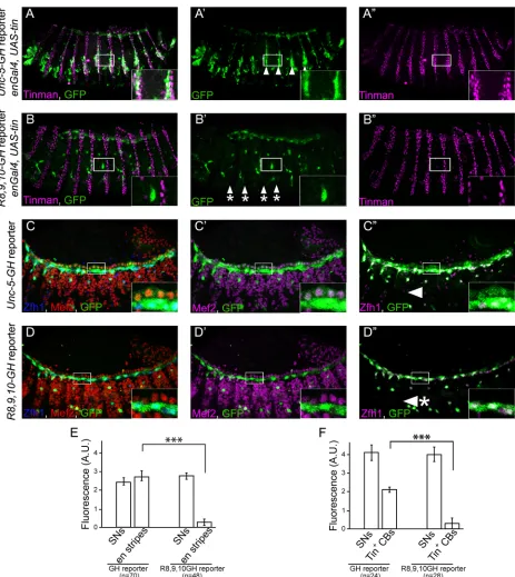
Fig 3. TheUnc-5-GHreporter is expressed in all cardioblasts and pericardial cells.(A) Schematic representation of the positions and the relative sizes of the dissected fragments from theUnc-5locus. Reporter constructs were generated by fusing each fragment to a GFP open reading frame. Examination of GFP expression in transgenic lines carrying any of these reporters indicates potential enhancer activity of each fragment. All reporters containing the smallest (bottom) fragment revealed activity in the DV. The smallest fragment (GH-reporter) was chosen for further examinations. (B-D”) We used specific markers to label CBs: Mef2, (B and B’, [29]), PCs: all PCs with Zfh1 (B and B”,[30,31]) or subsets with Eve and Odd (C-C”, [32,33]); and a Svp-LacZ reporter for labelling a set of Tin-negative myocardial cells, also known as Seven Up (Svp)-positive myocardial cells (SMCs) (D-D”, [17,34]). GFP expression (green) is present in all CBs (B’; magenta) and PCs (B”; magenta). (C–C”) Odd- (C’; magenta) and Eve-positive (C”; magenta) PCs express GFP (green) driven by
Unc-5-GHenhancer fragment. (D–D”)Unc-5-GHreporter also drives expression in Tin-positive CBs and Svp myocardial cells (SMC). (E-E”) Correlation
between GH enhancer expression pattern and endogenousUnc-5mRNA expression in the DV was examined by in situ hybridization. Colocalization of Tau-Myc expression pattern (labeled in green), driven by the enhancer (Unc-5 GH-Gal4), andUnc-5mRNA (magenta) indicates perfect overlap between the two. XZ an YZ sections are presented at the bottom and right of the main panel (E) and white lines indicate their location. CB cardioblast, PC pericardial cell. All panels are dorsal views with anterior to the left. A magnification of the regions delineated by insets is shown for each panel. All embryos are stage 15.
tin
is required for activation of the
Unc-5
DV enhancer in cardioblasts
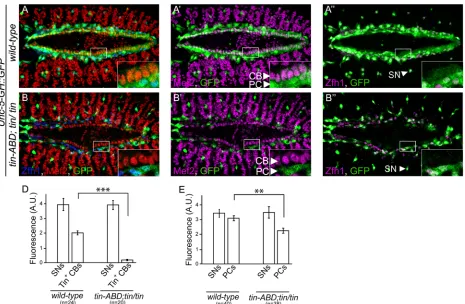
[image:6.612.40.505.287.591.2]Sincetinis required forUnc-5expression in the DV (Fig 1B and 1C). We speculated thattin might exert its regulation through the unique DV enhancer we identified within theUnc-5 genomic locus. Therefore, we examined GFP expression driven by theUnc-5-GHenhancer in tin-ABD;tin346mutant embryos at later stages of embryonic cardiogenesis (Fig 4). GFP expres-sion driven by the enhancer was virtually absent in CBs fromtin-ABD;tin346embryos (from 1.98 ± 0.128 inwild-typeCBs to 0.173 ± 0.04 intin-ABD;tin346CBs,P<6 x 10⁻¹⁴,Fig 3D). However, GFP expression was still present in PCs (Fig 4B–4B”), where it was slightly reduced (from 3.4 ± 0.2 inwild-typeto 2.47 ± 0.21 intin-ABD;tin346,P<6 x 10⁻⁴,Fig 3E). Importantly, GFP expression from the GH enhancer in a GFP-positive subpopulation of sensory neurons (SNs) wheretinis not expressed nor required was not affected intin-ABD;tin346mutants (3.86 ± 0.4 inwild-typeand 3.82 ± 0.29 intin-ABD;tin346,Fig 4A”and 4B”[arrowheads], D, E). Furthermore, SMCs, where Tin is not normally expressed, accordingly, still expressed GFP in tin-ABD;tin346mutants (S1A–S1A”Fig). These results indicate thattinspecifically regulates
Fig 4. TheUnc-5heart enhancer element is regulated bytin in vivo.(A–A”) Reporter gene (green) is expressed in all CBs and PCs inwild-typeembryos.
(B–B”) Intin-ABD;tin346/tin346mutant embryos, wheretinis only absent in the DV, reporter gene expression is almost absent in CBs while it is only partially
downregulated in PCs. Note the unchanged GFP expression in sensory neurons (arrowheads in A”and B”) inwild-typeand mutant backgrounds.
Quantification of GFP expression in CBs (D) or PCs (E). Genotypes of embryos are indicated on the X axis and fluorescence on the Y axis. GFP expression in SNs is used as internal control and their fluorescence is not affected intin-ABD;tin346/tin346mutant background (3.86±0.4 s.e.m and 3.82±0.29 s.e.m, in
wild-typeortinmutants respectively). However, the signal is significantly (P<6 x 10⁻¹⁴) reduced in CBs, from 1.98±0.128 to 0.173±0.04 (D). PCs show a
slight reduction in signal (E), from 3.4±0.2 inwild-typeto 2.47±0.21 intin-ABD;tin346(P
<6.2 x 10⁻⁴). All panels are dorsal views with anterior to the left. A
magnification of the regions delineated by insets is shown for each panel. All embryos are aged from early to late stage 15. CB cardioblast, PC pericardial cell, SMC Svp positive myocardial cells, SN sensory neuron.
expression through theUnc-5-GHenhancer in CBs at the developmental stage when they fuse to form the heart lumen.
Tin binds the Unc-5 DV enhancer at three conserved Tin-binding
elements
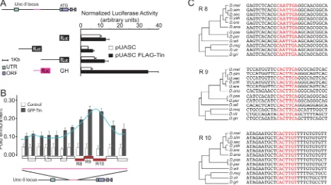
[image:7.612.40.508.334.596.2]To determine if Tin is sufficient to induce transcription through theUnc-5-GHenhancer we fused differentUnc-5enhancers [28] to renilla luciferase ORF and co-transfected them withtin inDrosophilaS2 cells. The only fragment responsive to Tin in this assay was the one containing the GH enhancer (Fig 5A). Therefore, this enhancer is not only under the control of Tin in vivo in the DV, but also responded to it in vitro. Our assay results also suggested that regulation of this enhancer was very likely mediated through a direct interaction with Tin. To test this hypothesis, we performed chromatin immunoprecipitation (ChIP) followed by qPCR using overlapping primers covering theUnc-5-GHenhancer. Our ChIP-qPCR results identified a unique enrichment peak near the 3’end of theUnc-5-GHenhancer covering three consecutive amplified regions (referred to as R8, R9, and R10,Fig 5B). Further analysis of the sequence within the peak revealed three potential binding sites on theUnc-5-GHenhancer that closely match the described consensus Tin-binding sequence [23,24,35]. These motifs are conserved inDrosophilaspecies with a divergence time>107years, highlighting their functional
Fig 5. TheUnc-5-GHenhancer element is directly regulated in vitro by Tin via multiple evolutionary conserved Tin-binding motifs.(A) Tin induces activation of theUnc-5-GHenhancer in S2R+ cells. Schematic of a few enhancers within the 5’region ofUnc-5used to make luciferase constructs used in the luciferase reporter assays. Luciferase activity was normalized to Firefly activity and the only construct presenting activity corresponds to theUnc-5-GH element (magenta). (B) ChIP analysis of theUnc-5-GHlocus in S2R+ cells transfected with pAct5C-GFP-tinman. The precipitated DNA was amplified by real-time qPCR using overlapping primers (boxes on the X axis of the graph) designed to fully cover the identified GH enhancer element (magenta line). Enrichments are presented as percentages of total input and error bars represent the standard deviation. ChIP signal is schematically outlined as a curve peaking at R8, R9, and R10. A schematic of theUnc-5locus is also illustrated below the graph. (C) Alignment of these regions against the 12 sequenced
Drosophilaspecies reveals complete evolutionary conservation of the Tin-binding motifs in R8, R9 and R10 regions ofUnc-5-GHenhancer (highlighted in
red).
relevance (Fig 5C). Thus, our ChIP data has identified evolutionary conserved Tin-binding sites within theUnc-5-GHenhancer that are likely required for its regulation by Tin.
Tinman regulates the Unc-5-GH enhancer in vitro through the conserved
Tin-binding elements
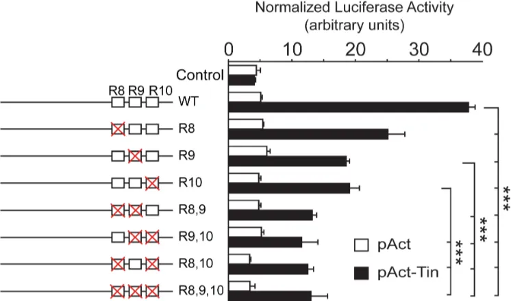
In order to determine the requirement of the identified sites to promote Tin-mediated tran-scription, we compared the transcriptional activity of thewild-type Unc-5-GHenhancer with constructs where each site is changed alone or in combinations (Fig 6). Our in vitro luciferase assay results revealed that mutagenesis of each site lead to reduced transcriptional activity, fur-ther confirming that Tin regulates the DV enhancer and its interaction with the conserved binding sites is required to induceUnc-5transcription.
Tinman activity in vivo is mediated through its binding elements on the
GH enhancer
[image:8.612.201.574.412.632.2]Given thattinis sufficient to induceUnc-5expression in ectodermal stripes (Fig 2A–2B’), if this regulation is mediated through the DV enhancer, it should also be sufficient to induce ectopic transcription from the GH reporter. As expected, misexpression of the reporter was observed in thetin-misexpressing ectoderm (Fig 7A–7A”and 7E). Thus,tinis sufficient to induceUnc-5expression from its endogenous locus or from a reporter containing the Unc-5-GHenhancer in vitro or in vivo. As theUnc5-GHenhancer is regulated directly by Tin in vitro (Fig 5) and in CBs in vivo (Fig 4), we reasoned that it might be mediated through the three identified Tin-binding sites in the GH enhancer (Fig 6). To verify this requirement, we misexpressedtininenstripes in the presence of a mutant reporter with all three binding sites mutated (R8,9,10-GH). While thewild-typereporter is ectopically expressed inenstripes our
Fig 6. The three Tin-binding motifs in theUnc-5-GHelement mediate induction ofUnc-5transcription by Tin in vitro.Mutating the three Tin-binding motifs (singly or in combination) results in reduction of the Tin transcriptional activity as observed in our luciferase assays. Each mutation on a Tin-binding site is represented as a red cross, over the corresponding site (represented as a box) at the left of the graph. Error bars represent the standard deviation and the significance of pairwise comparisons is indicated by*** (p<0.005).
Fig 7. The three Tin-binding motifs inUnc-5-GHelement mediate induction ofUnc-5transcription by Tin in vivo.Ectopic expression oftinin
engrailedstripes results in the induction of theUnc-5-GHenhancer in a striped pattern (A-A”; arrowheads in A’). Tin expression in ectodermal stripes is
labeled with anti-Tin antibody (magenta). Anti-GFP antibody was used to reveal the expression of the reporter (green). As expected, embryos carrying the
R8,9,10-GHmutant reporter display little or no GFP induction in the stripes (B-B”; arrowhead-asterisks, and E). (C-C”) Thewild-type Unc-5-GHenhancer
induces expression of the GFP reporter (green) in all CBs and PCs. Mef2 (red, D or magenta, D’) and Zfh1 (blue, D or magenta D”) antibodies are used to reveal CBs or PCs, respectively. TheR8,9,10-GHenhancer generates a GFP expression pattern similar to that of thewild-type Unc-5-GHenhancer in
tin-ABD; tin346/tin346embryos (Fig 4B–4B”) with near complete loss of GFP expression in Tin-positive CBs (D’and F) and a reduction of expression in PCs (D”).
(E and F) Quantification of GFP expression by the mutatedUnc-5enhancer (R8,9,10-GH) in ectodermal stripes (E) and CBs (F). Genotypes are indicated on the X axis and fluorescence intensities on the Y axis. For all quantifications GFP expression in sensory neurons (SNs) was used as internal control, as the fluorescence in these cells is not affected. In E, fluorescence is significantly reduced (p<1.2 x 10⁻¹⁴) inengrailedstripes of embryos with the mutant reporter
results revealed little or no activity in embryos with theR8,9,10-GHmutant reporter (compare
Fig 7A–7A”withFig 7B–7B”and 7E). Therefore, the ability of Tin to regulateUnc-5in vivo, in the ectoderm, is strictly dependent on the conserved Tin-binding sites identified in vitro. Based on these observations we predicted that Tin regulatesUnc-5through a direct binding to these sites also in CBs. Indeed, GFP expression from theR8,9,10-GHmutant enhancer was also absent from CBs (compareFig 7C–7C”withFig 7D–7D”and 7F), indicating that these sites are required by Tin to regulateUnc-5in CBs. As our internal control we also determined that reporter expression was not affected in cells that never express nor require Tin such as SNs (Fig 7E and 7F). Together, our results demonstrate thatUnc-5is regulated bytinin cardioblasts through three evolutionarily conserved Tin-binding sites.
Discussion
Cardiac mesoderm specification is strongly dependent on the combined actions of several tran-scription factors including theDocfamily of T-box transcription factors andtin[2,23,24]. Early mesodermal expression ofUnc-5is also dependent on the combined actions oftinand Doc[23] andUnc-5cardiogenic mesoderm enhancers are bound bytin,DocandPnr[24]. However, at later stages of cardiogenesis their expression pattern segregates;tinis restricted to CBs and becomes the major regulator in these cells whileDocexpression is restricted to SMCs (reviewed in [2]). One oftinfunctions in CBs is to repressDoc, and consequently activate only tindependent genes [17]. Our results show that at this developmental stage CB-specific expres-sion of Unc-5 is strictly dependent ontin(Fig 1). Intin-ABD;tin346/tin346mutants all CBs ectopically expressDoc[17]; however, it does not seem sufficient to promoteUnc-5expression on them (Fig 1) or through theUnc-5-GHheart enhancer (Fig 4). In SMCs wheretinis not expressed butUnc-5is (Fig 3)Unc-5is still expressed intinmutants. It is very likely that its expression in these cells is dependent onDocandsvp. In fact, cardiac mesoderm specification is strongly dependent on the combined action of several transcription factors including theDoc family of T-box transcription factors andtin[2]. At this stageUnc-5expression is dependent on both,tinandDoc[23]. Thus,tinspecific regulation ofUnc-5in CBs when the tubular DV assembles could represent a mark of the original cardiogenic transcriptional code owing to its developmental lineage. It would be interesting to determine if Doc is regulating Unc-5 in SMCs to confirm the segregation the expression pattern of the transcriptional regulators is reflected functionally. Enhancer regulation in CBs, where expression is virtually absent intinmutants, contrasts with that of PCs where is still moderately active (Fig 4) indicating a partial require-ment fortin. Interestingly, some PCs expresseve, a known regulator ofUnc-5in motoneurons [28,36].tinmay work combinatorially witheveand other regulators in PCs as shown for Unc-5regulation byevein motoneurons [37].
It has been recently shown that Unc-5 receptor’s role during heart morphogenesis is to pre-serve the luminal space between opposing CB membranes during heart tube lumen formation [7,8]. Accordingly,Unc-5and theUnc-5-GHreporter are expressed during tubulogenesis (Fig 3A–3E”) and its expression in CBs is strictly dependent ontin(Fig 1). Thus, there is a perfect match betweenUnc-5expression in CBs andtinregulation. Furthermore, the elimination of the Tin-binding sites in the DV enhancer renders it unresponsive to Tin in vitro (Fig 6) and in
R8,9,10-GHreporter (with unchanged SNs’fluorescence of 2.52±0.192 s.e.m and 2.8±0.177 s.e.m, respectively)]. (F) GFP fluorescence is also
significantly reduced (p<7 x 10⁻¹⁸) in CBs from 1.98±0.127 s.e.m. for thewild-type Unc-5-GHreporter to 0.28±0.052 s.e.m. for theR8,9,10-GHmutant reporter. In F, fluorescence in SNs is not affected with unchanged SNs’fluorescence of 3.75±0.44 s.e.m inUnc-5-GHembryos and 3.79±0.36 s.e.m in
R8,9,10-GHembryos, respectively. All panels are lateral views of stage 14–15 embryos with dorsal side up and anterior to the left. A magnification of the
regions delineated by insets is shown for each panel.
vivo (Fig 7). Therefore, our results strongly suggest thatUnc-5is specifically regulated by tin-man, through a direct interaction with three evolutionary conserved sites within its regulatory region at later stages of DV tubulogenesis.
Given the high degree of conservation on the molecular pathways controlling heart mor-phogenesis in vertebrates [38,39] the NK2, family of transcription factors is a very likely candi-date to drive this process, in part, through a direct regulation of guidance receptors.
Materials and Methods
Genetics
The following stocks were used:Tin-ABD;tin346/TM3,eve-lacZ, svp-lacZ[17],en-Gal4,tupisl-1/ Cyo,pnr1/TM3, pan3/Dp(2;4)eyD, AlpeyD:eyD,Unc-5 GH-GFP,Unc-5GH-Gal4 (described below),TinC-Gal4[40], andUAS-tau-Myc.
Generation of constructs
Unc-5locus dissection was carried out by PCR-amplifications using genomic DNA as template to amplify overlapping fragments of random sizes. The PCR products were cloned using TOPO TA Cloning (Invitrogen), sequenced and recombined into destination vectors: pGate-way-nlsVenus-attBand/orpGateway-Gal4and integrated into the attP2 site [41]. The pGate-way-Rlucvector was used for luciferase assays. PCR amplifiedtinwas cloned into pActC-GFP or pAct5C-FLAG plasmids to generate GFP-Tin and FLAG-Tin used in ChIP or luciferase assays, respectively. For site directed mutagenesis of Tin binding sites the most conserved nucleotides within the CACTTGA consensus motif, the“CA”dinucleotide and the first“T” [24], were mutated to“GT”and“A“, respectively. The following primers were used for muta-genesis:CACGGTATAGAGGCAACGGandCCGTTGCCTCTATACCGTGfor R8,GTTCGTCTACA GGGCAGTCACandGTGACTGCCCTGTAGACGAACfor R9, andTGCTGTCTAGTTTTGTGTGT TCTGandCAGAACACACAAAACTAGACAGCAfor R10.
Immunohistochemistry and mRNA in situ hybridization
Embryo collection, immunohistochemistry and in situ hybridization were performed as previ-ously described [28]. Reporter gene (GFP) expression in DV was quantified at embryonic stages 14–16 in different genetic backgrounds. The following antibodies were used: Mef2 (1:2000), Zfh1 (1:1500), Odd (1:1000) [33], Tin (1:1000) [27], chicken anti-GFP (1:1500) (ab13970; Abcam). Eve (1:50), c-Myc 9E10 (1:50),β-gal (1:50) were purchased from DHSB. Secondary antibodies: Alexa 555, Alexa 488-conjugated (Invitrogen) and Cy5-conjugated (Jackson ImmunoResearch Laboratories).Unc-5in situ hybridization was performed with digoxigenin-labelled probes as previously described [21]. HRP-conjugated anti-digoxigenin (Roche) followed by incubation with Cy3-labelled tyramide (PerkinElmer), as substrate, was used for detection of the hybridized probes.
Statistical analysis
Statistical significance of alterations in luciferase activity levels for reporters with different mutations or fluorescence intensity in different samples were calculated using one-tailed t-test for pair-wise comparisons and histograms were generated using Microsoft Excel 2013.
Chromatin immunoprecipitation
ChIP was performed and analyzed essentially as described previously [42]. In summary, extracts from S2R+ cells transfected with eitherpAct5C-GFP-TinorpAct5C(as mock control) were fixed in 1% formaldehyde for 10 minutes at room temperature and then lysed. Following shearing the chromatin by sonication, lysates were incubated with rabbit anti-GFP (ab290; Abcam) for 2 hours at 4°C followed by incubation with protein A-sepharose (P9424; Sigma) for an additional 2 hours. Beads were then washed and the immunoprecipitated material were eluted at 70°C overnight. Phenole-chloroform DNA extraction was performed the next day to purify the precipitated DNA. The immunoprecipitated DNA was subsequently quantified by real-time qPCR.
Luciferase reporter Assays
S2R+ cells were used for luciferase assays. Approximately 105cells were transfected (using FuGENE1HD Transfection Reagent, E2311) with the transcription factor plasmid, Rluc con-struct, and PolIII-Fluc (as internal control). Cells were analyzed for luciferase activity 36 hours post transfection using the Dual-Glo Luciferase Kit (Promega) according to manufacturer’s instructions. Samples were assessed in triplicate.
Supporting Information
S1 Fig. Reporter expression in SMCs.Intin-ABD; tin346/tin346mutant background a few CBs maintain reporter gene expression (inset in A’, arrowhead). LacZ co-staining, in the presence of LacZ reporter, (blue in A or magenta in A”) indicates that these are Tin-negative, Svp-positive CBs (SMCs).
(TIF)
Acknowledgments
We would like to thank Hanh Nguyen, Manfred Frasch, Matthew Wolf, James Skeath, Brian McCabe, for fly stocks and reagents. This work was supported by SFI grants 07/IN.1/B913 and 08/ RFP/NSC1617 and a New Foundations Award from the IRC to JPL and a Trinity research award to JA.
Author Contributions
Conceived and designed the experiments: JA AD SGK JPL. Performed the experiments: JA NN JJC-A AD. Analyzed the data: JA NN JJC-A AD SGK JPL. Contributed reagents/materials/ analysis tools: JA NN JJC-A AD SGK JPL. Wrote the paper: JA SGK JPL.
References
1. Harvey RP, Lai D, Elliott D, Biben C, Solloway M, Prall O, et al. Homeodomain factor Nkx2-5 in heart development and disease. Cold Spring Harb Symp Quant Biol. 2002; 67:107–14. Epub 2003/07/16. PMID:12858530.
3. Qian L, Liu J, Bodmer R. Slit and Robo control cardiac cell polarity and morphogenesis. Curr Biol. 2005; 15(24):2271–8. doi:10.1016/j.cub.2005.10.037PMID:16360689.
4. MacMullin A, Jacobs JR. Slit coordinates cardiac morphogenesis in Drosophila. Dev Biol. 2006; 293 (1):154–64. doi:10.1016/j.ydbio.2006.01.027PMID:16516189.
5. Santiago-Martinez E, Soplop NH, Kramer SG. Lateral positioning at the dorsal midline: Slit and Round-about receptors guide Drosophila heart cell migration. Proc Natl Acad Sci U S A. 2006; 103(33):12441– 6. doi:10.1073/pnas.0605284103PMID:16888037; PubMed Central PMCID: PMC1567898.
6. Santiago-Martinez E, Soplop NH, Patel R, Kramer SG. Repulsion by Slit and Roundabout prevents Shotgun/E-cadherin-mediated cell adhesion during Drosophila heart tube lumen formation. J Cell Biol. 2008; 182(2):241–8. doi:10.1083/jcb.200804120PMID:18663139; PubMed Central PMCID: PMC2483515.
7. Albrecht S, Altenhein B, Paululat A. The transmembrane receptor Uncoordinated5 (Unc5) is essential for heart lumen formation in Drosophila melanogaster. Dev Biol. 2011; 350(1):89–100. doi:10.1016/j. ydbio.2010.11.016PMID:21094637.
8. Macabenta FD, Jensen AG, Cheng YS, Kramer JJ, Kramer SG. Frazzled/DCC facilitates cardiac cell outgrowth and attachment during Drosophila dorsal vessel formation. Dev Biol. 2013; 380(2):233–42. doi:10.1016/j.ydbio.2013.05.007PMID:23685255; PubMed Central PMCID: PMC4137861. 9. Engelkamp D. Cloning of three mouse Unc5 genes and their expression patterns at mid-gestation.
Mech Dev. 2002; 118(1–2):191–7. PMID:12351186.
10. Medioni C, Bertrand N, Mesbah K, Hudry B, Dupays L, Wolstein O, et al. Expression of Slit and Robo genes in the developing mouse heart. Dev Dyn. 2010; 239(12):3303–11. doi:10.1002/dvdy.22449 PMID:20941780; PubMed Central PMCID: PMC2996720.
11. Fish JE, Wythe JD, Xiao T, Bruneau BG, Stainier DY, Srivastava D, et al. A Slit/miR-218/Robo regula-tory loop is required during heart tube formation in zebrafish. Development. 2011; 138(7):1409–19. doi: 10.1242/dev.060046PMID:21385766; PubMed Central PMCID: PMC3050667.
12. Mommersteeg MT, Andrews WD, Ypsilanti AR, Zelina P, Yeh ML, Norden J, et al. Slit-roundabout sig-naling regulates the development of the cardiac systemic venous return and pericardium. Circ Res. 2013; 112(3):465–75. doi:10.1161/CIRCRESAHA.112.277426PMID:23255421.
13. Kodo K, Nishizawa T, Furutani M, Arai S, Yamamura E, Joo K, et al. GATA6 mutations cause human cardiac outflow tract defects by disrupting semaphorin-plexin signaling. Proc Natl Acad Sci U S A. 2009; 106(33):13933–8. doi:10.1073/pnas.0904744106PMID:19666519; PubMed Central PMCID: PMC2728998.
14. Toyofuku T, Yoshida J, Sugimoto T, Yamamoto M, Makino N, Takamatsu H, et al. Repulsive and attrac-tive semaphorins cooperate to direct the navigation of cardiac neural crest cells. Developmental biol-ogy. 2008; 321(1):251–62. doi:10.1016/j.ydbio.2008.06.028PMID:18625214.
15. Cripps RM, Olson EN. Control of cardiac development by an evolutionarily conserved transcriptional network. Dev Biol. 2002; 246(1):14–28. doi:10.1006/dbio.2002.0666PMID:12027431.
16. Oka T, Xu J, Molkentin JD. Re-employment of developmental transcription factors in adult heart dis-ease. Semin Cell Dev Biol. 2007; 18(1):117–31. Epub 2006/12/13. doi:10.1016/j.semcdb.2006.11.012 PMID:17161634; PubMed Central PMCID: PMC1855184.
17. Zaffran S, Reim I, Qian L, Lo PC, Bodmer R, Frasch M. Cardioblast-intrinsic Tinman activity controls proper diversification and differentiation of myocardial cells in Drosophila. Development. 2006; 133 (20):4073–83. doi:10.1242/dev.02586PMID:16987868.
18. Harris R, Sabatelli LM, Seeger MA. Guidance cues at the Drosophila CNS midline: identification and characterization of two Drosophila Netrin/UNC-6 homologs. Neuron. 1996; 17(2):217–28. PMID: 8780646.
19. Mitchell KJ, Doyle JL, Serafini T, Kennedy TE, Tessier-Lavigne M, Goodman CS, et al. Genetic analy-sis of Netrin genes in Drosophila: Netrins guide CNS commissural axons and peripheral motor axons. Neuron. 1996; 17(2):203–15. PMID:8780645.
20. Keleman K, Dickson BJ. Short- and long-range repulsion by the Drosophila Unc5 netrin receptor. Neu-ron. 2001; 32(4):605–17. PMID:11719202.
21. Labrador JP, O'Keefe D, Yoshikawa S, McKinnon RD, Thomas JB, Bashaw GJ. The
homeobox transcription factor even-skipped regulates netrin-receptor expression to control dorsal motor-axon projections in Drosophila. Curr Biol. 2005; 15(15):1413–9. doi:10.1016/j.cub.2005.06.058 PMID:16085495.
23. Jin H, Stojnic R, Adryan B, Ozdemir A, Stathopoulos A, Frasch M. Genome-wide screens for in vivo Tin-man binding sites identify cardiac enhancers with diverse functional architectures. PLoS Genet. 2013; 9(1):e1003195. doi:10.1371/journal.pgen.1003195PMID:23326246; PubMed Central PMCID: PMC3542182.
24. Junion G, Spivakov M, Girardot C, Braun M, Gustafson EH, Birney E, et al. A transcription factor collec-tive defines cardiac cell fate and reflects lineage history. Cell. 2012; 148(3):473–86. doi:10.1016/j.cell. 2012.01.030PMID:22304916.
25. Bodmer R, Jan LY, Jan YN. A new homeobox-containing gene, msh-2, is transiently expressed early during mesoderm formation of Drosophila. Development. 1990; 110(3):661–9. PMID:1982429. 26. Bodmer R. The gene tinman is required for specification of the heart and visceral muscles in
Drosoph-ila. Development. 1993; 118(3):719–29. PMID:7915669.
27. Azpiazu N, Frasch M. tinman and bagpipe: two homeo box genes that determine cell fates in the dorsal mesoderm of Drosophila. Genes Dev. 1993; 7(7B):1325–40. PMID:8101173.
28. Zarin AA, Daly AC, Hulsmeier J, Asadzadeh J, Labrador JP. A GATA/homeodomain transcriptional code regulates axon guidance through the Unc-5 receptor. Development. 2012; 139(10):1798–805. doi:10.1242/dev.070656PMID:22461564.
29. Bour BA, O'Brien MA, Lockwood WL, Goldstein ES, Bodmer R, Taghert PH, et al. Drosophila MEF2, a transcription factor that is essential for myogenesis. Genes Dev. 1995; 9(6):730–41. PMID:7729689. 30. Lai ZC, Rushton E, Bate M, Rubin GM. Loss of function of the Drosophila zfh-1 gene results in abnormal
development of mesodermally derived tissues. Proc Natl Acad Sci U S A. 1993; 90(9):4122–6. PMID: 8097886; PubMed Central PMCID: PMC46458.
31. Su MT, Fujioka M, Goto T, Bodmer R. The Drosophila homeobox genes zfh-1 and even-skipped are required for cardiac-specific differentiation of a numb-dependent lineage decision. Development. 1999; 126(14):3241–51. PMID:10375513.
32. Ward EJ, Coulter DE. odd-skipped is expressed in multiple tissues during Drosophila embryogenesis. Mech Dev. 2000; 96(2):233–6. PMID:10960790.
33. Ward EJ, Skeath JB. Characterization of a novel subset of cardiac cells and their progenitors in the Dro-sophila embryo. Development. 2000; 127(22):4959–69. PMID:11044409.
34. Gajewski K, Choi CY, Kim Y, Schulz RA. Genetically distinct cardial cells within the Drosophila heart. Genesis. 2000; 28(1):36–43. PMID:11020715.
35. Xu X, Yin Z, Hudson JB, Ferguson EL, Frasch M. Smad proteins act in combination with synergistic and antagonistic regulators to target Dpp responses to the Drosophila mesoderm. Genes Dev. 1998; 12(15):2354–70. PMID:9694800; PubMed Central PMCID: PMC317052.
36. Zarin AA, Asadzadeh J, Labrador JP. Transcriptional regulation of guidance at the midline and in motor circuits. Cell Mol Life Sci. 2014; 71(3):419–32. doi:10.1007/s00018-013-1434-xPMID:23917723. 37. Zarin AA, Asadzadeh J, Hokamp K, McCartney D, Yang L, Bashaw GJ, et al. A transcription factor
net-work coordinates attraction, repulsion, and adhesion combinatorially to control motor axon pathway selection. Neuron. 2014; 81(6):1297–311. Epub Feb 20th. doi:10.1016/j.neuron.2014.01.038PMID: 24560702; PubMed Central PMCID: PMC4128230.
38. Wolf MJ, Amrein H, Izatt JA, Choma MA, Reedy MC, Rockman HA. Drosophila as a model for the iden-tification of genes causing adult human heart disease. Proc Natl Acad Sci U S A. 2006; 103(5):1394–9. doi:10.1073/pnas.0507359103PMID:16432241; PubMed Central PMCID: PMC1360529.
39. Neely GG, Kuba K, Cammarato A, Isobe K, Amann S, Zhang L, et al. A global in vivo Drosophila RNAi screen identifies NOT3 as a conserved regulator of heart function. Cell. 2010; 141(1):142–53. doi:10. 1016/j.cell.2010.02.023PMID:20371351; PubMed Central PMCID: PMC2855221.
40. Lo PCH, Frasch M. A role for the COUP-TF-related gene seven-up in the diversification of cardioblast identities in the dorsal vessel of Drosophila. Mechanisms of Development. 2001; 104:49–60. doi:10. 1016/S0925-4773(01)00361-6PMID:11404079
41. Fish MP, Groth AC, Calos MP, Nusse R. Creating transgenic Drosophila by microinjecting the site-spe-cific phiC31 integrase mRNA and a transgene-containing donor plasmid. Nat Protoc. 2007; 2
(10):2325–31. doi:10.1038/nprot.2007.328PMID:17947973.