R E S E A R C H A R T I C L E
Open Access
Changes in plasma CXCL4 levels are
associated with improvements in lung
function in patients receiving
immunosuppressive therapy for systemic
sclerosis-related interstitial lung disease
Elizabeth R. Volkmann
1*, Donald P. Tashkin
1, Michael D. Roth
1, Philip J. Clements
1, Dinesh Khanna
2, Daniel E. Furst
1,
Maureen Mayes
3, Julio Charles
3, Chi-Hong Tseng
1, Robert M. Elashoff
4and Shervin Assassi
3Abstract
Background:Increased circulatory levels of the chemokine CXCL4 have been associated with the presence of
interstitial lung disease (ILD) in an observational study of patients with systemic sclerosis (SSc). The purpose of the present study was to evaluate the relationship between baseline CXCL4 level and extent of ILD in the context of a randomized controlled trial and to determine whether changes in CXCL4 levels in response to immunosuppression are associated with future progression of SSc-ILD.
Methods:A total of 142 SSc-ILD patients from Scleroderma Lung Study (SLS) II were randomized in a double-blind, parallel-arm trial, to receive mycophenolate (MMF) for 2 years or oral cyclophosphamide (CYC) for 1 year followed by 1 year of placebo. Plasma CXCL4 levels were measured at baseline, 12 months, and 24 months in SLS II participants (N= 136) and at a single time point in healthy controls (N= 67). A mixed-effects model evaluated the relationship between change in CXCL4 levels and SSc-ILD progression. The primary outcome was the course of the forced vital capacity.
Results:Baseline CXCL4 levels were significantly higher in SSc-ILD patients compared with healthy controls (2699 ± 1489 ng/ml vs 2233 ± 1351 ng/ml (mean ± SD);P= 0.019). However, no significant correlations were identified between CXCL4 levels and extent of ILD at baseline, as measured by the forced vital capacity, diffusing capacity of carbon monoxide, or radiographic extent of ILD. Plasma CXCL4 decreased significantly from baseline to 12 months in all patients (CYC:P< 0.001; MMF:P= 0.006) with no between-treatment differences (CYC vs MMF). Patients with the largest decline in CXCL4 levels during the first 12 months had an improved course of forced vital capacity %-predicted from 12 to 24 months (P= 0.040), even after adjusting for baseline disease severity and treatment arm assignment.
(Continued on next page)
* Correspondence:[email protected]
1Department of Medicine, University of California, David Geffen School of Medicine, Los Angeles, CA, USA
Full list of author information is available at the end of the article
(Continued from previous page)
Conclusions:Levels of CXCL4 were higher in patients with SSc-ILD compared with controls and decreased in all
patients treated with immunosuppressive therapy. While CXCL4 levels were not correlated with extent of ILD at baseline, changes in CXCL4 at 12 months predicted future progression of SSc-ILD from 12 to 24 months. These findings suggest that intermediate-term changes in CXCL4 may have predictive significance for long-term progression of SSc-ILD in patients receiving immunosuppressive therapy.
Trial registration:ClinicalTrials.gov NCT00883129. Registered 16 April 2009.
Keywords:Systemic sclerosis, Pulmonary fibrosis, Chemokines, Immunosuppression
Background
Although interstitial lung disease (ILD) occurs in the ma-jority of patients with systemic sclerosis (SSc) and is also the leading cause of death in SSc [1], ILD progression rates vary considerably in this patient population [2].
Identifying specific factors that may predict response to immunosuppression is central to improving our ability to determine which SSc patients may derive the greatest benefit from ILD-targeted therapy. While nu-merous studies have examined baseline factors that are associated with ILD progression [3–9], relatively few studies have examined whether these factors are associated with a positive response to immunosup-pression. In the Scleroderma Lung Study (SLS) I (com-paring cyclophosphamide (CYC) with placebo), the severity of reticular infiltrates on baseline high-resolution computed tomography (HRCT) was associ-ated with improved responsiveness to CYC therapy [10]. However, to our knowledge, no studies have ex-amined responsiveness to therapy with mycophenolate (MMF) in SSc-ILD.
Biomarkers may have clinical application for predict-ing response to immunosuppression in patients with SSc-ILD. Several studies have identified potential circu-lating biomarkers associated with accelerated SSc-ILD progression, including interleukin (IL)-6 [11], C-reactive protein (CRP) [12], monocyte chemoattractant protein-1 (MCP-1) [13], CC chemokine ligand 18 (CCL18) [14, 15], and CXCL4 [16]. However, these markers have not been consistently shown to predict relevant clin-ical outcomes across a variety of treatments and SSc populations. Furthermore, all of the studies assessing the aforementioned candidate biomarkers were ob-servational by design and consequently included pa-tients who exhibited diverse phenotypic expressions of SSc, received a variety of immunosuppressant therapies, and were followed for varying periods of time [11–16].
Of these potential biomarkers, the chemokine, CXCL4, may play an important role in perpetuating profibrotic ac-tivity in the SSc-ILD disease state. An observational study found that CXCL4 levels were higher in SSc patients with
ILD compared with those without ILD in a discovery co-hort, as well as in two independent replication cohorts [16]. In a subgroup of patients from this study (N= 79), SSc patients with high baseline CXCL4 levels had acceler-ated progression of ILD as measured by a decline in the diffusing capacity for carbon monoxide (DLCO) [16]. Notably, this study also found that CXCL4 levels were remarkably higher in SSc patients compared with healthy controls (270-fold higher) [16].
The present study aimed to evaluate whether the chemokine CXCL4 is associated with the extent of ILD in a well-characterized cohort of SSc patients, all of whom had clinically significant ILD. Unlike previous studies of this nature, the present study analyzed CXCL4 at multiple time points in concert with the sim-ultaneous collection of extensive clinical, physiologic, and radiographic data. A secondary aim was to deter-mine whether changes in CXCL4 levels can predict im-provements in lung function in patients receiving MMF or CYC in the setting of a randomized controlled clinical trial (RCT).
Methods
Study participants
Participants in the SLS II trial (N= 142) were all adults, aged 18–75 years, who exhibited either limited or diffuse cutaneous SSc [17] and active ILD as demonstrated by restrictive ventilatory impairment (forced vital capacity (FVC) <80% but ≥45% predicted), exertional dyspnea (Grade ≥2 on the Magnitude of Task component of the Mahler Baseline Dyspnea Index (BDI)) [18], and the presence of any ground glass opacity (GGO; hazy opacity through which normal lung markings can be discerned) on HRCT. Key exclusion criteria included pulmonary hypertension; clinically significant abnormalities on HRCT not attributable to SSc; and smoking within the past 6 months or evidence of significant airflow obstruc-tion. Complete details of the SLS II population have been reported previously [19].
matched for age, ethnicity, and gender to SLS II partic-ipants in a 1:2 ratio.
SLS II study design
Enrolled patients were randomly (1:1 block) assigned in a double-blind, parallel-arm manner to receive either oral CYC for 1 year followed by 1 year of placebo or MMF for 2 years. SLS II was a superiority study con-ducted at 14 university hospitals throughout the USA (for the list of sites see Ethics approval and consent to participate) and recruited patients between November 2009 and January 2013. For complete details of the SLS II protocol, please see the supplementary Web appendix accompanying the main SLS manuscript [19]. There were no significant changes in the protocol involving the design, outcomes, treatment, or eligibility criteria during the trial. Baseline measurements included the following physiological variables: spirometry (FVC and forced ex-pired volume in 1 second (FEV1)), lung volumes
(func-tional residual capacity (FRC), residual volume (RV), and total lung capacity (TLC) by whole-body plethysmogra-phy or helium dilution), hemoglobin-adjusted single-breath DLCO, and the ratio of DLCO to alveolar volume (DL/VA). The FVC (primary SLS II endpoint) and DLCO (secondary SLS II endpoint) were measured every 3 months, and the TLC was measured every 6 months during the trial. HRCT thoracic imaging was obtained at baseline and at 24 months and a computer-aided design (CAD) scoring system was employed to provide quanti-tative measures of different patterns of ILD as described previously [19, 20]. The Quantitative ILD (QILD) score was the sum of all abnormally classified scores, including scores for quantitative lung fibrosis (QLF, linear reticular markings), GGO, and honeycomb changes (clustered air-filled cysts with dense walls). Scores were calculated as the percentage of voxels for both the whole lung (WL), including both lungs, and for the lobe of maximal involvement (LM).
CXCL4 measurement
SLS II plasma samples were collected at the baseline, 12-month, and 24-month study visits and were immedi-ately processed on-site on the day of collection, stored at
−70 °C, and shipped on dry ice to the central repository at the University of Texas, Houston. Samples from healthy controls collected at the University of Texas, Houston, were handled in the same manner except that no shipping was required. Validated, commercially avail-able ELISA kits (Human CXCL4/PF4 Quantikine Kit; R&D Systems) were used to assess CXCL4 levels. All as-says were performed in duplicate and the coefficient of variance was <20%. Technicians performing the assays were blinded to the study arm assignment. CXCL4 data
were logarithmically transformed to correct for data skewness.
Statistical analysis Baseline characteristics
Summary statistics were generated for baseline charac-teristics. A two-sample ttest was used to compare con-tinuous variables and a chi-square test was used to compare categorical variables. Pearson correlations were performed to examine the relationship between CXCL4 levels and baseline measures of extent of ILD, as mea-sured by the FVC, DLCO, QILD, and QLF, as well as the relationship between baseline CXCL4 levels and platelet counts.
Relationship between change in CXCL4 and progression of SSc-ILD
To determine whether a change in plasma CXCL4 might correlate with or predict the progression of SSc-ILD, we first plotted the course of measured plasma CXCL4, by treatment arm, from baseline to 24 months and deter-mined the corresponding magnitude of change for indi-vidual subjects from baseline to 12 and 24 months. Pearson correlations were initially performed to examine the relationship between the change in CXCL4 levels from baseline to 12 months and the absolute change for the primary outcome (FVC %-predicted) and key sec-ondary outcomes (DLCO %-predicted and HRCT mea-sures of the extent of ILD).
Following these initial analyses, a mixed-effects model was generated, the outcome of which was the course of FVC %-predicted measured in 3-month increments from 12 to 24 months (primary outcome). The model in-cluded the following covariates: change in CXCL4 from baseline to 12 months, baseline FVC %-predicted, base-line extent of quantitative fibrosis/ILD, treatment arm, and a time trend. The covariate of baseline extent of quantitative fibrosis/ILD was defined as the first princi-pal component from a principrinci-pal component analysis of the following variables: QILD-LM, QILD-WL, QLF-LM, and QLF-WL.
All tests were two-sided, and all analyses were per-formed using SAS 9.2 (SAS Institute, Inc., Cary, NC, USA).
Results
Baseline characteristics
median 1720 (IR 1224, 2874); P= 0.019) (Fig. 1). There were no significant differences in baseline CXCL4 levels between treatment arms, between limited or diffuse SSc patients, between men and women, or between ethnicities (allP≥0.2). There were also no significant correlations be-tween baseline CXCL4 and age, or disease duration, as measured by the onset of the first Raynaud’s phenomenon symptom (r=−0.10,P= 0.24), or as measured by the onset of the first non-Raynaud’s phenomenon symptom attribut-able to SSc (r= 0.040, P= 0.64). Similarly, among the healthy controls there were no significant differences in CXCL4 levels between men and women or between eth-nicities, and nor was there any correlation between CXCL4 levels and age.
CXCL4 correlations
At baseline there were no significant correlations be-tween plasma CXCL4 levels and any of the physiologic or radiographic measures of extent or severity of SSc-ILD (Table 2). There were also no significant correla-tions between plasma CXCL4 and extent of SSc-ILD at 12 or 24 months (data not shown).
There was no significant correlation between baseline CXCL4 and baseline mRSS among all participants (r= 0.10;P= 0.24), or among patients with diffuse cutaneous sclerosis (N= 80;r= 0.059;P= 0.60).
At baseline, CXCL4 levels demonstrated a weak but signifi-cant correlation with the platelet count (r= 0.320,P< 0.001).
Plasma levels of CXCL4 levels decrease in response to immunosuppression
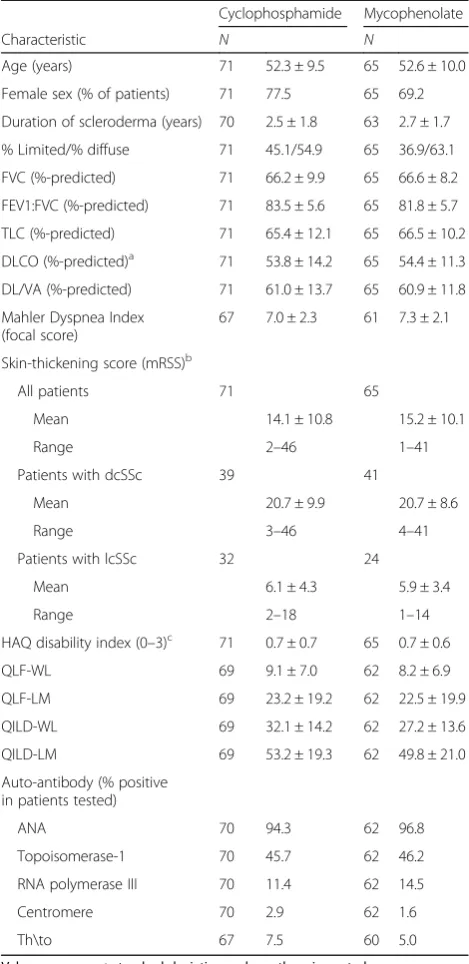
[image:4.595.56.291.110.593.2]Among the 136 patients with baseline plasma CXCL4 levels measured, 103 patients had 12-month CXCL4 Table 1Baseline characteristics of SLS II participants with
CXCL4 measurements at baseline
Cyclophosphamide Mycophenolate
Characteristic N N
Age (years) 71 52.3 ± 9.5 65 52.6 ± 10.0
Female sex (% of patients) 71 77.5 65 69.2
Duration of scleroderma (years) 70 2.5 ± 1.8 63 2.7 ± 1.7
% Limited/% diffuse 71 45.1/54.9 65 36.9/63.1
FVC (%-predicted) 71 66.2 ± 9.9 65 66.6 ± 8.2
FEV1:FVC (%-predicted) 71 83.5 ± 5.6 65 81.8 ± 5.7
TLC (%-predicted) 71 65.4 ± 12.1 65 66.5 ± 10.2
DLCO (%-predicted)a 71 53.8 ± 14.2 65 54.4 ± 11.3
DL/VA (%-predicted) 71 61.0 ± 13.7 65 60.9 ± 11.8
Mahler Dyspnea Index (focal score)
67 7.0 ± 2.3 61 7.3 ± 2.1
Skin-thickening score (mRSS)b
All patients 71 65
Mean 14.1 ± 10.8 15.2 ± 10.1
Range 2–46 1–41
Patients with dcSSc 39 41
Mean 20.7 ± 9.9 20.7 ± 8.6
Range 3–46 4–41
Patients with lcSSc 32 24
Mean 6.1 ± 4.3 5.9 ± 3.4
Range 2–18 1–14
HAQ disability index (0–3)c 71 0.7 ± 0.7 65 0.7 ± 0.6
QLF-WL 69 9.1 ± 7.0 62 8.2 ± 6.9
QLF-LM 69 23.2 ± 19.2 62 22.5 ± 19.9
QILD-WL 69 32.1 ± 14.2 62 27.2 ± 13.6
QILD-LM 69 53.2 ± 19.3 62 49.8 ± 21.0
Auto-antibody (% positive in patients tested)
ANA 70 94.3 62 96.8
Topoisomerase-1 70 45.7 62 46.2
RNA polymerase III 70 11.4 62 14.5
Centromere 70 2.9 62 1.6
Th\to 67 7.5 60 5.0
Values are mean ± standard deviation, unless otherwise noted
a
Adjusted for hemoglobin
b
Scores for skin thickening (mRSS) can range from 0 to 51, with higher scores indicating more severe thickening
c
Scores for HAQ Disability Index can range from 1 to 3, with higher numbers indicating greater disability
FVCforced vital capacity,FEV1forced expired volume in 1 second,TLCtotal lung capacity,DLCOdiffusing capacity of the lung for carbon monoxide,DL/VA ratio of DLCO to alveolar volume,mRSSmodified Rodnan skin score,dcScc diffuse cutaneous systemic sclerosis,lcSSclimited or cutaneous systemic sclerosis,HAQHealth Assessment Questionnaire,QLFquantitative extent of lung fibrosis on high-resolution chest computed tomography,QILD quantita-tive extent of total interstitial lung disease (including fibrosis, honeycomb, and ground glass opacity),WLwhole lung,LMlobe of maximal involvement ANAAnti-nuclear antibody,Th/ToTh/To ribonucleoprotein antibody
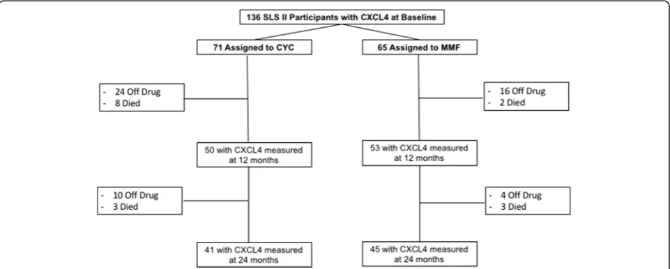
[image:4.595.304.539.498.694.2]measurements (CYC = 50; MMF = 53), and 86 patients
had 24-month CXCL4 measurements (CYC = 41;
MMF = 45) (Fig. 2). CXCL4 levels were measured in all patients who returned for the follow-up study visits at 12 and 24 months.
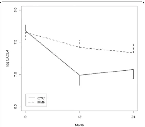
The majority of patients in both treatment arms expe-rienced a decrease in CXCL4 levels from baseline to 12 months (CYC: 76% (37/49); MMF: 67% (35/52)) and from baseline to 24 months (CYC: 70% (28/40); MMF: 68% (30/44)). Plasma CXCL4 levels declined significantly in the CYC and MMF arms (P< 0.001 andP= 0.006, re-spectively). Similarly, CXCL4 levels showed a significant decrease from baseline to 24 months in the CYC and MMF arms (P< 0.001 and P= 0.011, respectively). Although participants in the CYC arm experienced nu-merically larger decreases in CXCL4 levels, there was no
statistically significant difference in the change in CXCL4 levels between treatment arms from baseline to 12 months (P= 0.120) or from baseline to 24 months (P= 0.380) (Fig. 3). While CXCL4 levels slightly in-creased from 12 to 24 months in the CYC arm (during the placebo phase), CXCL4 levels continued to decline from 12 to 24 months in the MMF arm.
Changes in plasma levels of CXCL4 levels may predict improved course of FVC
As reported previously [19], the course of the FVC in SLS II improved significantly from baseline to 24 months in both the CYC and MMF treatment arms, with no between-treatment difference. Of note, FVC measure-ments were obtained in 51 and 59 participants at 12 months and in 50 and 53 participants at 24 months for the CYC and MMF arms, respectively. When changes in the plasma CXCL4 levels at 12 months were correlated with changes in the FVC (bivariate analysis), there was a trend for a negative correlation between the change in CXCL4 from baseline to 12 months and the change in FVC from baseline to 18 months (r=−0.203;
P= 0.063), 21 months (r=−0.212; P= 0.062), and 24 months (r=−0.182; P= 0.082), but not at 12 months (r=−0.070;P= 0.499) or 15 months (r=−0.048;P= 0.662).
[image:5.595.57.290.109.222.2]There were no significant correlations between the change from baseline in CXCL4 at 12 months and the change from baseline in DLCO at 12, 15, 18, 21, or 24 months (all P> 0.3). There was also no significant correlation between the change from baseline in CXCL4 at 12 months and the change from baseline in QLF-LM/ WL or QILD-LM/WL at 24 months (allP> 0.4).
Table 2Baseline CXCL4 levels are not correlated with extent of ILD in SLS II subjects
Surrogate ILD measurements Pearson correlation (r) Pvalue
TLC% predicted 0.123 0.154
FVC% predicted 0.089 0.304
DLCO% predicted 0.082 0.345
QLF-LM −0.027 0.763
QILD-LM −0.063 0.473
QLF-WL −0.034 0.703
QILD-WL −0.022 0.799
Values are mean ± standard deviation, unless otherwise noted ILDinterstitial lung disease,SLSScleroderma Lung Study,FVCforced vital capacity,TLCtotal lung capacity,DLCOdiffusing capacity of the lung for carbon monoxide,QLFquantitative extent of lung fibrosis on high-resolution chest computed tomography,QILDquantitative extent of total interstitial lung disease (including fibrosis, honeycomb, and ground glass opacity),WLwhole lung,LMlobe of maximal involvement
Fig. 2Disposition of SLS II study participants with regard to CXCL4 measurement during the 24-month trial. FVC measurements were obtained in 51 and 59 participants at 12 months and in 50 and 53 participants at 24 months for the CYC and MMF arms, respectively.CYCcyclophosphamide,
[image:5.595.58.539.501.694.2]To further explore the relationship between the change in CXCL4 and the course of the FVC, a mixed-effects model was generated and the results demon-strated that the change in CXCL4 from baseline to 12 months was associated with the course of the FVC (as a continuous variable) from 12 to 24 months (esti-mate −1.232; 95% CI −2.405, −0.060; P= 0.040), after controlling for treatment arm assignment, a time trend, baseline quantitative extent of ILD/fibrosis, and baseline FVC. A similar statistical approach was used in the ana-lysis of the primary and secondary outcomes in SLS II [19]. The results demonstrated that every 1-unit de-crease in the log CXCL4 level was associated with a 1.23% increase in the FVC. In other words, patients who experienced the greatest decline in CXCL4 levels during the first 12 months of the study had an improved course of FVC from 12 to 24 months (Table 3).
Given the significant correlation between CXCL4 levels and platelet count at baseline, baseline platelet count was added to the mixed-effects model. Adding baseline platelet count to the model did not affect the significant relationship between change in CXCL4 levels and the outcome variable. However, the baseline platelet count was not independently associated with the course of the FVC (P= 0.163), and for this reason this variable was not included in the final model.
Because not all patients had CXCL4 measurements at 12 months, the final model included 97 patients with SSc-ILD. Notably, there were no significant differences
in baseline characteristics (i.e., age, gender, diffuse/lim-ited, FVC, DLCO, MRSS, QILD-WL/LM, QLF-WL/LM) between the 97 patients included in the final mixed-effects model and the 39 patients who were not included.
Discussion
In a well-characterized cohort of patients with SSc-ILD, CXCL4 levels decreased significantly in response to im-munosuppressive therapy with both CYC and MMF. Furthermore, a greater change in CXCL4 from baseline to 12 months led to improvements in SSc-ILD at 24 months as measured by the FVC %-predicted. These findings suggest that intermediate-term changes in CXCL4 may have predictive significance for long-term progression of SSc-ILD in patients receiving immuno-suppressive therapy.
These findings are consistent with the purported pathobiological role of CXCL4 in moderating fibrotic and inflammatory processes. In addition to the previ-ously reported association with various dimensions of SSc [16], CXCL4 levels have also been elevated in liver fibrosis [21] and in patients with antiphospholipid syn-drome [22]. In a recent study, CXCL4 blocked the induction of the anti-inflammatory enzyme heme oxygenase-1. Furthermore, the product of heme oxy-genase enzymatic activity, bilirubin, showed an inverse correlation with CXCL4 levels in plasma samples of SSc patients [23]. While our understanding of the role of CXCL4 in the development of SSc-ILD is evolving, the present manuscript provides further support for its role in the pathogenesis of SSc.
This study further sheds light on plausible biological mediators/targets of the two immunosuppressant agents under study (i.e. MMF and CYC). Although CXCL4 levels were not correlated with any of the baseline surro-gate markers of ILD severity, CXCL4 levels significantly declined in both treatment arms in response to im-munosuppression. Furthermore, the decline in CXCL4 paralleled the improvements appreciated in the course
Fig. 3CXCL4 levels decreased significantly in response to immunosuppression in each treatment arm from baseline to 12 months (CYC:P< 0.001; MMF:P= 0.006) and from baseline to 24 months (CYC:P< 0.001; MMF:P= 0.011). There was no between-treatment difference in the reduction in CXCL4 from baseline to 12 months or from baseline to 24 months.CYCcyclophosphamide,
[image:6.595.57.292.86.291.2]MMFmycophenolate
Table 3Multivariable analysis examining the relationship between change in CXCL4 from baseline to 12 months and the course of FVC from 12 to 24 months (N= 97)
Baseline covariate Estimate 95% CI Pvalue
ΔCXCL4 −1.232 −2.405,−0.060 0.040
Baseline FVC 0.972 0.807, 1.138 <0.0001
Baseline QILD/QLFa 0.297 −0.496, 1.089 0.464
Treatment arm −0.361 −3.182, 2.460 0.802 a
[image:6.595.304.538.121.191.2]of the FVC over 24 months as reported in the main SLS II manuscript [19]. Although there was no significant difference in the degree to which CXCL4 decreased be-tween treatment arms, the sample size may have limited our ability to detect a difference because CXCL4 levels appeared to decrease to a greater extent in the CYC arm compared with the MMF arm within the first 12 months of treatment (Fig. 3). Interestingly, CXCL4 levels de-clined most dramatically during the first 12 months of therapy in both study arms. The patients assigned to CYC received placebo in the second year, which may ex-plain the slight upward trend in CXCL4 levels during the second year of the study. However, the CXCL4 levels did not return to those observed at the baseline visit after 2 years in the CYC arm. This finding may in part be related to the use of alternate therapy during the lat-ter period. Of the 15 subjects in SLS II who withdrew from or failed treatment but returned for final study out-come measures, 12 indicated that they were switched to potentially disease-modifying therapy (MMF, intravenous CYC, rituximab, tocilizumab, intravenous immunoglobu-lin, or azathioprine) and these alternative therapies may have influenced the results [19]. In the MMF arm, CXCL4 levels continued to decline during the second year of the study, likely related to continuous use of im-munosuppression in this arm.
Interestingly, we did not observe significant correla-tions between baseline CXCL4 levels and extent of ILD as measured by the FVC, DLCO, TLC, or extent of quantitative radiographic fibrosis at baseline, 12 months, or 24 months. While a prior study [16] demonstrated that patients with high CXCL4 were more likely to have fibrosis on HRCT, this prior observational study in-cluded SSc patients both with and without ILD and at varying disease stages. By contrast, the present study only included patients with ILD at a relatively early stage of disease (mean disease duration of 2.6 years from the onset of the first non-Raynaud’s symptom attributable to SSc), nearly all of whom already had evidence of fibrosis on HRCT [19]. Our findings suggest that CXCL4 may be a more useful predictive biomarker for response to immunosuppression in patients with early SSc-ILD. Whether CXCL4 correlates with severity of ILD at later stages of SSc remains uncertain. Furthermore, because our cohort did not evaluate SSc patients without ILD, we could not assess whether CXCL4 levels can distin-guish SSc patients with and without ILD.
We did observe a significant but weak correlation be-tween CXCL4 levels and platelet levels. However, the platelet level did not demonstrate predictive significance for FVC progression in the mixed-effects model. Import-antly, precautions were taken to limit platelet activation, which can significantly affect CXCL4 levels, including using strict caution to minimize venostasis during the
blood draw with butterfly needles and immediately cen-trifuging the specimens. Of note, the prior study in this area [16] did not assess platelet levels.
Consistent with the work of van Bon et al. [16], we ob-served an increase in plasma CXCL4 levels in SSc-ILD patients compared with healthy controls. However, the magnitude of this difference was strikingly smaller in the present study (1.6-fold increase in our study vs 277-fold increase in the previous study). The observed difference in CXCL4 levels between SSc-ILD patients and healthy controls in the present report is consistent with the re-ported fold increases of other cytokines and chemokines in SSc plasma samples [12, 24]. While several possible explanations for this disparity exist, we note that we used a different ELISA assay in the present study than in the previous study (R&D Systems/Quantikine in the present study vs R&D Systems/DuoSet in the previous study). The assay we utilized has been fully validated for the measurement of CXCL4 in complex fluids such as serum and plasma [25].
There are notable strengths of the present study. First, this is the first study to measure CXCL4 levels at mul-tiple time points in patients with early, symptomatic SSc-ILD. Second, our initial measurement of CXCL4 oc-curred prior to the initiation of immunosuppressive therapy for SSc-ILD; thus, the reported baseline levels of CXCL4 are likely closely related to the underlying dis-ease state without any interference from disdis-ease- disease-modifying therapies (patients were ineligible for SLS II if they had received CYC or MMF for >8 weeks ever in the past, or they had received intravenous CYC more than twice ever in the past, or were taking CYC/MMF/any medication with potential disease-modifying activity within 30 days of randomization). Third, unlike prior ob-servational studies in this field, this cohort had uniform follow-up measurements with likely less missing data. Fourth, our analyses controlled for treatment assign-ment. Finally, this study was conducted at 14 centers and included patients from varying ethnic backgrounds, thereby increasing the generalizability of our findings.
Future studies may consider measuring CXCL4 at even earlier time points as a means of determining treatment response prior to 1 year. While we report that the change in CXCL4 at 1 year was associated with im-proved SSc-ILD progression at 2 years, additional studies would be useful to determine whether changes in CXCL4 at earlier time points might have similar predict-ive value. If the latter were found, it might be useful from a clinical standpoint to determine whether a pa-tient is responding to a particular therapy at 3 or 6 months, to avoid the unfruitful continuation of an in-effective therapy.
Conclusions
While CXCL4 was not associated with extent of SSc-ILD at baseline, this chemokine decreased significantly in response to immunosuppressive therapy. Moreover, the change in CXCL4 levels was associated with future progression of SSc-ILD. Although the exact role of CXCL4 in the pathobi-ology of SSc-ILD is uncertain, these findings support the potential predictive capability of this chemokine for deter-mining response to immunosuppressive therapy.
Abbreviations
CAD:Computer-aided design; CCL18: CC chemokine ligand 18; CRP: C-reactive protein; CYC: Cyclophosphamide; DL/VA: Ratio of DLCO to alveolar volume; DLCO: Diffusing capacity for carbon monoxide; FEV1: Forced expired
volume in 1 second; FRC: Functional residual capacity; FVC: Forced vital capacity; GGO: Ground glass opacity; HAQ: Health Assessment Questionnaire; HRCT: High-resolution chest computed tomography; IL: Interleukin; ILD: Interstitial lung disease; LM: Lobe of maximal involvement; MCP-1: Monocyte chemoattractant protein-1; MMF: Mycophenolate; mRSS: Modified Rodnan skin score; QILD: Quantitative extent of total interstitial lung disease (including fibrosis, honeycomb, and ground glass opacity); QLF: Quantitative extent of lung fibrosis on HRCT; RV: Residual volume; SLS: Scleroderma Lung Study; SSc: Systemic sclerosis; TLC: Total lung capacity; WL: Whole lung
Acknowledgements
The authors thank the patients for their participation in SLS II.
Funding
This work was supported by grants from the NHLBI/NIH: R01 HL089758 (DPT) and R01 HL089901 (RME), NIH/NIAMS K23AR061436 (SA), Rheumatology Research Foundation (SA), NIH/NIAMS K24 AR063120 (DK), and the Scleroderma Foundation (ERV). The study drug (MMF) and matching placebo were supplied at no charge through Drug Supply Grant # CEL539 from Hoffmann-La Roche/ Genentech.
Availability of data and materials
The datasets for the current study are available from the corresponding author upon reasonable request.
Authors’contributions
ERV made substantial contributions to study conception and design, as well as analysis and interpretation of data, drafting the article, and approval of the final manuscript. DPT, MDR, PPC, DK, DEF, and MM made substantial contributions to study conception and design, revising the manuscript critically for important intellectual content, and approval of the final manuscript. JC and C-HT made substantial contributions to the analysis and interpretation of data, revising the manuscript critically for important intellectual content, and approval of the final manuscript. RME and SA made substantial contributions to study conception and design, the analysis and interpretation of data, revising the manuscript critically for important intellectual content, and approval of the final manuscript.
Competing interests
No nonfinancial conflicts of interest exist for any of the authors. Financial conflicts of interest are as follows.
ERV, PPC, RME, C-HT, and JC all report nothing to disclose.
DPT reports personal fees from EMD Serono, and nonfinancial support from Genentech, during the conduct of the study.
MDR reports nonfinancial support from Hoffmann-La Roche/Genentech, during the conduct of the study.
DEF reports grant/research support from AbbVie, Actelion, Amgen, BMS, Gilead, GSK, Novartis, Pfizer, Roche/Genentech, and UCB, and consultant work with AbbVie, Amgen, BMS, Cytori, Janssen, Gilead, GSK, NIH, Novartis, Pfizer, Roche/Genentech, UCB Speaker's Bureau (CME ONLY) AbbVie, Actelion, and UCB during the course of the study.
DK reports personal fees from Bayer, grants from Bristol-Myer Squibb, personal fees from Cytori, personal fees from EMD Serono (Merck), personal fees from Forward, grants and personal fees from Genentech/Roche (InterMune), and personal fees from Lycera during the course of the study. MM reports grants from InterMune, grants from Roche/Genentech, grants from Bayer, grants from University of Michigan, grants from Boehringer-Ingelheim, grants from DOD, grants from Corbus, and grants from Cytori during the course of the study.
SA reports grants and personal fees from Boehringer Ingelheim, grants from Genentech, grants and personal fees from Biogen Idec, and grants from Bayer HealthCare during the course of the study.
Consent for publication Not applicable.
Ethics approval and consent to participate
This study was approved by all of the local institutional review boards for the 14 participating SLS II sites (see following list), and all patients underwent informed consent.
Boston University UCLA
Georgetown University Johns Hopkins University
Medical University of South Carolina National Jewish Health
Northwestern University Rutgers University UCSF
University of Illinois University of Michigan University of Texas, Houston University of Utah University of Minnesota
Author details
1Department of Medicine, University of California, David Geffen School of Medicine, Los Angeles, CA, USA.2Department of Medicine, University of Michigan Medical School, Ann Arbor, MI, USA.3Department of Medicine, University of Texas Health Science Center at Houston, Houston, TX, USA. 4Department of Biomathematics, University of California, Los Angeles, CA, USA.
Received: 16 September 2016 Accepted: 6 December 2016
References
1. Tyndall AJ, Bannert B, Vonk M, et al. Causes and risk factors for death in systemic sclerosis: a study from the EULAR Scleroderma Trials and Research (EUSTAR) database. Ann Rheum Dis. 2010;69:1809–15.
2. Wells AU. Interstitial lung disease in systemic sclerosis. Press Med. 2014;43: e329–43.
3. Morgan C, Knight C, Lunt M, et al. Predictors of end stage lung disease in a cohort of patients with scleroderma. Ann Rheum Dis. 2003;62:146–50. 4. Plastiras SC, Karadimitrakis SP, Ziakas PD, et al. Scleroderma lung: initial
5. Nihtyanova SI, Schreiber BE, Ong VH, et al. Prediction of pulmonary complications and long-term survival in systemic sclerosis. Arthritis Rheumatol. 2014;66:1625–35.
6. Steen VD, Medsger Jr TA. Severe organ involvement in systemic sclerosis with diffuse scleroderma. Arthritis Rheum. 2000;43:2437–44.
7. Goh NSL, Desai SR, Veeraraghaven S, et al. Interstitial lung disease in systemic sclerosis. A simple staging system. Am J Respir Crit Care Med. 2008;177:1248–54.
8. Moore OA, Goh N, Corte T, et al. Extent of disease on high-resolution compute tomography lung is a predictor of decline and mortality in systemic sclerosis-related interstitial lung disease. Rheumatology (Oxford). 2013;165:378–81.
9. Assassi S, Sharif R, Lasky RE, et al. GENISOS Study Group. Arthritis Res Ther. 2010;12:R166.
10. Roth MD, Tseng CH, Clements PJ, et al. Predicting treatment outcomes and responder subsets in scleroderma-related interstitial lung disease. Arthritis Rheum. 2011;63:2797–808.
11. De Lauretis A, Sestini P, Pantelidis P, et al. Serum interleukin 6 is predictive of early functional decline and mortality in interstitial lung disease associated with systemic sclerosis. J Rheumatol. 2013;40:435–46.
12. Liu X, Mayes MD, Pedroza C, et al. Does C-reactive protein predict the long-term progression of interstitial lung disease and survival in patients with early systemic sclerosis? Arthritis Care Res. 2013;65:1375–80.
13. Wu M, Pedroza C, Salazar G, et al. Plasma MCP-1 and IL-10 levels predict long-term progression of interstitial lung disease in patients with early systemic sclerosis. Arthritis Rheum. 2013;65:S742.
14. Tiev KP, Hua-Huy T, Kettaneh A, et al. Serum CC chemokine ligand-18 predicts lung disease worsening in systemic sclerosis. Eur Respir. 2011;38:1355–60. 15. Schupp J, Becker M, Günther J, et al. Serum CCL18 is predictive for lung disease
progression and mortality in systemic sclerosis. Eur Respir J. 2014;43:1530–2. 16. van Bon L, Affandi AJ, Broen J, et al. Proteome-wide analysis and CXCL4 as
a biomarker in systemic sclerosis. N Engl J Med. 2014;370:433–43. 17. Masi T. Subcommittee for Scleroderma Criteria of the American
Rheumatism Association Diagnostic and Therapeutic Criteria Committee. Preliminary criteria for the classification of systemic sclerosis (scleroderma). Arthritis Rheumatol. 1980;23:581–90.
18. Mahler DA, Weinberg DH, Wells CK, et al. The measurement of dyspnea. Contents, interobserver agreement and physiologic correlates of two new clinical indexes. Chest. 1984;85:751–8.
19. Tashkin DP, Roth MD, Clements PJ, et al., for the Scleroderma Lung Study II Investigators. Mycophenolate mofetil versus oral cyclophosphamide in scleroderma-related interstitial lung disease: Scleroderma lung study II (SLS-II), a double-blind, parallel group, mmunomodu controlled trial. Lancet Resp Med. 2016. [Epub ahead of print]
20. Kim HJ, Li G, Gjertson D, et al. Classification of parenchymal abnormality in scleroderma lung using a novel approach to denoise images collected via a multicenter study. Acad Radiol. 2008;15:1004–16.
21. Zaldivar MM, Pauels K, von Hundelshausen P, et al. CXC chemokine ligand 4 (Cxcl4) is a platelet-derived mediator of experimental liver fibrosis. Hepatology. 2010;51:1345–53.
22. Patsouras MD, Skiara MP, Grikak EP, et al. Elevated expression of platelet-derived chemokines in patients with antiphospholipid syndrome. J Autoimmun. 2015;65:30–7.
23. van Bon L, Cossu M, Scharstuhl A, et al. Low heme oxygenase-1 levels in patients with systemic sclerosis are associated with an altered Toll-like receptor response: another role for CXCL4? Rheumatol (Oxford). 2016 [Epub ahead of print].
24. Gourh P, Arnett FC, Assassi S, et al. Plasma cytokine profiles in systemic sclerosis: associations with autoantibody subsets and clinical manifestations. Arthritis Res Ther. 2009;11:R147.
25. R&D Systems: Quantikine ELISA Human CXCL4/PF4 Immunoassay. https:// resources.rndsystems.com/pdfs/datasheets/dpf40.pdf (2016). Accessed 13 Sep 2016.
• We accept pre-submission inquiries
• Our selector tool helps you to find the most relevant journal • We provide round the clock customer support
• Convenient online submission • Thorough peer review
• Inclusion in PubMed and all major indexing services • Maximum visibility for your research
Submit your manuscript at www.biomedcentral.com/submit