© 2017, IRJET | Impact Factor value: 6.171 | ISO 9001:2008 Certified Journal | Page 1522
“LUNG CANCER DETECTION USING IMAGE PROCESSING TECHNIQUES”
Shradha fule
Student, M.Tech 1st year, Dept. of Electronics and Communication Engineering, Anjuman College of Engineering &
Technology, Nagpur, Maharashtra, India
---***---Abstract – As per the technical evolution and latest trend
taken into consideration, we have decided to make research over biomedical term i.e. Lungs cancer detection. Recently, image processing techniques are widely used in several medical areas for image improvement in earlier detection and treatment stages. There are various types of cancers i.e. lungs cancer, Breast cancer, blood cancer, throat cancer, brain cancer, tongs cancer, mouth cancer etc. Lung cancer is a disease of abnormal cells multiplying and growing into a tumor. Cancer cells can be carried away from the lungs in blood, or lymph fluid that surrounds lung tissue. In this project we access cancer image into MATLAB collected from different hospitals where present work is going on and this available image was color image we have access that image into MATLAB and followed conversion. Image quality and accuracy is the core factors of this research, image quality assessment as well as improvement are depending on the enhancement stage where low pre-processing techniques is used based on Gabor filter within Gaussian rules. The segmentation and enhancement procedure is used to obtain the feature extraction of normal and abnormal image. Relying on general features, a normality comparison is made. In this research, the main detected features for accurate images comparison are pixels percentage and mask-labelling.
Key Words: Enhancement, Segmentation,Feature extraction, Binarization , Filtering etc.
1.INTRODUCTION –
Lung cancer is of disease of abnormal cells multiplying and growing into a tumor. Cancer cells can be carried away from the lungs in blood, or lymph fluid that surrounds lung tissue. Lymph flows through lymphatic vessels, which drain into lymph nodes located in the lungs and in the centre of the chest. Lung cancer often spreads toward the centre of the chest because the natural flow of lymph out of the lung is towards the centre of the chest. Metastasis occurs when a cancer cell leaves the site where it began and moves into a lymph node or to another part of the body through the blood streams. Cancer that starts in the lung is called a primary lung cancer. There are various types of lung cancer such as Carcinoma, Adenocarcinoma and Squamous cell carcinomas. The rank order of cancers for both males and females among Jordanians in 2008 indicated that were 356 cases of lung cancer accounting for (7.7%) of all newly diagnosed cancer cases in 2008. Lung cancer affected 297 (13.1%) males and 59 (2.5%) females with a male to female ratio of 5:1 with a lung cancer ranked second among males and 10th among
females. It consists of few stages. The first stage starts with taking a collection of CT images (normal and abnormal) from the available database from IMBA home. The second stage applies the several techniques of image enhancement, to get a best level of quality ofclearness. The third stage applies image segmentation algorithms which play a effective role in
image processing stages, and thefourth stage obtains the
general features from enhanced segmented image which
gives indicator of normality or abnormality of images. Lung cancer is the most dangerous and widespread cancer in the world according to stage of discovery of the cancer cells in the lungs, so the process early detection of the disease plays a very important and essential role to avoid the serious advance stages to reduce its percentage of distribution.
1.1Objective
(a)To study the medical image processing under the concepts of MATLAB
(b)Enhancement of image using Gabor filter
(c)Image segmentation using Marker-Controlled Watershed Segmentation Approach
(d)Obtain the general features of the enhanced segmented image using Binarization.
1.2 Flow Chart
© 2017, IRJET | Impact Factor value: 6.171 | ISO 9001:2008 Certified Journal | Page 1523 1.3 Images used for Database
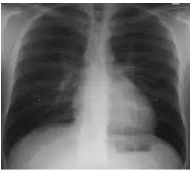
The presented work uses a set of digital images consisting of 20 small-cell types of lung cancer images, 20 non-small-cell types of lung cancer images, a total of 40 images (samples) each of 200 X 200 pixels in size. The images are obtained by collecting from private hospitals, and browsing the public database JSRT (Japanese Society of Radiological Technology) from internet. The digitized images are stored in the JPEG format with a resolution of 8 bits per plane. All images are stored as 200 X 200 X 256 JPEG raw data.
2. Image Enhancement
Image enhacement is the process of sharpening or smoothen the image. It improves the image quality and remove the noise from the image.It provides the better input for the digital image processing. Image enhancement belongs to image preprocessing methods. Objective of image enhancement – process the image (e.g. contrast improvement, image sharpening ,…) so that it is better suited for further processing or analysis
Image enhancement techniniques classified into two main parts:
1.Spatial domain methods- which directly operates on a pixel of a digital image.
2.Frequency domain method-which operates on a fourior transform of a image. it is the low level processing technique. Image enhancement methods are based on image quality. No mathematical criteria are used for optimizing processing results. In the image enhancement stage following three techniques are used: Gabor filter, Auto-enhancement and Fast Fourier transform techniques
2.1 Gabor filter
The Gabor filter was originally introduced by Dennis Gabor; we used it for CT images. In image processing a Gabor filter, named after a dennis gabor, is a linear filter used for texture analysis, which means that it basically analyses whether there are any specific frequency content in the image in specific directions in a localized region around the point or region of analysis In the spatial domain, a 2D Gabor filter is a Gaussian kernel function modulated by a sinusoidal plane wave.The Gabor function is a very essential tool in computer visibility and image processing, especially for texture analysis, due to its optimum locali ation property in both spatial and frequency domain.Image representation based on the Gabor function produce an excellent local and multiscale decomposition in terms of logons that are simultaneously localization in space and frequency domains.A Gabor filter is linear filter whose impulse response is defined by a harmonic function multiplied by a Gaussian function. Because of the multiplication-convolution property, the Fourier transform of Gabor filter’s impulse response is the convolution of
Fouriertransform of the harmonic function and the Fourier
transform of Gaussian function.Frequency and orientation representations of Gabor filters are similar to those of the human visual system, and they have been found to be particularly appropriate for texture representation and discrimination. In the spatial domain, a Gabor filter is a Gaussian kernel function modulated by a sinusoidal plane wave. The filter has a real and an imaginary component representing orthogonal directions. The two components may be formed into a complex number or used individually.
Before
After applying gabor filter
3.2 Fast fourior transform
[image:2.595.337.529.232.405.2]© 2017, IRJET | Impact Factor value: 6.171 | ISO 9001:2008 Certified Journal | Page 1524 3.3 Auto-enhancement
[image:3.595.332.530.92.294.2]Auto enhancement methos is based on the subjective observation and stastical operation.In this operation such as mean and variance are calculated. The enhancement percentage in this research was equal to 38.025%.
Table 1
Gabor filter Auto-enhancement FFT filter
80.735% 38.025% 27.51%
3.Image Segmentation
Image segmentation is the process of distribute a digital image into various segments such as sets of pixels, also known as super-pixels. The main aim of segmentation is to change the representation of an image into something that is easier to analyze . In image segmentation used to locate objects,edges and boundaries in images. image segmentation include the process of assigning a label to every pixel in an image so that pixels with the same label share certain characteristics.
The output of image segmentation is a set of segments that collectively cover the entire image, or a set of edges and boundaries extracted from the image.some of the pixels in a perticuar region are similar with some characteristic or property, such as color, intensity, or texture. Adjacent regions are different with respect to the same characteristic.there are two techniques used for image segmentation that are thresholding and watershed masking approach.
3.1Thresholding Approach
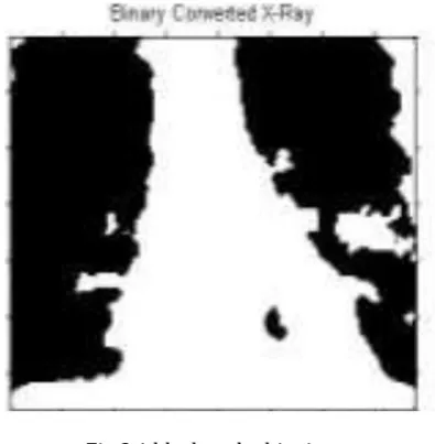
Thresholding is one of the most powerful tools for image segmentation. The segmented image obtained from thresholding has the advantages of smaller storage space, fast processing speed and ease in manipulation, compared with gray level image which usually contains 256 levels. In this we have a gray scale image given for a thresholding procedure it converts the rgb image into a binary image i.e black and white image which has only two shades i.e black and white which represent the level 0 and 1 only.the threshold value for this will be lies between 0 and 1 because it has only two levels., after achieving the threshold value; image will be segmented based on it.
Fig 3.1 black and white image
3.2Marker-Controlled Watershed Segmentation
Marker watershed segmentation technique indicate the presence of objects or background at specific image locations Marker-Controlled Watershed Segmentation Approach has two types: External associated with the background and Internal associated with the object of interest. Image segmentation using the Watershed transform works well if we can find or “mark” foreground objects and background locations, to find “catchment basins” and “watershed ridge lines” in an image by treating it as a surface where the light pixels are high and dark pixels are low.According to the
experimental subjective assessment during the
segmentation stage Marker-Controlled Watershed
Segmentation approach has more accuracy and quality than Thresholding approach
4.Feature Extraction
Feature plays a very important role in the area of image processing. Before getting features, various image preprocessing techniques like binarization , thresholding, normalization,masking approach etc. are applied on the sampled image. After that, feature extraction techniques are applied to get features that will be useful in classifying and recognition of images.
4.1 Binarization
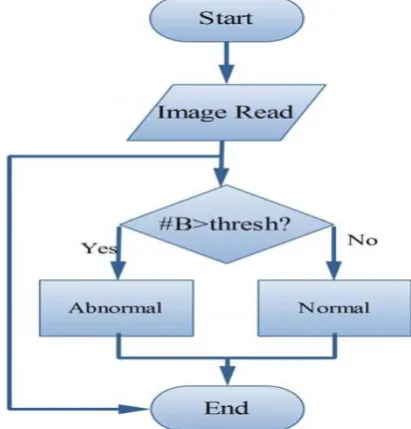
© 2017, IRJET | Impact Factor value: 6.171 | ISO 9001:2008 Certified Journal | Page 1525 otherwise, if the number of the black pixels is smaller than
[image:4.595.61.267.134.349.2]the specified value of a threshold, it indicates that the image is abnormal.
Fig 4.1flowchart for white and black pixel
4.2 Masking approach
Masking approach depends on the fact that the masses are appeared as white connected areas inside ROI (lungs), as they increase the percent of cancer presence increase. The appearance of solid blue colour indicates normal case while appearance of RGB masses indicates the presence of cancer; the TAR of this method is (85.7%) and FAR has (14.3%). If we Combining Binarization and Masking together this will take a decision that whether the report is normal or abnormal according to the mentioned assumptions in the previous two approaches, we can say that the if the number of black pixel is greater than the number of white pixel indicate that the report is normal otherwise the report is abnormal.
Fig 4.2masked X-ray image
5. PROPOSED PLAN OF WORK
STEP-1: Collect the lung cancer images from respective cancer hospital
STEP-2: Access one particular image into MATLAB with the help of command
STEP-3: Enhancement process
Image enhancement is to improve the interpretability or perception of information included in the image for human viewers, or to provide better input for other automated image processing techniques.We are using Gabor filter for image enhancement process
STEP-4: Segmentation process
Segmentation divides the image into its constituents regions or objects. It has many useful applications for the medical professional such as visualization and volume estimation of object of interest, detection of abnormalities, tissue qualification and classification, and more. We are using Marker-Controlled Watershed Segmentation Approach for segmentation
STEP-5: Features extraction
To know normality or abnormality of the images this process
is usedWe are usingbinarization and masking for feature
extraction
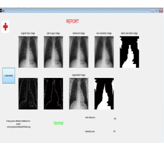
[image:4.595.53.262.581.741.2]© 2017, IRJET | Impact Factor value: 6.171 | ISO 9001:2008 Certified Journal | Page 1526 Fig 5.1result for detected image
Fig 5.2 result for normal image
6. Advantages
(a) Early detection of cancer greatly increases the chances for successful treatment.
(b) With the use of this treatment is often simpler and more likely to be effective.
(c)The proposed systems are more efficient and give the better result.
(d)Provides better image quality and accuracy
7. Applications
[1]It is widely used in many medical areas for early detection of cancer .so the the proper treatment will be provided to the patient.
[1] Febr Mokhled S. AL-TARAWNEH, “Lung Cancer Detection Using Image Processing Techniques”, Leonardo Electronic Journal of Practices and Technologies, June 2012.
[2] Muhammad Usman, Muhammad Shoaib and Mohamad Rahal, “Lung Cancer Detection Using Digital Image Processing”, PIERS Proceedings, Stockholm, Sweden, Aug. 12-15, 2013.
[3] Anita Chaudhary and Sonit Sukhraj Singh, “Multi-resolution Analysis Technique for Lung Cancer Detection in Computed Tomographic Images”, International Journal of Research in Engineering & Applied Sciences, IJREAS Volume 2, Issue 2 January 2012
[2]This image processing technique can also be used to detect other cancer such as breast cancer and tumor in our body part.
8.Conclusion
An image processing technique is built to detect diseases at early stage of cancer so the patient can take the treatment at early stages.The time factor is major factor to discover the abnormal tissue in target x-ray images.The accuracy and the quality of image is one the major core factor of this research.Image quality as well as image enhancement stage were adopted as low pre-processing techniques based on gabor filter.This technique is efficient for segmentation stage so the region of interest for feature extraction obtaining.on the basis of general features a normality and abnormality comparison is made.the main feature for detection of accurate image comparison are pixel percentage and mask labeling which gives us the indication that the process of detection this disease plays a very important and essential role to avoid serious stages and to reduce its percentage distribution in the world. To obtain more accurate results we three stages: Image Enhancement stage, Image Segmentation stage and Features Extraction stage.
ACKNOWLEDGEMENT
The success of any work depends on efforts of many individuals. We would like to take this opportunity to express our deep gratitude to those who extended their support and have guided us to complete this project work. I like to thank Prof.Mohd.Nasiruddin (HOD) for providing us the necessary information about topic. I would again like to thank Prof.Dr. Sajid Anwar, Principal of the College, for providing us the necessary help and facilities we needed. I express my thanks to all the staff members of Electronics & communication Engineering department who have directly or indirectly extended their kind co-operation in the completion of my reaserch paper.
[image:5.595.36.297.319.549.2]