Biology Dissertations Department of Biology
12-16-2019
Histone Deacetylase-linked Repression and Metabolically-linked
Histone Deacetylase-linked Repression and Metabolically-linked
Derepression of Adenovirus Persistent Infection of Lymphocytes
Derepression of Adenovirus Persistent Infection of Lymphocytes
Megan Dickherber Georgia State University
Follow this and additional works at: https://scholarworks.gsu.edu/biology_diss
Recommended Citation Recommended Citation
Dickherber, Megan, "Histone Deacetylase-linked Repression and Metabolically-linked Derepression of Adenovirus Persistent Infection of Lymphocytes." Dissertation, Georgia State University, 2019. https://scholarworks.gsu.edu/biology_diss/227
DEREPRESSION OF ADENOVIRUS PERSISTENT INFECTION OF LYMPHOCYTES
by
MEGAN LOUISE DICKHERBER
Under the Direction of Charlese Garnett-Benson, PhD
ABSTRACT
Adenovirus (AdV) infection is ubiquitous in the human population and causes acute
infection in the respiratory and gastrointestinal tracts. In addition to lytic infections in epithelial
cells, AdV can persist in a latent form in mucosal lymphocytes, and nearly 80% of children contain
viral DNA in the lymphocytes of their tonsils and adenoids. Reactivation of latent AdV is thought
to be the source of deadly viremia in pediatric transplant patients. Adenovirus latency and
reactivation in lymphocytes is not well-studied, though immune cell activation has been reported
to promote productive infection from latency. In lymphocytes, programs of gene expression during
both resting and activated states have been shown to be regulated in part by chromatin remodelers
and co-repressors, including Class I and II histone deacetylases (HDACs), Class III HDACs
nucleus, viral gene expression is potentially regulated by these same cellular chromatin-modifying
mechanisms and responsive to immunoactivation of the host lymphocyte. In this doctoral work,
we show that enzymatic activity of Class I HDACs and sirtuins, but not Class II HDACs, contribute
to the repression of viral early and late genes during persistent infection. We also show that
modulation of cellular NAD+/NADH can de-repress adenovirus gene expression in
persistently-infected lymphocytes. In contrast, disrupting the NAD-dependent CtBP repressor complex
interaction with PxDLS-containing binding partners paradoxically alters AdV gene expression.
DEREPRESSION OF ADENOVIRUS PERSISTENT INFECTION OF LYMPHOCYTES
by
MEGAN LOUISE DICKHERBER
A Dissertation Submitted in Partial Fulfillment of the Requirements for the Degree of
Doctor of Philosophy
in the College of Arts and Sciences
Georgia State University
Copyright by Megan Louise Dickherber
DEREPRESSION OF ADENOVIRUS PERSISTENT INFECTION OF LYMPHOCYTES
by
MEGAN LOUISE DICKHERBER
Committee Chair: Charlese Garnett-Benson
Committee: Margo Brinton
Casonya Johnson
Electronic Version Approved:
Office of Graduate Studies
College of Arts and Sciences
Georgia State University
DEDICATION
I dedicate my doctoral work to my husband, Tony, and my two children, Marcus and Cela.
To my husband, Tony – You have been by my side through successes and failures,
believing in me even when I did not. You have always been in the trenches with me, willing to
hear a practice presentation or be a proof-reader or discuss my project, no matter what time of day
or night. We share the drive to challenge ourselves and to give back to the world some of our
blessings. You are the love of my life.
To my children, Marcus and Cela – You are the sunshine in my every day and inspire me
ACKNOWLEDGEMENTS
I would like to thank my advisor, Dr. Charlie Benson, for taking me under her wing and
guiding me through my doctoral work. I have not met a teacher or mentor as committed to her
students as she has been. She viewed every interaction as an opportunity to grow and to help us
reach the next level of accomplishment. Her passion for science and love of learning, and her
fearlessness towards new challenges have been truly inspiring.
I would like to thank Dr. Margo Brinton for her guidance and patient feedback over the
last several years and multiple classes I took from her. She was always available to talk through
challenges or ideas and give expert insight into how to tackle them. She had high standards for
students in her classes and gave the support to help them get there. Her unabashed love of science
(and especially RNA) made learning with her fun.
I would also like to thank Dr. Casonya Johnson for serving on my committee and her
continued support for navigating the doctoral program and thinking “big-picture” about what my
long-term goals are. Her way of getting right to the heart of the matter in a kind and supportive
TABLE OF CONTENTS
ACKNOWLEDGEMENTS ... II
LIST OF TABLES ... VIII
LIST OF FIGURES ... IX
LIST OF ABBREVIATIONS ... XI
1 INTRODUCTION ... 1
1.1 Human Adenoviruses ... 1
1.2 Viral Structure, Life Cycle, and Tropism ... 3
1.2.1 Lytic Infection ... 3
1.2.2 Persistent Infection ... 4
1.3 Chromatin Structure of AdV Genome ... 6
1.3.1 AdV Genome is Episomal ... 7
1.3.2 AdV Genome Organization and Overview of Transcriptional Program ... 7
1.3.3 Chromatin Structure during Lytic Infection ... 8
1.3.4 Chromatin Structure of AdV Vectors ... 11
1.3.5 Chromatin Structure during Persistent Infection ... 12
1.4 Transcriptional Regulation of Viral Gene Expression ... 12
1.4.1 Viral E1A-289R ... 13
1.4.2 Histone Acetyltransferases (HATs): p300 and Tip60 ... 17
1.4.4 Class III HDACs – Sirtuins ... 18
1.4.5 Co-repressive C-terminal Binding Proteins (CtBPs) ... 19
1.5 Overarching Hypothesis ... 20
References ... 21
2 NAD-LINKED MECHANISMS OF GENE DE-REPRESSION AND A NOVEL ROLE FOR CTBP IN PERSISTENT ADENOVIRUS INFECTION OF LYMPHOCYTES ... 33
2.1 Abstract ... 33
2.2 Background ... 34
2.3 Materials and Methods ... 38
2.4 Results ... 44
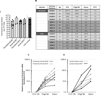
2.4.1 Viral transcription in persistently-infected lymphocytes is repressed compared to lytically-infected cells but relative amounts across viral transcripts are similar .. 44
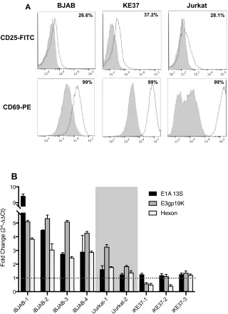
2.4.2 Cellular activation of infected lymphocyte cell lines upregulates viral gene expression ... 47
2.4.3 Infection with adenovirus can reduce the NAD+/NADH ratio and PMA/ionomycin stimulation shifts this ratio in lymphocytic cell lines ... 48
2.4.4 Direct modulation of the NAD+/NADH ratio can upregulate viral gene expression in persistently-infected cells ... 51
2.4.6 Inhibition of CtBP-E1A interaction upregulates E1A 13S expression in T
lymphocyte cell lines ... 54
2.5 Discussion ... 58
2.6 Conclusion ... 67
References ... 67
3 CONTRIBUTIONS OF CLASS I, II, AND III HDACS TO VIRAL GENE REPRESSION DURING ADENOVIRUS PERSISTENT INFECTION ... 75
3.1 Background ... 75
3.2 Materials and Methods ... 79
3.3 Results ... 84
3.3.1 Class I HDACs are ubiquitously expressed, but Class II HDACs are differentially expressed across cell-line models of infection ... 84
3.3.2 Enzymatic activity of Class I HDACs, but not Class II, is involved in repression of AdV genes in persistently-infected lymphocyte cell lines. ... 86
3.3.3 Activating Class III HDACs (sirtuins) with resveratrol upregulates E1A expression. ... 88
3.3.4 Inhibiting SIRT1 and SIRT2 with sirtinol upregulates E3 and hexon in persistently-infected cell lines. ... 89
3.3.5 Long-term sirtuin inhibition with nicotinamide does not prevent establishment of persistent infection. ... 91
3.5 Conclusion ... 96
References ... 96
4 LIMITATIONS OF CURRENT APPROACHES FOR THE STUDY OF ADENOVIRUS PERSISTENT INFECTION ... 101
4.1 Background ... 101
4.2 Materials and Methods ... 102
4.3 Results: Part A - Determining the Frequency of AdV-Infected Cells in a Persistently-Infected Cell Culture ... 105
4.3.1 Ad5GFP establishes persistent infection in BJAB and Jurkat cell lines, but GFP expression is repressed alongside AdV genes ... 107
4.3.2 Repressed GFP and hexon genes respond to histone deacetylase (HDAC) inhibition and phorbol 12-myristate 13-acetate (PMA) ionomycin (Iono) treatment in persistently-infected Jurkat cells ... 109
4.4 Conclusions: Part A - Determining the Frequency of AdV-Infected Cells in a Persistently-Infected Cell Culture ... 111
4.5 Results: Part B - Detection of Viral Proteins in Low Abundance ... 112
4.5.1 E1A proteins are not detectable in persistently infected KE37 and BJAB cell lines due to non-specific binding of anti-E1A antibody ... 113
4.5.2 E2 DNA-binding protein is detectable in persistently-infected T cell lines ... 115
4.6 Conclusions: Part B - Detection of Viral Proteins in Low Abundance ... 116
References ... 117
5 CONCLUSIONS AND FUTURE STUDIES ... 120
5.1 Discussion ... 120
References ... 126
APPENDICES ... 130
Cell viability and density with different treatment drugs ... 130
Appendix A – Nicotinamide treatment ... 130
Appendix B – Trichostatin A (TSA) treatment ... 131
Appendix C – Tacedinaline treatment ... 132
Appendix D – TMP195 treatment ... 133
Appendix E – NSC95397 treatment ... 134
Appendix F – Tubacin treatment ... 135
Appendix G – Resveratrol treatment ... 136
LIST OF TABLES
Table 1-1. Major protein products and their functions from AdV transcription units. ... 9
Table 1-2. Transcription factor binding sites and regulatory elements in E1A-responsive viral
promoters. ... 16
LIST OF FIGURES
Figure 1-1. Structure of an Adenovirus Particle. ... 3
Figure 1-2. Comparison of events and tropism of lytic and persistent infection. ... 6
Figure 1-3. Genomic organization of AdV-C5. ... 8
Figure 1-4. Adenovirus E1A major isoforms. ... 13
Figure 1-5. E1A-289R transactivation of AdV early genes. ... 14
Figure 1-6. Conserved regions of E1A-289R and their cellular binding partners. ... 15
Figure 1-7. Overarching Hypothesis ... 21
Figure 2-1.Characterization of viral genome quantities and transcriptional repression in persistently-infected lymphocytes. ... 45
Figure 2-2.Cell stimulation with PMA and Ionomycin upregulates viral gene expression in infected lymphocytic cell lines. ... 49
Figure 2-3. PMA and ionomycin treatment increases NAD+/NADH ratio in lymphocyte cell lines. ... 50
Figure 2-4. Viral gene expression is responsive to the NAD+/NADH ratio. ... 53
Figure 2-5. Epithelial cells and lymphocytic cells differ in CtBP2 expression. ... 55
Figure 2-6. CtBP-binding inhibitor, NSC95397, differentially impacts AdV gene expression across lymphocytic and epithelial cell lines. ... 56
Figure 3-1. Regulation of HDAC function during infection with DNA viruses. ... 77
Figure 3-2. Class I and II HDAC protein levels in cell line models of AdV infection. ... 85
Figure 3-3. Inhibitors of Class I and II HDACs. ... 86
Figure 3-5. Treatment with HDAC6 inhibitor (Class IIb) does not alter viral gene expression. .. 88
Figure 3-6. Activating sirtuins with resveratrol upregulates E1A and down-regulates hexon in
persistently-infected lymphocytic cell lines. ... 90
Figure 3-7. Inhibition of SIRT1 and SIRT2 with sirtinol upregulates viral gene expression. ... 90
Figure 3-8. Long-term sirtuin inhibition with NAM does not prevent establishment of persistent
infection. ... 93
Figure 4-1. Percent of cells expressing GFP and hexon during acute infection with Ad5GFP in
lymphocyte cell lines. ... 108
Figure 4-2. Treatment with HDAC inhibitor TSA increases number of cells expressing GFP
reporter gene in persistently-infected Jurkat cells. ... 110
Figure 4-3. Treatment with PMA/Ionomycin increases number of cells expressing GFP and
hexon in persistently-infected Jurkat cells. ... 110
Figure 4-4. Immunoblot for E1A in persistently-infected lymphocytes. ... 114
Figure 4-5. E2 DNA-binding protein is detectable in persistently-infected T cells. ... 116
LIST OF ABBREVIATIONS
ACTB b-actin
ADP Adenovirus Death Protein AdV Adenovirus
ALL Acute Lymphoblastic Leukemia APC Allophycocyanin
BSA Bovine Serum Albumin
CAR Coxsackie and Adenovirus Receptor ChIP Chromatin immunoprecipitation CMV Cytomegalovirus
CR Conserved Region
CtBP C-terminal Binding Protein DBF DNA binding factor
DBP AdV DNA-binding protein DDR DNA damage response Dpi Days post infection
DMEM Dulbecco’s modified Eagle medium DNMT DNA methyltransferase
EBV Epstein-Barr Virus
EIF1 Eukaryotic translation initiation factor 1 FISH Fluorescence in situ hybridization FCS Fetal Calf Serum
FITC Fluorescein Isothiocyanate
GAPDH Glyceraldehyde-3-phosphate Dehydrogenase GFP Green fluorescent protein
HAT Histone acetyltransferase HDAC Histone Deacetylase HDACi HDAC inhibitor HPV human papilloma virus HRP Horseradish Peroxidase
HPRT1 Hypoxanthine Phosphoribosyltransferase 1 HPV Human papilloma virus
HSCT Hematopoietic Stem Cell Transplant HSV Herpes simplex virus
iBJAB Infected BJAB cells iKE37 Infected KE37 cells Iono Ionomycin
ITR Inverted terminal repeat
KSHV Kaposi’s sarcoma-associated herpesvirus LCR Long coding region
MEF2 myocyte enhancer binding factor 2
MIEP CMV major immediate early gene promoter MLP AdV major late promoter
NAD+ Nicotinamide adenine dinucleotide
NADH Nicotinamide adenine dinucleotide (NAD) + hydrogen (H) NAM Nicotinamide
PARP Poly(ADP-ribose) polymerase PE Phycoerythrin
PFU Plaque-forming unit PKR Protein kinase R
PBS Phosphate-buffered saline PMA Phorbol 12-myristate 13-acetate PRC Polycomb repressive complex RCA Rolling circle amplification RPMI Roswell Park Memorial Institute
RT-qPCR Reverse transcription – quantitative polymerase chain reaction RV Resveratrol
SDS-PAGE Sodium dodecyl sulfate polyacrylamide gel electrophoresis
SF Serum-free
shRNA Short hairpin RNA siRNA Silencing RNA
TAF-1b Template activating factor 1b TBST Tris-buffered Saline with Tween Tip60 Tat-interacting protein 60
TCC Tethered chromosome capture
TET Ten-eleven translocation family proteins TLR Toll-like receptor
TPL Tripartite leader TSA Trichostatin A
1 INTRODUCTION
This doctoral thesis focuses on mechanisms of transcriptional regulation of adenovirus
(AdV) persistent infection in lymphocytes. In this introductory chapter, a brief overview of the
human adenoviruses will be given with rationale for the study of AdV Species C in connection
with persistent infection. An overview of the sequential steps occurring in the lytic infection of
epithelial cells will then be compared to what is known for persistent infection described in cell
lines models and primary lymphocytes. Because our studies have focused on mechanisms of gene
expression regulation, a review of what is known of viral chromatin structure in lytic and persistent
infection and in the context of adenoviral vector transgene delivery will be given. This chapter will
conclude with a review of known mechanisms of transcriptional regulation of the adenovirus
genome, with rationale for the overarching hypothesis for this work.
1.1 Human Adenoviruses
Human adenoviruses were discovered in 1953 by Rowe et al. as the cytopathogenic
agent present in cultured adenoid tissues (1) and independently by Hilleman et al. as a cause of an
acute respiratory disease outbreak among military personnel (2). Since then, the viruses of the
family Adenoviridae have been found in all major classes of vertebrates, with the genus
Mastadenovirus containing mammalian adenoviruses including the seven species (A-G) found in
human hosts (3). Currently there are more than 85 types of human AdV that have been identified
through serotyping and genomic analysis (4-6). Human AdV infections cause a variety of different
diseases including conjunctivitis (A, B, E), gastroenteritis (A, F, G), and respiratory disease (B, C,
E) (3).
Infections with Species C (types 1 and 2) were among the AdVs most reported in a 2017
symptomatic respiratory infections (8). AdV-C infections predominantly occur in very young
children and are a major cause of respiratory disease in pediatric patients (9-12). In a study of
extracted tonsils or adenoids, approximately 80% of patients under age 19 contained AdV-C (types
1, 2, 5, and 6) DNA in lymphocytes of those tissues, most in a non-replicating state (10). In
addition, AdV-C are responsible for more than half of adenovirus infections and severe disease in
immunocompromised patients (6,11). Pediatric patients receiving hematopoietic stem cell
transplants (HSCT) are at significant risk for developing disseminated AdV infection, with
AdV-related post-transplantation mortality between 3.2 and 6.0%, or approximately 100 children per
year in the U.S. (13,14). These infections can result from de novo exposure to the virus, but
reactivation of latent adenovirus from the patient’s own tissues is the predominant cause for the
most severely immunocompromised patients (14). The mechanisms allowing the virus to persist
and the conditions inducing reactivation of the virus are almost entirely unknown. This gap in
knowledge has proven to be a critical barrier to preventing AdV-related disease in transplant
recipients.
As AdV-C is the predominant species associated with the latent or persistent infection
of mucosal tissues of the tonsils, adenoids, and gastrointestinal tract (10,15), the mechanisms
governing the persistent AdV-C infection in lymphocytes are the focus of this work. Our study
will be the first work to investigate and describe how cellular transcriptional regulators are
involved in maintenance of persistent adenovirus infection in lymphocytes. Understanding the
mechanisms for adenoviral persistence may provide novel targets for therapies to prevent
1.2 Viral Structure, Life Cycle, and Tropism
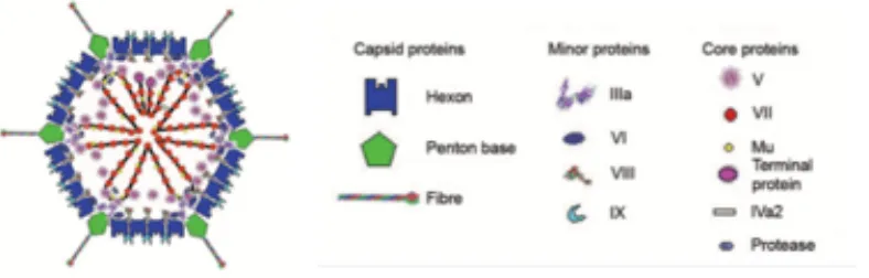
AdVs are 90nm, non-enveloped viruses with 36kB linear, double-stranded DNA genomes
(3). The viral DNA is condensed into the viral particles by basic viral protein µ and packaged by
major core protein VII (16) (Figure 1-1). The capsid of the virus consists largely of a hexon protein
[image:21.612.92.486.244.370.2]icosahedral shell with penton base proteins attached to protruding fiber proteins at the vertices (16)
(Figure 1-1). No host-cell proteins have been found to be associated with the viral particles (3).
Figure 1-1. Structure of an Adenovirus Particle.
Adapted from (16).
1.2.1 Lytic Infection
Epithelial cells are the primary target for AdV, and adsorption of the virus occurs when the
fiber protein attaches to the cellular protein coxsackie-adenovirus receptor (CAR) on the
membrane of the host cell. A secondary attachment via the RGD-motif in the penton base protein
to an avb3/5 integrin triggers clathrin-mediated endocytosis and the initial stages of disassembly of
the virus (16-19). Following release from the endosomes, the partially uncoated virus is transported
by microtubules to the nuclear pore (3). The viral genome, bound only to core protein VII, enters
the host cell nucleus where it is quickly chromatinized through association with cellular histones
(20).
Upon expression of viral early genes and virally associated (va) RNAs I and II, the cellular
altered to generate metabolic precursors and to drive the cell into S-phase (21,22). Following
replication of the viral genome, the late stage of infection begins with inhibition of cellular protein
translation and production of viral structural proteins. Viral particles assemble in the nucleus, and
the host cell is lysed to release thousands of viral progeny (Figure 1-2) (3,22).
1.2.2 Persistent Infection
Despite the some-what ironic fact that AdVs were discovered through persistent infection
of adenoids (1), the steps leading to the establishment of persistent AdV infection and mechanisms
of reactivation are currently almost entirely unknown. Early epidemiological studies showed that
AdV could be detected intermittently in patient fecal samples for months to years after resolution
of symptoms, indicating the presence of some viral reservoir in the host (23). As mentioned above,
it is now understood that the persistent adenovirus infection occurs in lymphocytes, and
predominantly T cells, of the tonsils, adenoids, and GI tract, with the majority of viral genomes
present in a non-replicating state (10,15).
Unlike infection of epithelial cells, species C adenoviruses infect lymphoid cells with
very different and less-studied infection kinetics. Infection of these cells begins with attachment
to the avb3/5 integrins alone, as most hematopoietic cells do not express CAR (18,24,25). While no
studies have reported on the mechanism of entry in lymphocytes, internalization may be governed
by different mechanisms than in epithelial cells (26), and other mechanisms such as caveolae- and
macropinocytosis-mediated endocytosis of adenovirus particles have been reported in various cell
types when CAR is not the primary receptor (27,28). The next steps of uncoating and transport of
the genome to the nucleus have also not been studied in lymphocytes, but unusually low expression
appeared normal, indicating that steps of the early life cycle in lymphocytes may deviate from
those in epithelial cells as early as post-internalization (25).
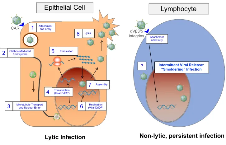
Persistent infection of lymphocytes typically does not progress to lysis but rather to a
non-lytic, “smoldering” infection in which replicating virus is rare (10,13,29). Immunoactivation of
infected primary lymphocytes has been shown to induce production of infectious virus, showing
that persistence and reactivation may be closely tied to the active state of the lymphocyte (10). In
order to circumvent the challenges of obtaining naturally-infected primary lymphocytes from
patient samples to study, lymphocytic cell-line models of persistent infection have been developed
which harbor viral DNA for months in culture (13,29). These models of persistent infection also
contain low levels of replicating virus, albeit at slightly higher levels than infected primary cells
(13,29), and release low amounts of virus detectable through extraction of DNA from culture
supernatants or application of supernatants onto permissive cell lines ((13) and our unpublished
results). Viability of these persistently-infected cell lines remains unaffected (13,29), indicating
either very few cells are lysing to release the viral particles, or an as-of-yet unidentified mechanism
for egress is occurring (Figure 1-2).
The persistent infection in these lymphocytic cell-line models has been shown to be
regulated, in part, by as-of-yet uncharacterized transcriptional controls not detectable in lytic
infection of epithelial cells (see Section 1.3.2 - AdV Genome Organization and Overview of
Transcriptional Program below for more detail). Murali et al. (2014) determined that the early
gene E3-Adenovirus Death Protein (ADP), which is critical for cell lysis, was repressed both
transcriptionally and post-transcriptionally in cells which maintain the persistent infection (30).
Krzywkowski et al. (2017) showed that in a persistently-infected B cell line very few individual
lytically-infected HeLa cells, even in cells with large amounts of viral DNA (31). Furuse et al.
(2013) determined vaRNAs I and II were expressed in comparable amounts between
persistently-infected B cells and lytically-persistently-infected epithelial cells, but in different relative proportions (32).
These few studies suggest a specific program of repression is needed to maintain the persistent
[image:24.612.111.474.237.470.2]infection, but further work is needed for more complete characterization.
Figure 1-2. Comparison of events and tropism of lytic and persistent infection.
1.3 Chromatin Structure of AdV Genome
Despite the large body of work describing the impact of adenovirus infection on cellular
chromatin, the role of chromatin in regulation of expression from the viral genome itself has been
less well-studied. The following sections will begin with an overview of the organization of the
viral genome, then document what has been reported on chromatin structure and transcriptional
regulation of viral gene expression in the context of lytic and persistent infections. Additionally,
regulation of transgene expression from adenoviral vectors will be discussed. Epithelial Cell
Lytic Infection Non-lytic, persistent infection
Attachment and Entry
Microtubule Transport and Nuclear Entry
Transcription (Host DdRP)
Replication (Viral DdDP) Clathrin-Mediated
Endocytosis 1
2
3
4 5
6 Translation
7 Assembly
Lysis 8 CAR
Attachment and Entry
Intermittent Viral Release: “Smoldering” Infection
?
Lymphocyte
1.3.1 AdV Genome is Episomal
Unlike the genomes of other DNA viruses which integrate into the host cell DNA (33-36),
adenovirus genomes, both wild-type and vector DNA, remain episomal except for extremely rare
instances (29,37-40). The mechanisms which prevent integration have not been identified, but this
characteristic of adenoviral DNA has promoted its use as a viral vector for transgene delivery (40).
Avoiding integration may be an evolutionary advantage for the virus, as integration of foreign
DNA triggers de novo methylation and gene silencing as part of an ancient cellular antiviral
defense (41-43). In vitromethylation of the integrated E1A promoter in HEK293 cells showed
reduced expression of E1A (44), but whether this is biologically relevant has not been determined
as no DNA methylation has been reported for the episomal adenovirus genome (45).
1.3.2 AdV Genome Organization and Overview of Transcriptional Program
The program of viral transcription in lytic infection is well-documented and is primarily
orchestrated through coordination by the E1A proteins. Historically, E1A has been reported the
first transcript expressed from the viral genome, which is alternatively spliced into two principle
variants, 13S and 12S, giving rise to the 289R and 243R proteins, respectively. Through a recent
study tracking viral transcription in normal lung fibroblasts, it is now understood that non-coding
vaRNAs I and II, which inhibit the dsRNA-sensor protein kinase R (PKR), are first expressed,
followed quickly by expression of E1A and E4 (21). The viral early genes, E1B, E2, and E3 are
next expressed which prepare the cell for viral DNA replication by blocking apoptosis, producing
the viral polymerase and essential replication proteins, and tamping down the host-cell immune
response, respectively (46). Following mass production of viral DNA, the late genes are expressed
genome and main transcription units are shown in Figure 1-3. The major proteins from the
[image:26.612.111.520.159.248.2]transcription units and their functions are shown in Table 1-1.
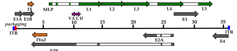
Figure 1-3. Genomic organization of AdV-C5.
The linear double-stranded DNA genome is depicted as a thin line, 5’ to 3’, with the inverted terminal repeats (ITR) at each end: lengths are marked in kbps. Transcription units are shown relative to their position and orientation. Early genes (gray bars): E1A, E1B, E2A/B, E3, and E4. Intermediate genes (orange bars): IX and Iva2. Late genes (green bars): L1-L5, produced from the major late promoter (MLP). Open rectangles denote introns. Late genes (L1-L5) produced from the MLP all contain the tripartite leader (TPL) at their 5’ ends. Modified from (47) and (48).
1.3.3 Chromatin Structure during Lytic Infection
While viral DNA interacts with several viral core proteins in the formation of infectious
particles (Figure 1-1), only protein VII remains associated with the viral genome from packaged
state in the viral capsid to the host cell nucleus (66). The interaction between VII and the viral
DNA serves multiple purposes. Protein VII is a core basic protein that was originally believed to
be essential for compacting the viral genome into the capsid (67). Recent work using viruses
lacking the late VII gene showed protein VII was not essential for encapsidation, but played a more
important role in pressurizing the core and genome release (68). Upon entry of the viral genome
into the nucleus, VII monomers are spaced across the DNA similarly to histones across cellular
DNA, a structure that protects the viral DNA from activating the DNA damage response (69).
Protein VII from the incoming viral particle remains associated with the viral chromatin 5" 10" 15" 20" 25" 30" 35"
ITR" ITR"
packaging(E1A"E1B" E3"
E4" E2A"
E2B" IVa2"
IX" MLP" L1" L2" L3" L4" L5"
Gene/ Transcription
Unit
Corresponding
Protein(s) Role in Viral Life Cycle
First early - E1A E1A-13s & 12s Early gene activation Induction of S-phase
Early – E3 E3gp19K Blocks MHC-I presentation
Late – L3 Hexon Structural - Capsid !
Table 1-1. Major protein products and their functions from AdV transcription units.
** denotes genes falling within the early gene region, but expressed at intermediate time points. References for all proteins from each gene as indicated.
Early
Gene Protein Function
Late
Gene Protein Function
E1A E1A-289R Activate early
genes
L1 52/55K Packaging
E1A-243R Induce cell cycle pIIIa Packaging
(49) (50)
E1B 19K Inhibit apoptosis L2 penton Capsid/attachment
55K Inhibit p53, Ub
pathway
pVII DNA condensation
IX** Capsid stability pV Secure DNA to
capsid
(51),(52),(52) pX (µ) DNA condensation
E2 DBP DNA replication (53),(54),(55),(56)
pTP Primes DNA
polymerase
L3 pVI Endosome lysis
pol Viral DNA
polymerase
hexon Capsid
Iva2** Packaging, ATPase protease Maturation of
structural proteins
(57),(58) (50),(59),(60)
E3 12.5K Unknown L4 100K ¯ translation
shut-off
6.7K Inhibit apoptosis 22K Packaging
gp19K MHC escape 33K Splicing, packaging
ADP Cell lysis/egress pVIII Hexon stabilization
RID-! ¯ TNF, Fas,
TRAIL
(61),(62),(58)
RID-b ¯ TNF, Fas,
TRAIL L5 fiber Attachment
(46), (30) (53)
E4 Orf1 Unknown
Orf2 Unknown
Orf3 ¯ host translation,
chromatin remodel
Orf4 Splicing viral RNA
Orf4-34K Packaging
Orf6 ¯ host translation,
Ub pathway
Orf6/7 mRNA export,
throughout the early phase of infection and is also important for E1A transactivation of early genes
(70).
The viral DNA/protein VII-chromatin structure that enters the nucleus must be quickly
remodeled to allow for efficient transcription of viral genes (71,72). Some protein VII is lost to
accommodate incorporation of cellular histones into the viral chromatin, and
replication-independent variant H3.3 and other core histones become localized throughout most promoter and
protein-coding regions (73,74). Although sites of high levels of protein VII binding on the viral
DNA correlate with gene repression (75,76), low levels of VII can enhance transcription, likely
through chromatin remodeling by interaction with template-activating factor 1𝛽 (TAF-1𝛽) and
nuclear phosphoprotein pp32 (members of the SET nucleosome assembly complex and inhibitor
of histone acetyltransferases [INHAT] complex, respectively) (70,72,73,77). Because increases in
acetylation of H3 can be detected as viral promoters become active, the remodeled viral chromatin
structure appears to be similar to cellular chromatin, subjecting it to regulation by the cellular
machinery (20).
Recent work has shown that interactions between the host and viral chromatin may also be
important to the program of viral gene expression. Studies using Hi-C analysis and tethered
chromosome conformation capture (TCC) to detect positioning of the viral chromatin show that
AdV DNA interacts predominately with the transcription start sites (TSSs) and enhancers of highly
active cellular chromatin (78,79). The viral genome associates with genes that are upregulated
during the course of infection, suggesting that the temporal changes in cellular gene expression
that occur may be linked with sequential expression of viral genes (79). Besides colocalization of
viral DNA and active cellular DNA, some additional interactions between the viral and host cell
H3K27me (78). This histone marker is usually associated with repression mediated by the
Polycomb repressive complex (PRC) (80). Interestingly, the PRC complex plays a role in
regulation of latency of other DNA viruses, but whether or not PRC-based repression is involved
in adenovirus lytic or persistent infection has not been determined (81).
1.3.4 Chromatin Structure of AdV Vectors
The AdV DNA backbone is commonly used as a vehicle for gene-therapy and oncolysis
because of the extensive knowledge available about the lytic life cycle and genomic elements and
the relatively low safety risk (82). However, it is clear that suboptimal, cell type-dependent
conditions are a common challenge that must be overcome to effectively use AdVs as a delivery
mechanism. In fact, in cells that are not the primary wild-type adenoviral targets, AdV vectors
lose long-term transgene expression, mirroring some aspects of the persistent infection in
lymphocytes (83). Mechanisms that have been reported for repression of gene expression in these
systems may be important to consider in understanding the full repertoire of regulators involved
in directing the fate of the viral genome for wild-type viruses.
Like wild-type viral genomes of lytic infection, AdV vectors also are associated with
protein VII and cellular histones early in infection (84,85). This association with histones has been
shown to be important for establishing the needed chromatin structure for transgene expression
(85). However, even in the absence of all functional viral proteins, over time, transgene expression
from adenovirus vectors can be diminished through remodeling of the vector chromatin induced
by innate immune responses to the presence of the foreign DNA. Histone deacetylase inhibitors
(HDACi) can upregulate expression of transgenes delivered in vectors of several different viral
backgrounds, showing that non-specific hypoacetylation may be a mechanism of defense (86).
reduced expression of a transgene through a decrease in the active chromatin mark H3K9Ac
relative to the heterchromatic mark H3K9me2 (85,87), but future work is needed to identify the
details of the pathway and the enzymes involved.
1.3.5 Chromatin Structure during Persistent Infection
The chromatin structure of the viral genome in persistent infection is largely unstudied.
Unlike AdV-vector DNA, which often has viral genes mutated or excised, wild-type genomes with
fully intact viral genes establish persistent infection (10). The normal progression of viral gene
expression is delayed in all lymphocyte cell-line models of infection relative to infection in
epithelial cells (29), but whether this is a result of suboptimal chromatin structure has not been
studied. Considering that both genomes of lytic infection and viral vectors associate with cellular
histones (20), and that viral transcripts can be detected in persistently infected cells (10,31,32), the
in-coming viral chromatin/VII structure is likely remodeled to include cellular histones at least to
some degree. We also recently reported that AdV gene expression in persistent infections is
responsive to treatment with HDAC inhibitor trichostatin A (TSA) (Wilms et al., submitted)
further supporting a role for histones and their modification in the chromatin structure. In a recent
study using a padlock probe-based rolling-circle amplification (RCA) to evaluate concurrent AdV
DNA and mRNAs in single epithelial or lymphocytic cells, viral DNA could not be detected in
lymphocytes expressing low amounts of viral mRNA (31). This was interpreted to be a result of
protein VII interference with probe binding (31), and suggests that at least some lymphocytes
contained viral genomes which lacked proper chromatin remodeling for gene expression.
1.4 Transcriptional Regulation of Viral Gene Expression
In additional to the chromatin structure itself, a second level of regulation of viral gene
recruitment of transcriptional complexes to promoters of the viral genes. This section will begin
by describing the activity of the best-known transcriptional regulator of AdV gene expression, the
viral E1A-289R protein. Then key cellular enzymes and proteins that have been found to play a
role in regulation of transcription of viral genes in lytic infections and expression of AdV vector
transgenes will be discussed. This section will conclude with the statement of hypothesis for this
dissertation, concerning transcriptional regulation of viral gene expression in persistent infection
in lymphocytes.
1.4.1 Viral E1A-289R
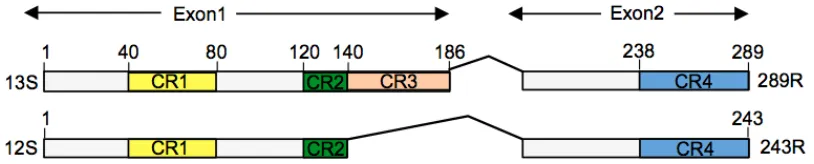
The AdV E1A-289 protein, the larger of the two E1A isoforms (Figure 1-4), is the
single-most important protein in transcriptional regulation of the viral genome owing to its function of
transactivating the full repertoire of early genes (88). E1A-289R has no intrinsic DNA-binding
capacity, but through interaction with cellular transcription factors recruits the basal transcriptional
[image:31.612.94.501.473.555.2]machinery and regulatory complexes to the early gene promoters (90). Essential to the
Figure 1-4. Adenovirus E1A major isoforms.
289R (translated from a 13S mRNA transcript) and 243R (translated from a 12S mRNA transcript). CR: regions of E1A that are conserved across many adenovirus serotypes. CR3 is unique to E1A-289R. Figure modified from (89).
transactivational function of E1A-289R is the interaction between CR3 and the MED23/Mediator
regulation including chromatin remodeling and mRNA processing (92). Mediator is thought to
establish enhancer-promoter loops, and also is important for transcription initiation (92). Figure
[image:32.612.108.507.183.305.2]1-5 depicts the E1A-289R/Mediator-regulated transactivation of the AdV early genes.
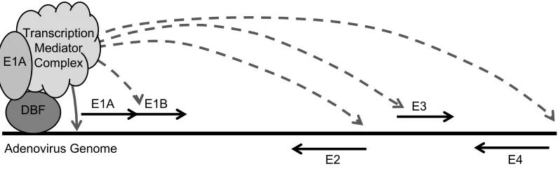
Figure 1-5. E1A-289R transactivation of AdV early genes.
The solid black line represents the viral genome (not drawn to scale), and the relative locations of the viral early genes are shown. The first viral protein produced is E1A-289R, which then auto-activates viral gene E1A (solid gray line). Similar E1A-containing complexes then move to promoters of and activate viral early genes E4, E3, E1B, and E2 (dashed gray lines) (93). DBF: any of several cellular DNA-binding factors.
While the CR3 region of E1A-289R recruits the mediator complex, the activity of this
complex has the potential to be further modified through the multitude of interactions E1A-289R
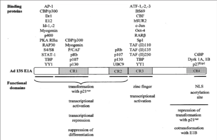
has with additional cellular proteins (49). Figure 1-6 shows many of the E1A-289R binding
partners, including transcription factors, histone acetyltransferases, co-repressors, transcriptional
machinery, and chromatin remodelers (modified from (90)). Binding sites for the transcription
factors that interact with E1A-289R, and many others, can be found in the promoters of the early
genes. Table 1-2 lists the transcriptional regulatory elements in each of the responsive viral
promoters, and the degree to which E1A-289R upregulates expression.
E1A E1B
E2
E3
E4 DBF
Transcription Mediator Complex E1A
Figure 1-6. Conserved regions of E1A-289R and their cellular binding partners.
Modified from (90).
In a very interesting new finding, E1A-289R transactivation of early genes E1B, E2 and
E3 was surprisingly found to be regulated in part by the interaction between E1A-289R and the
E4orf3 protein (109). E4orf3 functions largely to silence the stress response induced by viral
infection through the non-specific induction of heterochromatin formation, inadvertently silencing
viral genomes as well (110). Soriano et al. (2019) found the interaction between E1A and E4orf3
serves to fine-tune transactivation of the early genes: E1A counteracted the induction of
heterochromatin formation on the viral genome induced by E4orf3, and E4orf3 interacted with
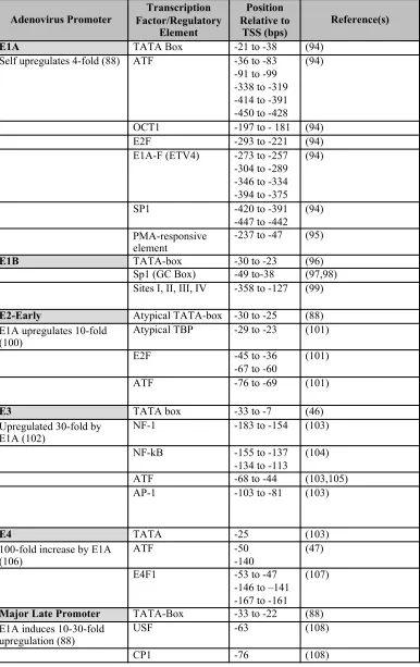
Table 1-2. Transcription factor binding sites and regulatory elements in E1A-responsive viral promoters.
Adenovirus Promoter Factor/Regulatory Transcription Element
Position Relative to
TSS (bps)
Reference(s)
E1A TATA Box -21 to -38 (94)
Self upregulates 4-fold (88) ATF -36 to -83
-91 to -99 -338 to -319 -414 to -391 -450 to -428
(94)
OCT1 -197 to - 181 (94)
E2F -293 to -221 (94)
E1A-F (ETV4) -273 to -257
-304 to -289 -346 to -334 -394 to -375
(94)
SP1 -420 to -391
-447 to -442 (94)
PMA-responsive element
-237 to -47 (95)
E1B TATA-box -30 to -23 (96)
Sp1 (GC Box) -49 to-38 (97,98)
Sites I, II, III, IV -358 to -127 (99)
E2-Early Atypical TATA-box -30 to -25 (88)
E1A upregulates 10-fold (100)
Atypical TBP -29 to -23 (101)
E2F -45 to -36
-67 to -60
(101)
ATF -76 to -69 (101)
E3 TATA box -33 to -7 (46)
Upregulated 30-fold by E1A (102)
NF-1 -183 to -154 (103)
NF-kB -155 to -137
-134 to -113
(104)
ATF -68 to -44 (103,105)
AP-1 -103 to -81 (103)
E4 TATA -25 (103)
100-fold increase by E1A (106)
ATF -50
-140
(47)
E4F1 -53 to -47
-146 to –141 -167 to -161
(107)
Major Late Promoter TATA-Box -33 to -22 (88)
E1A induces 10-30-fold upregulation (88)
USF -63 (108)
1.4.2 Histone Acetyltransferases (HATs): p300 and Tip60
Histone acetyltransferase activity of p300, recruited through interaction with E1A-289R,
has recently been shown to be critical to the chromatin remodeling at AdV early gene promoters
(111). p300-induced increases in active marks H3K18ac and H3K27ac were seen at promoters for
E2, E3, and E4. Interestingly, an increase in these active marks was also noted for the E1A
promoter, suggesting a role for chromatin structure in regulation of this gene, despite speculation
that chromatinization was less relevant for this promoter (112). p300-dependent acetylation had
different effects on transcription at each viral promoter; initiation of transcription was dependent
on acetylation at the E2 promoter, but post-initiation steps were affected at the E3 and E4
promoters.
Tip60 (also known as KAT5), originally isolated and named as a cellular HIV-Tat
interacting protein, is a MYST family lysine acetyltranferase which acts as a transcriptional
regulator (113). Despite the known association between histone acetylation and active gene
expression, Tip60 appears to act in a repressive role in the AdV transcriptional program. In a study
by Gupta et al. (2013), Tip60 knock-down released repression of all AdV early genes, with a 3- to
4-fold increase in E1A expression, but had no impact on the major late promoter (MLP) (hexon
and fiber) (75). Further, Tip60 was found enriched at the TSS along with AdV protein VII and
unexpectedly, increased H4Ac at repressed E1A promoters. These findings together show that
carefully-regulated histone acetylation is important for optimal expression of AdV genes.
1.4.3 Class I and II HDACs
Despite the importance of HATs in AdV transcriptional regulation, little has been reported
about the role for their enzymatic counterparts, the histone deacetylases (HDACs). Perhaps this is
genes might be more relevant. For long-term persistent infections or retention of AdV vectors,
HDACs may have a larger impact on gene expression.
Silencing of transduced genes through the chromatinization of the AdV DNA vector
backbone is a common challenge facing AdV-based oncolytic viral therapies (85). While HDAC
inhibitors (HDACi) have been reported to increase the efficiency of oncolytic viral therapy through
the increased expression of viral receptors on the cell surface (114), HDACi also have been
reported to increase expression of genes delivered in the AdV vector (115,116). Various Class I
and II HDACi have been used successfully to upregulate transgene expression in numerous cell
types and lines from a range of host species, including primary bovine or rat muscle cells (117),
human thyroid carcinoma cell lines (116), mouse neuroblastoma cells, HeLa cells, human
neuroblastoma cells, primary human dendritic cells, keratinocytes, and primary rat astrocytes (86),
and rat neurons (118). The broad diversity of vector constructs and the wide array of cellular
backgrounds in which Class I and II HDACi increase gene expression suggest HDACs act in a
non-specific manner to silence foreign DNA.
We have recently reported that HDACs play a role in persistent infection with intact viral
genomes as well; TSA upregulates transcription of several viral genes in persistently-infected
lymphocytes (Wilms et al., manuscript submitted). The work in this dissertation provides
additional insight for the involvement of Class I and II HDACs in persistent AdV infection, and
shows that activity is targeted to specific viral genes (Chapter 3).
1.4.4 Class III HDACs – Sirtuins
Sirtuins (SIRT1-7), also called Class III HDACs, are a family of nicotinamide adenine
dinucleotide (NAD+)-dependent histone deacetylases with a variety of other enzymatic activities
function in many aspects of essential cellular processes such as chromatin remodeling and gene
expression, apoptosis, and DNA repair (120). The NAD+-dependence of sirtuins also links function
to the metabolic state of the cell (119). Relevant to AdV transcriptional regulation and as yet
uninvestigated, SIRT1 has been shown to deacetylate and thereby negatively regulate the HAT
activity of p300 (120), which could in turn negatively regulate expression of AdV genes (111).
Sirtuins have been shown to have broad anti-viral activity, as siRNA knock-down of all
seven sirtuins increases viral titers for both RNA and DNA viruses, including 1.5- to 3-fold
increases in AdV-C5 titers (119). In the same vein, activation of sirtuins through resveratrol
treatment inhibits adenovirus DNA replication of wild-type and vector viruses (118,121,122), but
the mode of action has not been described. This doctoral work shows that sirtuins contribute to
repression of AdV genes in persistently-infected lymphocytes (Chapter 3).
1.4.5 Co-repressive C-terminal Binding Proteins (CtBPs)
The CtBP family of transcriptional corepressors was discovered through their high affinity
binding to AdV E1A proteins (123,124). Mammalian cells express both CtBP1 its homolog CtBP2
(collectively known as CtBP) which can form homo- and hetero- tetrameric complexes, the
assembly and stability of which are dependent on NAD(H) binding (125-128). CtBP complexes
can recruit many different chromatin modulators including Class I HDACs 1 and 2, histone
methyltransferases, E3 ligases and other transcriptional regulators into large transcriptionally
repressive complexes at the promoters of genes ((129), reviewed in (130)). As a result of the
dependence on NAD(H) binding, CtBP has been reported to function as an NAD(H) sensor and
therefore a link between metabolic state and transcriptional regulation (131-133).
The role of CtBP in the AdV lytic life cycle is complex, acting in a repressive or a
E1A-289R and -243R-mediated transactivation of viral and cellular genes, respectively (134,135).
CtBP1 and 2 have long been known to suppress the ras-cooperative transformative activity of
E1A, but also appear to enhance E1A transcriptional regulation, possibly through suppression of
the interferon response (123,136-138). Both E1A-289R and -243R interact with high affinity to
both CtBP1 and 2 through a PLDLS-motif located in the shared conserved region 4 (CR4) at the
C-terminal end of the E1A proteins (Figure 1-4). E1A-289R has an additional CtBP-interaction
domain located in the CR3 region unique to this isoform (134), implicating CtBP in regulation of
early genes. Of note, NADH was found to facilitate binding of CtBP to E1A at 1000-fold lower
concentration than NAD+, suggesting that the NAD+/NADH ratio in the cell may affect the
formation of CtBP-E1A protein complexes (131). We show in this doctoral work that CtBP may
have an additional role in repression of E1A expression in a persistent AdV infection in
lymphocytes (Chapter 2).
1.5 Overarching Hypothesis
The overall aim of this work is to identify chromatin modifiers and transcriptional
regulators, known to play a role in repression of AdV gene expression in lytic infection or from
vectors, that may contribute to the transcriptional repression needed to establish a persistent
infection in lymphocytes. Specifically, I hypothesized that Class I, II HDACs, sirtuins, and CtBPs
all play a role in transcriptional repression of persistent infection. I further hypothesized that this
regulation, based on the NAD+ dependence of sirtuins and CtBPs, is inextricably linked to the
dramatic shifts in metabolic state of resting and activatedlymphocytes (depicted in Figure 1-7).
Chapter 2 of this work describes experiments done to establish the link between metabolism and
viral gene expression in persistently-infected lymphocytes, and the role of CtBP in repression of
specific viral genes. Chapter 4 describes additional work to address some technological challenges
to the study of persistent AdV infection. Chapter 5 summarizes major findings and identifies
[image:39.612.108.497.181.480.2]remaining questions for further study.
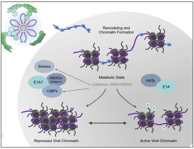
Figure 1-7. Overarching Hypothesis
AdV DNA (shown in purple) enters the nucleus with protein VII (shown in blue). The viral chromatin is remodeled by cellular histones and subject to transcriptional regulatory machinery in the cell. Repression of the viral chromatin, through interaction with cellular proteins dependent on the concentration of metabolic cofactors, is linked to the metabolic state of the lymphocyte. Modified from (139).
References
1. Rowe WP, Huebner RJ, Gilmore LK, et al. Isolation of a cytopathogenic agent from human adenoids undergoing spontaneous degeneration in tissue culture. Proceedings of the Society for Experimental Biology and Medicine 1953;84:570–573.
Remodeling and Chromatin Formation
HDACs
(Class I)
Sirtuins
CtBPs
HATs E1A Metabolic State
Cofactors: NAD+/NADH
Repressed Viral Chromatin Active Viral Chromatin E1A?
Ac Ac
Ac
2. Hilleman MR, Werner JH. Recovery of new agent from patients with acute respiratory illness. Proceedings of the Society for Experimental Biology and Medicine 1954;85:183– 188.
3. Berk AJ. Adenoviridae. In: Knipe DM, Howley PM, editor(s). Fields Virology, 6th Edition. Philadelphia, PA: Lippincott Williams & Wilkins; 2013. p. 1704–1731.
4. Tian X, Wu H, Zhou R. Molecular evolution of human adenovirus type 16 through multiple recombination events. Virus Genes 2019;:1–10.
5. Yang J, Mao N, Zhang C, et al. Human adenovirus species C recombinant virus continuously circulated in China. Sci Rep 2019;:1–8.
6. Dhingra A, Hage E, Ganzenmueller T, et al. Molecular Evolution of Human Adenovirus (HAdV) Species C. Sci Rep 2019;:1–13.
7. Binder AM, Biggs HM, Haynes AK, et al. Human Adenovirus Surveillance — United States, 2003–2016. Morbidity and Mortality Weekly Report 2017;66:1039–1042.
8. Scott MK, Chommanard C, Lu X, et al. Human Adenovirus Associated with Severe Respiratory Infection, Oregon, USA, 2013–2014. Emerg. Infect. Dis. 2016;22:1044– 1051.
9. Edwards KM, Thompson J, Paolini J, et al. Adenovirus infections in young children. [Internet]. Pediatrics 1985;76:420–424.
10. Garnett CT, Talekar G, Mahr JA, et al. Latent Species C Adenoviruses in Human Tonsil Tissues. Journal of Virology 2009;83:2417–2428.
11. Lion T. Adenovirus Infections in Immunocompetent and Immunocompromised Patients. Clinical Microbiology Reviews 2014;27:441–462.
12. Thounaojam AD, Balakrishnan A, Mun AB. Detection and Molecular Typing of Human Adenoviruses Associated with Respiratory Illnesses in Kerala. Jpn J Infect Dis 2016;69:500–504.
13. Markel D, Lam E, Harste G, et al. Type dependent patterns of human adenovirus persistence in human T-lymphocyte cell lines. J. Med. Virol. 2014;86:785–794.
14. Lion T. Adenovirus Infections in Immunocompetent and Immunocompromised Patients. Clinical Microbiology Reviews 2014;27:441–462.
15. Kosulin K, Geiger E, Vécsei A, et al. Persistence and reactivation of human adenoviruses in the gastrointestinal tract. Clinical Microbiology and Infection 2016;22:381.e1–381.e8.
17. Mathias P, Wickham T, Moore M, et al. Multiple adenovirus serotypes use alpha v integrins for infection. [Internet]. Journal of Virology 1994;68:6811–6814.
18. Burckhardt CJ, Suomalainen M, Schoenenberger P, et al. Drifting Motions of the Adenovirus Receptor CAR and Immobile Integrins Initiate Virus Uncoating and Membrane Lytic Protein Exposure. Cell Host and Microbe 2011;10:105–117.
19. Wiethoff CM, Nemerow GR. Adenovirus membrane penetration_ Tickling the tail of a sleeping dragon. Virology 2015;479-480:591–599.
20. Giberson AN, Davidson AR, Parks RJ. Chromatin structure of adenovirus DNA throughout infection. Nucleic Acids Research 2012;40:2369–2376.
21. Crisostomo L, Soriano AM, Mendez M, et al. Temporal dynamics of adenovirus 5 gene expression in normal human cells. PLoS ONE 2019;14:e0211192–18.
22. Prusinkiewicz MA, Mymryk JS. Metabolic Reprogramming of the Host Cell by Human Adenovirus Infection. Viruses 2019;11:141–21.
23. Fox JP, Brandt CD, Wassermann FE, et al. The virus watch program: a continuing surveillance of viral infections in metropolitan New York families. VI. Observations of adenovirus infections: virus excretion patterns, antibody response, efficiency of surveillance, patterns of infections, and relation to illness. Am. J. Epidemiol. 1969;89:25– 50.
24. Wickham TJ, Filardo EJ, Cheresh DA, et al. Integrin alpha-v/beta-5 Selectively Promotes Adenovirus Mediated Cell Membrane Permeabilization. The Journal of Cell Biology 1994;127:257–264.
25. McNees AL, Mahr JA, Ornelles D, et al. Postinternalization Inhibition of Adenovirus Gene Expression and Infectious Virus Production in Human T-Cell Lines. Journal of Virology 2004;78:6955–6966.
26. Garcia-Perez BE, la Cruz-Lopez De JJ, Castaneda-Sanchez JI, et al. Macropinocytosis is responsible for the uptake of pathogenic and non-pathogenic mycobacteria by B lymphocytes (Raji cells). BMC Microbiology 2012;12:1–1.
27. Lee C-H, Kasala D, Na Y, et al. Enhanced therapeutic efficacy of an adenovirus-PEI-bile-acid complex in tumors with low coxsackie and adenovirus receptor expression. Biomaterials 2014;35:5505–5516.
28. Kälin S, Amstutz B, Gastaldelli M, et al. Macropinocytotic uptake and infection of human epithelial cells with species B2 adenovirus type 35. Journal of Virology 2010;84:5336– 5350.
30. Murali VK, Ornelles DA, Gooding LR, et al. Adenovirus Death Protein (ADP) Is Required for Lytic Infection of Human Lymphocytes. Journal of Virology 2014;88:903– 912.
31. Krzywkowski T, Ciftci S, Assadian F, et al. Simultaneous Single-Cell In Situ Analysis of Human Adenovirus Type 5 DNA and mRNA Expression Patterns in Lytic and Persistent Infection. Journal of Virology 2017;91:e00166–17.
32. Furuse Y, Ornelles DA, Cullen BR. Persistently adenovirus-infected lymphoid cells express microRNAs derived from the viral VAI and especially VAII RNA. Virology 2013;447:140–145.
33. Xu M, Zhang W-L, Zhu Q, et al. Genome-wide profiling of Epstein-Barr virus integration by targeted sequencing in Epstein-Barr virus associated malignancies. Theranostics 2019;9:1115–1124.
34. Pantry S, Medveczky P. Latency, Integration, and Reactivation of Human Herpesvirus-6. Viruses 2017;9:194–12.
35. Tu T, Budzinska M, Shackel N, et al. HBV DNA Integration: Molecular Mechanisms and Clinical Implications. Viruses 2017;9:75–18.
36. Joo J, Omae Y, Hitomi Y, et al. The association of integration patterns of human papilloma virus and single nucleotide polymorphisms on immune- or DNA repair-related genes in cervical cancer patients. Sci Rep 2019;:1–9.
37. Ross PM. Cellular and adenovirus dl312 DNA metabolism in cycling or mitotic human cultures exposed to supralethal gamma radiation. The Journal of Cell Biology 1989;109:1993–2002.
38. Harui A, Suzuki S, Kochanek S, et al. Frequency and Stability of Chromosomal Integration of Adenovirus Vectors. Journal of Virology 1999;73:6141–6146.
39. Stephen SL, Montini E, Sivanandam VG, et al. Chromosomal integration of adenoviral vector DNA in vivo. Journal of Virology 2010;84:9987–9994.
40. Athanasopoulos T, Munye MM, Yáñez-Muñoz RJ. Nonintegrating Gene Therapy Vectors. Hematology/Oncology Clinics of North America 2017;31:753–770.
41. Doerfler W. Epigenetic consequences of foreign DNA insertions: de novo methylation and global alterations of methylation patterns in recipient genomes. Rev. Med. Virol. 2011;21:336–346.
43. Walsh CP, Chaillet JR, Bestor TH. Transcription of IAP endogenous retroviruses is constrained by cytosine methylation. Nature Genetics 1998;20:116–117.
44. Hsu C-C, Li H-P, Hung Y-H, et al. Targeted methylation of CMV and E1A viral promoters. Biochemical and Biophysical Research Communications 2010;402:228–234.
45. Kammer C, Doerfler W. Genomic sequencing reveals absence of DNA methylation in the major late promoter of adenovirus type 2 DNA in the virion and in productively infected cells. FEBS Letters 1995;362:301–305.
46. Lichtenstein DL, Toth K, Doronin K, et al. Functions and Mechanisms of Action of the Adenovirus E3 Proteins. Int Rev Immunol 2004;23:75–111.
47. Tauber B, Dobner T. Molecular regulation and biological function of adenovirus early genes: the E4 ORFs. Gene 2001;278:1–23.
48. Leppard K. E4 gene function in adenovirus, adenovirus vector and adeno- associated virus infections. Journal of General Virology 1997;78:2131–2138.
49. King CR, Zhang A, Tessier TM, et al. Hacking the Cell: Network Intrusion and Exploitation by Adenovirus E1A. mBio 2018;9:159–18.
50. Pérez-Berná AJ, Mangel WF, McGrath WJ, et al. Processing of the l1 52/55k protein by the adenovirus protease: a new substrate and new insights into virion maturation. Journal of Virology 2014;88:1513–1524.
51. Radke JR, Grigera F, Ucker DS, et al. Adenovirus E1B 19-kilodalton protein modulates innate immunity through apoptotic mimicry. Journal of Virology 2014;88:2658–2669.
52. Hung G, Flint SJ. Normal human cell proteins that interact with the adenovirus type 5 E1B 55 kDa protein. Virology 2017;504:12–24.
53. Benevento M, Di Palma S, Snijder J, et al. Adenovirus composition, proteolysis, and disassembly studied by in-depth qualitative and quantitative proteomics. The Journal of Biological Chemistry 2014;289:11421–11430.
54. Pérez-Berná AJ, Marion S, Chichón FJ, et al. Distribution of DNA-condensing protein complexes in the adenovirus core. Nucleic Acids Research 2015;43:4274–4283.
55. Takahashi E, Cohen SL, Tsai PK, et al. Quantitation of adenovirus type 5 empty capsids. Analytical Biochemistry 2006;349:208–217.
56. Liu H, Jin L, Koh SBS, et al. Atomic Structure of Human Adenovirus by Cryo-EM Reveals Interactions Among Protein Networks. Science 2010;329:1038–1043.
58. Ahi YS, Hassan AO, Vemula SV, et al. Adenoviral E4 34K protein interacts with virus packaging components and may serve as the putative portal. Sci Rep 2017;:1–8.
59. Graziano V, Luo G, Blainey PC, et al. Regulation of a Viral Proteinase by a Peptide and DNA in One-dimensional Space. Journal of Biological Chemistry 2013;288:2068–2080.
60. Reddy VS, Nemerow GR. Structures and organization of adenovirus cement proteins provide insights into the role of capsid maturation in virus entry and infection. Proceedings of the National Academy of Sciences of the United States of America 2014;111:11715– 11720.
61. Koyuncu OO, Dobner T. Arginine methylation of human adenovirus type 5 L4 100-kilodalton protein is required for efficient virus production. Journal of Virology 2009;83:4778–4790.
62. Guimet D, Hearing P. The adenovirus L4-22K protein has distinct functions in the posttranscriptional regulation of gene expression and encapsidation of the viral genome. Journal of Virology 2013;87:7688–7699.
63. Katsanis N, Fisher EMC. A Novel C-Terminal Binding Protein (CTBP2) Is Closely Related toCTBP1,an Adenovirus E1A-Binding Protein, and Maps to Human Chromosome 21q21.3. Genomics 1998;47:294–299.
64. Sohn S-Y, Hearing P. Mechanism of Adenovirus E4-ORF3-Mediated SUMO Modifications. mBio 2019;10:265–14.
65. Flint SJ. Viral Moulds and Cement: How Interactions among Human Adenovirus Hexons and Their Protein IX Cement May Buttress Human Adenovirus Particles. Journal of Molecular Biology 2017;429:2752–2754.
66. Chatterjee PK, Vayda ME, Flint SJ. Identification of Proteins and Protein Domains that Contact DNA Within Adenovirus Nucleoprotein Cores by Ultraviolet Light Crosslinking of Oligonucleotides 32P-labelled in Vivo. J Mol Bio 1986;188:23–37.
67. Mirza MA, Weber J. Structure of adenovirus chromatin. Biochimica et Biophysica Acta (BBA) - Gene Structure and Expression 1982;696:76–86.
68. Martín-González N, Hernando-Pérez M, Condezo GN, et al. Adenovirus major core protein condenses DNA in clusters and bundles, modulating genome release and capsid internal pressure. Nucleic Acids Research 2019;47:9231–9242.
69. Karen KA, Hearing P. Adenovirus core protein VII protects the viral genome from a DNA damage response at early times after infection. Journal of Virology 2011;85:4135–4142.
71. Matsumoto K, Nagata K, Ui M, et al. Template Activating Factor I, a Novel Host Factor Required to Stimulate the Adenovirus Core DNA Replication*. The Journal of Biological Chemistry 1993;268:10582–10587.
72. Okuwaki M, Nagata K. Template Activating Factor-I Remodels the Chromatin Structure and Stimulates Transcription from the Chromatin Template. Journal of Biological Chemistry 1998;273:34511–34518.
73. Komatsu T, Haruki H, Nagata K. Cellular and viral chromatin proteins are positive factors in the regulation of adenovirus gene expression. Nucleic Acids Research 2011;39:889– 901.
74. Komatsu T, Nagata K. Replication-Uncoupled Histone Deposition during Adenovirus DNA Replication. Journal of Virology 2012;86:6701–6711.
75. Gupta A, Jha S, Engel DA, et al. Tip60 degradation by adenovirus relieves transcriptional repression of viral transcriptional activator EIA. Oncogene 2013;32:5017–5025.
76. Avgousti DC, Herrmann C, Kulej K, et al. A core viral protein binds host nucleosomes to sequester immune danger signals. Nature 2016;535:173–177.
77. Seo S-B, McNamara P, Heo S, et al. Regulation of Histone Acetylation and Transcription by INHAT, a Human Cellular Complex Containing the Set Oncoprotein. Cell 2001;104:119–130.
78. Li H, Kalhor R, Li B, et al. Specific Virus-Host Genome Interactions Revealed by Tethered Chromosome Conformation Capture. bioRxiv 2017;10:e0146007–40.
79. Moreau P, Cournac A, Palumbo GA, et al. Tridimensional infiltration of DNA viruses into the host genome shows preferential contact with active chromatin. Nature Communications 2018;:1–14.
80. Wiles ET, Selker EU. H3K27 methylation: a promiscuous repressive chromatin mark. Current Opinion in Genetics & Development 2017;43:31–37.
81. Lieberman PM. Epigenetics and Genetics of Viral Latency. Cell Host and Microbe 2016;19:619–628.
82. Kaufmann JK, Nettelbeck DM. Virus chimeras for gene therapy, vaccination, and oncolysis: adenoviruses and beyond. Trends in Molecular Medicine 2012;18:366–377.
83. St George JA. Gene therapy progress and prospects: adenoviral vectors. Gene Therapy 2003;10:1135–1141.