Transforming growth factor beta 1 : Role in the
progression of chronic renal failure.
KHALIL, Mahmoud Salah.
Available from Sheffield Hallam University Research Archive (SHURA) at: http://shura.shu.ac.uk/19437/
This document is the author deposited version. You are advised to consult the publisher's version if you wish to cite from it.
Published version
KHALIL, Mahmoud Salah. (2002). Transforming growth factor beta 1 : Role in the progression of chronic renal failure. Doctoral, Sheffield Hallam University (United Kingdom)..
Copyright and re-use policy
See http://shura.shu.ac.uk/information.html
REFERENCE
Fines are charged at
50p
per hour
1 g JUN 2003
0 1 r»rrjggj
0 3 DEC 2003
*-ProQuest Number: 10694318
All rights reserved INFORMATION TO ALL USERS
The quality of this reproduction is dependent upon the quality of the copy submitted. In the unlikely event that the author did not send a com plete manuscript and there are missing pages, these will be noted. Also, if material had to be removed,
a note will indicate the deletion.
uest
ProQuest 10694318
Published by ProQuest LLC(2017). Copyright of the Dissertation is held by the Author. All rights reserved.
This work is protected against unauthorized copying under Title 17, United States C ode Microform Edition © ProQuest LLC.
ProQuest LLC.
789 East Eisenhower Parkway P.O. Box 1346
Transforming growth factor beta 1: Role in the progression of
chronic renal failure
Mahmoudi Salah Khalil
A thesis submitted in partial fulfilment of die requirements of
Sheffield Half am University
For die degree of Doctor of Philosophy
October 2002
Abstract
TGF-pi plays ait important role in the pathogenesis of experimental and clinical glomerulosclerosis and tubufointerstitial fibrosis. Associations have been described between polymorphisms of cytokine and growth factor genes and susceptibility to. or progression of, an increasing number of diseases. In this study, single nucleotide polymorphisms (SNPs) in the TGFB1 gene were investigated as possible markers for the progression of chronic renal failure (CRF). One hundred and forty two
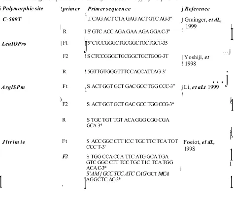
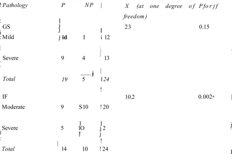
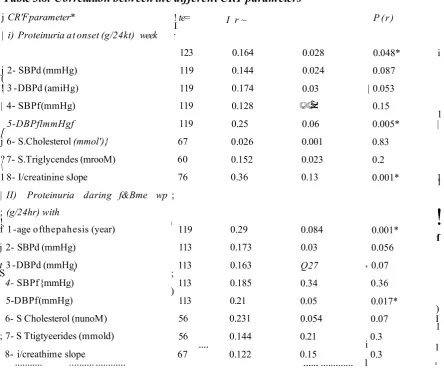
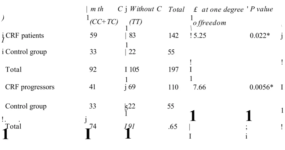
Caucasian patients with CRF were screened for four TGFB1 SNPs: T-509C in the promoter region; ArglSPr© and Leu I OPro in exon 1 and Thr263Ile in exon 5. There were significant differences between CRF patients and controls in allele frequencies of two of the SNPs (LeelOPr© and C-509T), indicating an association with susceptibility to CRF, We also observed a significant association between rate of progression of CRF (the slope of the reciprocal of serum creatinine v time) and genotype, both at codon 25 (odds ratio 3.77, 95% confidence interval, 2.2 - 6, p <0.001) and at the -509 promoter site (odds ratio 1.67, 95% confidence interval 1.1-2.5), p <0.005) in patients with primary nephropathy (excluding PKD). Genotype at codon 25 was also associated with severity of proteinuria (p= 0.038), plasma TGF-{31 protein levels (p = 0.01), and the severity of glomerulosclerosis (p<0.05). Genotype at C-509T was associated with the level of renal tubular TGF-pl immunostaining (p = 0,9006) and with renal interstitial inflammatory cellular infiltration (p=0.015). There was a highly significant correlation between the degree of cellular infiltration in renal tissues and tubular TGF-pi immunostaining.
ACKNOWLEDGEMENTS
Egyptian Embassy: For giving me the chance and the financial support to undertake this thesis.
Dr, A Blake in ore; For her guidance m directing my work, for her valuable supervision, teaching me the fundamentals o f molecular genetics and her continuing advice throughout my research
Professor A Megvid El Nafcas: For his guidance and his endless encouragement in the pursue of my research and his invaluable supervision of the thesis.
Dr. P Watson: for his useful guidance for a part of the genetics work.
Dr, N Quinton: For her kindness m teaching me the principles of laboratory work.
Pathology7 team {Northern General Hospital): For their kindness in supplying me with the tissue sections and information about some patients.
This thesis is dedicated to my wife and my daughters who shared with
me the stressful situations, w hich I faced during my years of research
ABBREVIATIONS
AAV: ANCA-associaled vasculitis ABC: avidin-biotin-peroxidase
ACE: angiotensin I converting enzyme ActR: activin receptor type
ADPKD: Autosomal dominant jwlycysfic kidney disease AEC: 3s amino- 9r etoyi-carffimazoie
AGT: angiotensinogen
ALK: activin receptor-like kinase
AMHR: anliHmnllerian hormone receptor Aug It angiotensin H
Apo: apoKpoprotem Arg: arginine
ARMS-PCR: amplification refectory mutation-screening polymerase chain reaction AT1R: angiotensin type 1 receptor
bFGF: basic fibroblast growth factor BMPs: bone moiphogenic proteins
CAPD: continuous ambulatory peritoneal dialysis GGN: crescentic gfomeralonepfaritis
GEN: chronic interstitial nephritis CrCl: creatinine clearance
CRD: chronic renal diseases
CREB: cAMP respond element binding protein CRF: chronic renal failure
CTGF: connective tissue growth fetor D*: coefficient of linkage disecpiiibriwa DBP: diastolic blood pressure
DBPd: diastolic blood pressure at diagnosis DBPf: diastolic blood pressure during follow up DM: diabetes mellf tits
DIN: diabetic nephropathy DMA: deoxyribonucleic Add
dMTPs: deoxynucleotide triprosphatcs
EcNOS: endothelial constitutive nitric oxide synthase ECM: extracellular collagenous matrix
EGF: epidermal growth fader
ELISA: enzyme linked Immunoassay ERK: extracellular signal-regulated Meases ESRD: end stage renal disease
ESRF: end stage renal ladine ETliendotbelin I
FGF: fibroblast growth factor
FSGS: focal and segmental glomerulosclerosis GDFs: growth and differentiation factors
GDNF: glial cell line-derived souotaDfduc factor GLUT: glucose transporter
GN: glomerulonephritis GS: glycine serine domain gTGF-pi: glomeruli TGF-pl
gTGF-pit-: glomeruli stained with TGF-pi gTGF-p 1-: glomeruli did not seined for TGF-p 1 HD: haemodialysis
ICI: interstitial cellular infiltration
ICI-: renal tissue without inflammatory ceftular infiltration ICH: renal tissue with inflammatoiy cellular infiltration. IGF-l: insulin-like growth feetor-l
IF: interstitial fibrosis IL-1: interleukin 1
ILIra: interleukin 1 teoepSor antagonist
IL1RN*2: interleukin 1 receptor antagonist allele 2 He: isoleucine
KLK: kallikrein
LAP: latency associated peptide LDL: low-density lipoproteins Leu: leucine
LLC: large latent complex
LTBP: latent precursor molecule fomdmg protein LTGF-p: latent tEecufsor molecule
MAP: mean arterial blood pressure MAPK: mitogen-activated protein kinase MCGN: mesangiocapillaiy gtomeralonepliritis MCP-1: monocyte ctono-attractenl protein 1 MDRD: modification of diet.in lend disease MH: mad homology
MIF-2: macrophage inhibitory factor-2 MIS: mullerian inhibitory- substance MMP: metaUoproteiimse
MN: membranous nepbiopa%
MRFIT: multiple Risk Factor Intervention Trial mRNA: messenger ribonucleic acid
MTHFR: melliyleneielraliydrofoMe reductase NGF: nerve growth factor
NO: nitric oxide
NOS: nitric oxide synthase
NP: non-progressive chronic renal Mure OU: obstructive uropatby
P: progressors chronic renal M ore PAl-1: plasminogen activator Mnbitor-l
PCR: polymerase chain reaction PDGF: platelet derived growth fetor PKD: polycystic kidney disease Pro: proline
RI: type 1 transforming growth factor beta receptor R lt type 2 transforming growth factor beta receptor RIH: type 3 transforming growth factor beta receptor
RANTES: regulated on activation, normal T expressed and secreted RRT: renal replacement therapy
SARA: Smad anchor for receptor activation SBPd: systolic blood pressure at diagnosis SBPf: systolic blood pressure during follow up l/Scr reciprocal o f serum creatinine
SLC: small latent complex
SLE: systemic lupus erythematosus
SMAD: signalling mother against decapentaplegic protein SNPs: single nucleotide polymorphisms
TBE: Tris borate EDTA
TpR: transforming growth factor beta receptor TGF-a: transforming growth fetor alpha TGF-pi: transforming growth fetor beta-1 Thn therionine
TIF: tubulointerstitial fibrosis
TIMP: tissue inMbifors o f metalioproteioase TNF-a: tumour necrosis factor alpha
tRNA: transferase ribomidiee add TSP-I: thntobospondinl
tTGF-pl: tubular TGF-pl
VC AM-1: vascular cell adhesion molecule-1 YEGF: vascular endotiidial gpowtfc fetor
Contents
Page
Abstract
IAcknowledgements
n
Dedication
m
Abbreviations
iv
Contents
vrn
Publications
xi
Contents
General introduction
i
1.1 Introduction 2
1.2 Chronic renal failure 2
1.2.1 Natural history of chronic renal failure 3 1.2.2 Factors influencing the progression of chronic renal failure 4 1.3 Mechanisms of progression of chrome renal failure 10
1.4 TGF-P 15
1.5 TGF-p activation 21
1.6 The role of TGF-p 21
1.7
Genetic aspects of chronic renal failure 361.8 TGF-pl and TGF-p gene 43
1.8.3 TGF-P 1 polymorphism and renal diseases 45
Aim of the thesis 47
Materials
and
Methods
48
2.1 Collection of blood samples 49
2.2 Genomic DNA extraction techniques 51
2.3 Polymorphisms analysis 52
2.4 Measurement of plasma TGF-p 1 57
2.5 Renal histology evaluation 57 2.6 Estimation of TGF-p 1 levels in renal tissue 57
2.7 Statistical analysis 58
Clinical data
623.1 Introduction 63
3.2 Methodology 65
3.3 Results 60
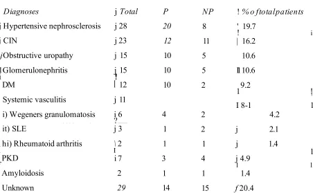
3.3.1 Clinical Observations 67
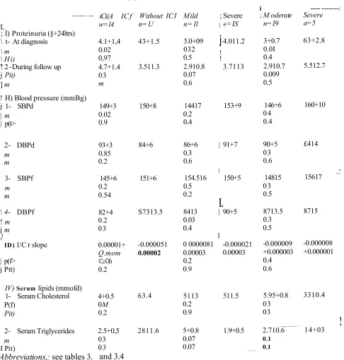
3.4 Factors affecting the progression of CRF 72 3.4.5 Correlations between the different parameters of CRF 77
3.5 Discussion 79
Investigation of four single nucleotide polymorphisms in the
83 84 88 90 90 91 104 114 125 128 131 135 136 138 139 EX
TGF-pl gene
4.1 Introduction4.2 Material and Methods 4.3 Results
4.3.1 Investigation ofDNA quality 4.3.2 C-509T
4.3.3 LeulGPr© 4.3.4 Arg25Pro 4.3.5 Thr263He
4.3.5 Linkage disequilibium studies 4.3.5 Discussion
Measurement of TGF-p l
5.3.1 Circulating TGF-pl 139
5.4 Renal immunoslainable TGF-pl 151
5.5 Discussion 166
Qeneral discussion
170
References
179
Appendix
220/- Abstracts a ad pasters:
1- Khalil, MLS.; Biakemore, A LE and El Nahas, A M (2001); Transforming growth factor beta-1 (TGF-pl) polymorphisms as a predictor of progressive renal insufficiency. Am Soc Nephrol: 12: 817A.
2- KhalH, M.S.; Blakemore, A,LF. and El Nahas, A.M. (2001); Transforming growth factor beta 1: role in the progression of chronic renal failure. The Renal Association.
II- Paper:
Salah, \L ; El Nahas, A M and Blakeniore, ALF. (2002); Transforming growth factor
beta-1 (TGF-pl) SNPs; genetic and phenotypic correlations in progressive renal insufficiency, (submitted to Kidney International).
III- Patentt
Khalil, M.S.; El Nahas, A.M. and Blakemore, A.l.F. (2001): Genetic markers of chronic renal failure (UK).
1.1 Introduction
Transforming growth factor beta-1 (TGF-pl) is a multifunctional growth factor implicated in the pathogenesis ofesqpaunenSal and ctinkal chronic renal M a e (CRF) (Okntia, 1990; Yamamoto, eit a i1993). It Ms potent fibrogenic propatks through the stimulation of synthesis of extracellular coiagenous matrix (BCM) and inhibition of its breakdown {Border and Noble, 1997)1 Although there are three types o f TGF-p (1,2 and 3), TGF-p i is the growth factor that has been implicated in fiferogenesis (Roberts and Sperm 1997). There is considerable experimental and clinical evidence pointing to an important role of TGF-pl in renal ffinrogpiesis (section 1.6.2) (Border and Noble,. 1994, 1997, Peters, ef &L„ 1999, Baste, 1999). However, a clear-cut association between TGF- p l and the profession of clinical nephropathies is lacking. In this thesis, the potential relationship between TGF-pl genetic polymorphisms and tire profession of human nephropathies is studied.
1.2 Chronic renal failure (CRF)
Chronic renal faftuie (CRF) is defined as irreversible, long-stMifcg loss of renal function. End stage renal failure (ESRF) refers to adv anced renal insufficiency when the glomerular filtration rate is below I© ml/min prior to the initiation of either dialysis or renal transplantation (El Nahas and Wiwearis, 1997). The patients complain of a wide range of symptoms including pom appetite, vomiting, bone pain, headache, insomnia,
itching, dry skin, malaise^ fatigue with light activity, muscle cramps, and change in mental alertness (El Nahas & Winearis, 1997). A survey of UK hospital biochemistry records revealed, that the prevalence o f CRF (plasma creatinine concentration >15© pmel/1) is 2058 adults per sniiioii population (prop) (UK Registry, 2001). The data also revealed the incidence of ESRF to fee around 78 pmp/year (UK Registry, 200]) Data from the UK registry suggest feat fee incidence of ESRF in the UK is currently .around 80-110 pmp/year (UK Registry, 2001) Corresponding data from the Untiled States (US) suggest an. incidence of 315 pmp/year; US prevalence is currently around 1217 pmp (USRDS, 2001% In addition* there as little doubt that the incidence o f ESRF is steadily7
increasing by aronnd 8-10% eweiy year (USRDS, 2001). One o f the major causes of ESRFintbeUS is diabetes meffitus (DM) (41.8 %). In Norway, type 1 DM progresses to ESRF in 40% of patients, while type 2 is thought to lead to renal involvement in 20% of patients (Type 2 DM is 10 times as common as Type 1) (Bergrem and Lea vested, 2001). In the US, the second most common cause of ESRF is hypertension (25.4%). Thirdly; chronic glomerulonephritis accounts for 21% of patients with end stage renal diseases (ESRD). Polycystic kidney disease and wrologic disease account for approximately 15% and 20% of cases of ESRD have no known cause (USRDS, 2001) On the other hand, m
the UK, the most common cause o f ESRF remains chronic gtomemlonephritis as the incidence and prevalence of diabetic nepferopatby remains below 20% (UK Registry' 2001).
L Z l Natural history o f chrome nm alfailure
Progression of CRF occurs following renal injury regardless of the underlying cause of nephropathy (For review see Locatdli and Del Vecchio, 2000). I1k reciprocal of serum creatifii.se (I/Sq) regression slope value against time (described below) determines the rate of decline of renal Rmctoon ^progression of CRF). When the pattern of the regression is a straight line this means that the rate of the progression of CRF occurs at a constant rate (Mitch, et aL, 1976, Rutherford, et aL, 1977, Bleyer, 1999). Generally speaking foe rate of decline of ienai rimctiom is constant, Implying that the process causing the decline of renal function is conthmom. Bl was repotted in the late seventies, that the progression of CRF from various diseases was ©marring at a constant rate (linear pattern of I/Sq- value against time) In foe majority of patients (Mitch, et aL, 1976 and Rufoerfotd, et aL,
1977), A non-linear -decline of renal inaction occurred in around 15% ©f patients (Shah and Levey, 1992). Further, some patients have a change in the regression line of the reciprocal of serum cim tm sm against time suggesting a spontaneous acceleration or slowing down of the rate of decline. These spontaneous changes are referred to as breakpoints in the slope which may be due to intercurrent events such as, infection, dehydration, poor Wood pressure control, or changes in the activity level, o f the underlying pathological processes which initiated the renal disease (Shah and Levey,
1992).
1.2.2 Factors influencing the progression of chronic renal failure
L2.2.1 Age
A ^ttig is associated with physiological changes in the kidneys, including a reduction in renal plasma flow (Riser,, et atti^ 19931, increase in filtration fraction (Baylis, et a t, 1990„ Flisher, et aL, 1993) and a decrease in renal size due to parenchymal reduction (Emamiait, et aL, 1993). Alas, fvim ay involution can be detected in the renal c o te with relative sparing of renal medulla (HoUenbeig, et aL, 1974). FurthenaMsre, 10 - 30% of the total glomeruli are sclerosed between the fourth and eighth decades of life (Kaplan, et aL,
1975). It is often assumed that GFR is nearly 50% at the age of SO compared to values after puberty (Kaysen and Myers, 1985) Ltndeznan and cowockers (1985) showed that creatinine clearance was not decreased in one third of healthy elderly people. Many studies have shown that the indcfence o f renal failure from various, renal diseases; increases- with age (Fees) et aL 1990, McGowen, 1990 and lungers, et a t, 1996). Berthoux and colleagues (1998) reported that renovascular diseases, and diabetes mellitus; (type 2) are the most common causes of ESRD' in the elderly. The same study also showed that primary gSomerolonephritis as common in the elderly as If constitutes about 12% of ESRD In the elderly . The prognosis of renal diseases in the elderly is also more severe than in the younger age group as they have, in general, a higher rate o f progression compared to younger patients with similar nephropathies (For review see Locatelli and Del Vecchio, K M ).
1.2*2.2 Gender
Regardless of the cause of CRF, ESRD as more common in males than females (For review, Locatelli and Del Vecchio, WOO). The USRDS study (2001) revealed that the Incidence of ESRD in men was 348 prop/year compared to 242 pmp/year in women (URSDS, 2001) Furthermore, the rate of decline in renal function is usually taster in males regardless, of die underlying nephropathy (Hannedouche, et aL, 1993) Some postulated that the decline o f renal function is faster in males due to a higher protein ingestion, larger muscle mass and increased creatinine generation (Levey, et aL 1989).
This is unlikely. Others argued that hormonal factors, including oestrogens, may play an important protective role in females with CRF (Velasquez and Bhatitena, 2001; For review, Neugartm, et aL„ 2000). Because the faster decline in renal function occurs only after puberty, it was assumed Jfeat sex hormones might piay a role in ft (ReckeiftofF, et al,
1997, Neugarten, et aL 2000). The slope of progression in males cordages with the mean arterial Wood pressure, which is generally Wgher in males. In females, ft correlates with the type of nephropathy (For review see Locatelli and Dei Vecchio, 2000)
/.2.Z J Race
In the US, African-Americans have a four-fold higher incidence of ESRD compared to Caucasians (USRDS, 2001). Freedman and colleagues (.1997) reported that the greater risk of ESRF in African-Americans might he due to a genetic predisposition. Tins may also be explained by higher levels of circulating fibrogenic growth factors in African- Americans compared to others (Suthanthiran, et aL, 2000). Also, it was reported that incidence of increased blood pressure levels in black individuals is twice as high as that in the white population (Coraoni, etaL, 1989). Die high blood pressure may contribute to the higher incidence of ESRF. Furthermore, in essential hypertension there is increased prevalence of mtrtaalbamhnina in black individuals (Summerson, et aL 1995). The prevalence of DM is twice as high in African-Americans as in Caucasians and. ESRD due to DM is 3-6 times more- eenKnesn in African-Americans (Carter, et dL, 1996). Also, ESRF was higher in Native Americans and Asian/Pacific Islanders than Caucasians (USRDS, 2001). The ineidance of CRF is increased in the first ami second-degree relatives of those with retail disorders (Ferguson, et aL, 1988). In Caucasians this relationship is weaker than in African-Americans. Pugh and colleagues (1988) found that Mexicati-Americans were also at a higher risk of developing ESRD than other populations. In the UK, reports suggest a higher prevalence of renal disease in souse Asian individuals although die rate of progression of the nephropathies was not found to be faster than Caucasians (For review. Wing and Jones, 2000, UK Registry, 2001).
1.ZZ4 Systemic hypertension
Systemic hypertension is one of the most important factors conteifeeting to the deterioration of renal function and elevated blood pressure, can fee a cause or a consequence of lena! mjury (For levsew, Adamczak, et <d.9 2002). The higher the blood pressure the faster the rate of decline of renal function and the progression of CRF (Tiemay, et a l, 1989). Brazy and. cowwkers (1989), showed that increase in mean diastolic Wood pressure (DBF) > 90 moiHg was associated with a greater rate of decline in the reciprocal of serum creatinine versus time compared to patients with CRF with a mean DBF < 90 mmHg). Furthermore, there is improvement in renal function with intensive antihypertensive therapy (For review, Locatelli and Bel Vecchio, 2000 and Adamczak, ei aL, 2002). It Ms even been suggested that a stabilization and even a regression of the progression of CRF cm fee achieved with aggressive antihypertensive therapy (For review, Dwmtrin ami Weir, 2000). The Modification of Diet in Renal Disease (MDRD) Study Group stowed that the lower the mean arterial blood pressure ( MAP) value, the slower the decline in GFR (Klahr, et a!.P 1994). This study implied that lower blood pressure targets should fee sought for patients with progressive CRF and high levels of proteinuria (Klahr, etttL, 1994). In those with protein excretion rate in excess of 3 g24h, it suggested that the MAP should to reduced to levels around 92 mmHg to obtain the same protective effect of renal function a MAP of 97 mmHg would provide those with less proteinuria (Klahr et @L, 1994, Peterson, et aL, 1995).
LZZ5 Prateifuiria
Proteinuria is an important pogprostie indicator of renal disease. Kineaid-Smith and Becker (1978) stowed that the presence of proteinuria predicted the progression of CRF in patients with chronic fwdtonqdnitisw The presence of proteinuria indicates poor prognosis in most cases of primary glomenilonephritis (Williams, et aL, 1988 and Cameron, 1989). El Mates and colleagues (1984) were the first to demonstrate that a reduction of proteinuria by dietary protein restriction predicted the renal functional response to the diet. Bjorek (1986) Apperloo (1994) and their colleagues showed that pharmacological interventions that slow the progression of CRF are associated with a reduction of proteinuria. More recently, a large body of experimental evidence has
suggested that pioteirama is not os% a marker of poor prognosis but may also be a mediator involve! in the progressive scarring process (Bruzzi, et aL, 1997, Eddy, 2001, Jafar, et aL 2001). Remuzzi ami Bertani (1998) postulated that the excessive filtration of macromolectiles, Including protein, into the glomerulus could accelerate sclerosis. Burton and Harris (1996) suggested that proteinuria might also cause tubular dysfunction and damage. Renal tubular damage caused by proteinuria can initiate inflammatory and fibrotie changes within the renal interstitram. The activation of proximal tubular cells by excessive exposure to proteins leads to their release of pro-inflammatory cytokines and chemokines as well as their release of proflbrotic growth factors such as platelet derived growth factor (PDGF) and TGF-p i (For review, Harris, 2000; Eddy, 2001; Waidle, 2001, 2002). It was also hypothesised that mesangial accumulation of proteins may produce mesangial cell injury and pnifisation and consequently increases production of mesagial matrix and gtomeralosclanwis (For review, Harris, 2000). Also, activation of tubular cells by proteinuria cm stimulate their release of extracellular matrix (ECM) components, accelerating interstitial flbrosis (For review see, Hams, 2000). Consequently, proteinuria is, along with systemic hypeftenstoo, thought to he one of the most significant risk factors in progressive CRF. Increasingly, attention is paid when hypertension is treated in patients with CRF to lower proteinuria as well (Jafar, et aL, 2001, Adamczak, et aL,
2002). Data from a European study suggested that the control of systemic hypertension without the concomitant reduction of proteinuria is ineffective (Locatelli, dtaL, 1996).
L2.2.6 Dxsiipidaemia
ft was postulated over twenty years ago by Moorhead and his colleagues (1982) that lipids could he toxic to both the glomeruli and the tubulointerstitium. Since, a growing body of experimental and dtnscal data has supported this hypothesis (Altman, et crl, 1999; Samuelsson, et d.., 1997). An association has Men shown between hypercholesterolaemia and die pogressaon of diabetic (Krowieskj, et d ,, 1994) and noncbabetic (Samuelsson. et aL 1997) nephropathies. Hyperl ipidaemia can induce glomerular toxicity through the accumulation of Jow-densily lipoproteins (LDL) as well as oxidized LDL in the mesangiuin, leading to structural and functional changes in
contribute to glomerular hypertension and sclerosis (Keane, at aL 1988, Keane, 2000). It was also concluded that hyperiqsdcmia activates mesangial cells and lead to mesangial matrix accumulation (Keane, et at, 1988). Harris and colleagues (1990) showed that a diet low in essential fitty acids protects against tubuiointeistitia! inilainniarion. Kees-Foti and co in M ’g t e (1994) showed that free fatty acids lead to the generation of a JipM chemotactic factor, which attracts monocytes, and initiates tubulointerstitial inflammation. So a high level of non-essential fatty acids may play a role in the inflammatory and fibrotie processes in the renal inftevstitnm. Further, it was advanced by some that the nephrotoxicity of pcoteinsmaialfouimnuria may be linked to their lipid-carrying capacity (For review, Harris, 2000). lipids rather than protein may fee toe culprit regarding tubulointerstitial Mlammatiem and fibrosis in heavy proteinuric states (Tor review, Harris, 2000). In experimental animals, there is a large number of experiments showing that the reduction of blood ipkJs levels is associated with a protective effect on kidney scarring (Keane, 2000, Praga, 2002). However, similar data is still Jacking in humans with CRF, although a recent review analysts suggested that lowering lipids In patients with CRF may be beneficial (Fried, et aL 2001).
1.2.2.7 Smoking
Smoking has a deleterious effect cm renal function, as evidenced by the development of mleroalbtmmusM and its progression to overt albuminuria in diabetic nephropathy (Chase, et aL 1991). Smoking of 15 packkyear in non-diabetic patients increases toe risk of development of ESRD by 5.8 fold (Orth, et aL 1998). Haemodialysed diabetic patients who smoked cigarettes had higher systolic Wood pressure and fibrinogen levels and had a higher incidence of myocardial infarction, than non-smokers (Biesenbach and Zazgomlk, 1996). A retrospective rouJticenrtre European case-control study stowed, that smoking is an independent risk facta? far ESRF in patients with both inflammatory and non inflammatory renal disease* Le. IgA gfomeralonephritis and polycystic kidney disease (Orth, et aL 2000). Smoking increases the severity of glomerulonephritis, particularly in men older than 40 and/or hypertensive patients (Stengel, et aL 2000, Regalado, et aL
(Walker, et a!., 1992), The calculated relative risk for ESRF was found to be 1.69 for smokers as compared to non-smokeis (Shqji, et ah, 2001). The deleterious effect of cigarette smoking may be through exacerbation of other risk factors such as high blood pressure, proteinuria or hjperlipadbemia. Moreover, progressive kidney failure is associated with decreased, diminution of nicorine by both renal and non-renal mechanisms (Molander, et aL, 2CB0). Furthermore, it was postulated that smoking induces renal damage by increasing blood pressure, alteration of intiarenaf hemodynamics and activation of tie sympathetic nervous system. Discontinuation of smoking improved renal prognosis, and is probably a very effective measure to retard progression o f renal failure CPiaga, 2002).
l,Z Z 8A lcokof
It was reported that cross-sectional data from the 1983 (National Health Interview Survey) showed that hypertensive women consumed less alcohol than non-hypertensive women (Laforge, et aL, 1990). On the other hand, the same authors reported that alcohol consumption was significant^7 associated with a greater risk of hypertension amongst men. Moreover, beer consumption and spirits consumption above three drinkxday were significant predictors of male hypertension (Laforge, et aL, 1990). The consumption of more than two alcoholic drinks per day is associated with an increased risk of ESRD and hypertension in the general population (Laforge, et ah, 1990). On the other hand, a lower intake of alcohol (< 2 dririksfday) did not appear to be harmful (Peroeger, et. ah, 1999, Parekh and Klag, 2001).. Trie mechanisms by which alcohol consumption leads to hypertension and, perhaps, renal disease are unknown.
1.2.2.9 Recreational drugs
Use of heroin and/or cocaine increases the risk of ESRD (Perneger, et. aL, 2001). After adjusting for patients" age, sex, race, socioeconomic status, and history of hypertension and diabetes, persons who had ever used heroin or other opiates (any amount) were at increased risk for ESRD (Perneger, et aL, 2001). There was also an increased risk for individuals who consumed crack cocaine, although it was difficult to dissociate it from
heroin consumption (Perneger, et <stLn2001). The same authors suggested that cocaine might cause accelerated h p sten sk n , acute renal failure from ihaMomyolysis, and progression of jse-exisilng renal disease.
13 Mechanisms of progression of cferwic renal failure
13.1 G1 om ero J osclerosis
13J 3 System ic am i glom erular hypertension
As previously discussed, systemic hypertension not only induces CRF but also induces a faster loss of ram! funtitkm in squired renal disuses and loss of renal function associated with iwimal ageing (Adamczak, et aL, 2002). In the subtotal nephrectomy model of CRF, the loss of function of some nephrons leads to hypertrophy of the remaining nephrons and decrease of vascular resistance (especially the afferent renal arterioles) due to impaired autoregotelion. (For review see Dworfcin and Weir, 2000) This process leads to an increase m the glomerular capillary7 pressure of the remaining nephrons (glomerular hypertension) (Anderson, et aL, 1986). It was postulated that there was a significant association between systemic hypertension aw! the increment of glomerular capillary pressure (For review see Dworkin and Weir, 2CWQ). The harmful effects of renal parenchymal disease, e.g. diabetes, mellitus and systemic hypertension, are a result of production or aDceasmaiiou of glomerular capillary hypertension, rather than induction of exteagtomerukr vascular injury or glomerular ischaemia. Histological examination reveals an increase of glomerular volume and glomerular sclerosis in hypertensive patients (For review see Dworkin and Weir, 2000). fa the presence of an underlying nephropathy, the glomerular response to systemic hypertension is impaired (For review see Dwaririn and Weir, 2000). This leads to a rise in gkimendar pressure and progression of gkxoefulossxdenxdsL Studies suggest that the control of glomerular capillary pressure protects against kidney deterioration with or without control of systemic hypertension (Anderson and Brenner, 1987, For review, Anderson 2000, Dworkin and Weir 2000).
Lowenstein and cow odm (1970) also argued that systemic hypertension leads to ischaemia due to renal vascular disease which, in turn, leads to a decrease in glomerular perfusion/ischaemia. Gkmenilasclerasis induces efferent arteriolar hypoperfusion and hypoxia (decreased oxygen delivery) of the tubular cells and their release of cytokines such as TGF-pl, platdet-derived growth factor (PDGF), endothebn and vascular endothelial growth factor (VEGF) lOrphamdes, etal.f 1997; Fine, e ta lf 1998).
Both systemic hypertension ard non-hypertensive injury? that cause loss of single nephron units results in hypertension in the remaining glomeruli (glomerular hypertension).. Glomerular hypertension cm lead to injury to the glomerular capillary' wall causing it to leak plasma proteins into the mine (Anderson and Brenner, 1987; Anderson, 2000).
13. L2 Rale o f proteinuria
Systemic and glomerular hypertension are both associated with proteinuria. Olson and colleagues (1985) demonstrated that glomerular hypertension leads to transudation of plasma proteins into the endothelial and subendotheliai spaces which can promote glomerular hyalioosis, thereby narrowing aid occluding glomerular capillaries. Increased trafficking and accumulation of proteinaeious molecules in the glomerular mesangium may also contribute to the pathogenesis of glomerulosclerosis (Bertani and Remuzri,
1998). Reimke and Klein (1989/ showed that the increment of glomerular permeability to protein affected not only the glomerular mesangium but also the glomerular epithelial cells leading to structural and functional changes and this further increased the passage of protein molecules across the glomerular capillaries (For review, Harris 2000, Anderson 2000).
13J 3 Renal hypertrophy
Loss of function of some of the nephrons leads to an increase in the size and cellular number of the remaining glomeruli (Wesson, 1989; .Anderson and Mey®; 1997). Several growth factors such as insulan-like growth factor-1 (IGF-1), epidermal growth factor (EOF), platelet-derived growth factor (PDGF) and TGF-pl are responsible for the
hypertrophy and hyperplasia of the remaining glomeruli (fine, et «a£, 1992). Glomerular hypertrophy has been t o forward as a likely pathway to glomerulosclerosis (Ichikawa, et aL, 1986). These authors argued that it was glomerular hypertrophy rather than gJomeruJar hypertension that initiated and perpetuated glomerulosclerosis (Ichikawa, et aL 1986). Moreover, growth factors associated with hypertrophy Might he also instrumental in the glomerulosclerosis (Fogo and Ichikawa, 1989).. Prominent amongst these growth factors, TGF-j31 is known to he a potent hypertrophic growth factor (Fine, et a l, 1992),. Increase in glomerular size (hypertrophy) along with an increased glomerular capillary pressure (hypertension) would lead to a significant increase in the glomerular capillaries' wall tension. Such an increased wait tension with the associated shear stress would initiate a cascade of events culminating in glomerulosclerosis (For review, Dwoikia and Weir, 2000). Initially, glomerular endothelium would fee most affected % the haemodynamic strain It would initiate a local inflammatory response. Glomerular Inflammation with the acctsnnlatkMi of leukocyte and monocytes has to n shown in immune and non-immune-mediated renal diseases (Erwig, et aL, 2000). Infiltration of the glomerular tuft by monocytes leads to their interaction with glomerular ceils especially mesangial cells (Mene, et &L 2002). Mesangial cells are activated and proliferate in response to the release by monocytes/foam cells of nffitogeoie growth factors such as PDGF (Johnson, 1994). The activation of mesangial cells has to n linked to the trans-differentiation of these cells into myofibroblasts (Johnson, et 1991). These cells have been shown to release ECM components including interstitial collagen III. Stretching of the glomeruli by hypertrophy and hypertension is also likely to lead to the stretching of glomerular epithelial cells (Rennke, 1994). This would lead to the denudemeot of areas of the basement membrane not covered by the stretched podoeytes (Kriz, et aL 2001). This would facilitate the leakage of proteins and otter macromolecules Furthermore, the activation o f epithelial cells within the glomerulus has been shown to be associated with their release of fibrogemc growth factors and BCM components. This would also exacerbate glomerulosclerosis (Kriz; 1996) Fibrogemc growth factors such as TGF-pi are likely to be involved in ail the stages of glomerulosclerosis including hypertrophy* inflammation and fibrosis (Basile, 1999)
In sum m ary, the faypotks^ for induction of glomerulosclerosis have implicated glomerular hyperfiltration, hypoperfusion, glomerular hypertension, glomerular hypertrophy; nephrotoxicity’ of proteinuria and the role of growth factors such as PDGF and TGF-pL
1.3.2 Tubulointerstitial fibrosis (T it')
Local and systemic cytokines play a role in the induction of TEL There are several cells responsible for the production of ECM (scar) in the kidneys. These cells are infiltrating macrophages (attracted by activated tubules as discussed above), tubular epithelial cells; peritubular capillary7 endothelial cels (after vasoconstriction or ischaemiaTiypoxia) and film>blastspencyTes These cells are resident kidney cells, migrating and transformed cells (For review, Jernigam and Eddy, 2000). The cells release fibrogeiric factors such as; TGF-pl, angiotensin II, endotMin I, TGF-a, PDGF, and FGF (For review, Jemigan and Eddy, 2000). Accumulation of ECM te d s to obliteration of peritubular capillaries and the death of tubular cells due to isctaemia (Bohle, et al., 1981; Seron, ef a t, 2001) and consequently, progressive renal insufficiency (Jemigan and Eddy, 2000; Eddy, 2001). There is a good correlation between die severity of tubulointerstitial fibrosis and renal function (Bohle, et aL„ 1994)..
1.3. Z I Composition andformation of int erstitialfibrosis
Interstitial fibrosis is the result o f excessive accumulation of ECM in the renal interstitium. Matrix is composed o f both normal interstitial proteins such as collagen (L III, ¥ , ¥11, XV) and proteins which are normally restricted to the basement membrane such as collagen IV and laimmn (For review, Jemigan and Eddy, 2000). Two enzymatic pathways regulate matrix protem turnover. The first due to the activation of matrix metalloproteinases (MMP) ami the second initiated by the generation of pSasmm from plasminogen (For review, Brantom and Kopp, 1999). Plasmin cam degrade fibrin, iibronectin, and laminim (Liotta, et a l, 1981), and also activate tire gelafimase class of MMPs (Wong, etaL, 1992). Furthermore, plasmin activates latent cottagenases and TGF- pl (Pollanen, et a t, 1991). Tissue and urokinase activators as well as plasminogen activator activate plasminogen. Imtersritiaj collagenases such as MMPs, degrade the
interstitial matrix eolkgees (collagen I, II, III), stromelysin family members degrade the basement membrane proteins (collagen IV, V) and other proteins such as ftbronectin, gelatinases degrade basement membrane proteins and elastm (Norman and Lewis, 1996). Fuithennore, membrane type MMPs (MT-MMP) which degrade collagen III, fibronectm, laminin, have been descnbed (Norman and Lewis, 1996). Tissue MMPs are activated by
membrane-bound MMP proteins and inactivated by tissue inhibitors of metallofroteinases (TIMP-1-3). Plasminogen activator inhibitor-! (PA1-I) inhibits the other coflagenolytic pathway by Inhibiting plasmin activ ation. During the course of renal scarring and interstitial fibrosis, there is a down-regulation of MMPs and npcegulation of then inhibitors, TIMPs and PAJ-I (For review, Jemigan and Eddy, 2000). These processes lead to decreased breakdown of deposited ECM and, consequently, increase in its accumulation in the scarred kidney. Transforming growth factor-p 1 has the capacity to directly inhibit MMPs and activate their inhibitors TIMPs and PAM. Therefore, TGF-^1 stimulation of ECM synthesis as well as Inhibition of its breakdown would lead to irreversible renal fibrosis (Border and Noble, 1994, 1997 andBasile 1999).
Figure 4 Mechanisms leading to progressive renal failure.
Pathways Leading To
Progressive Renal Failure
Renal growth factor & cytokine
scbvstion
Renat injury t
Nephron m ass
I
Transdifferentiat*an \ \
of renal cells to X \
fibroblast Influx of \
p h c n © ty pe m o n c c yt es and m acrophages
t
Fibrogenesrs --- ► a
-Progressive Loss of Filtration Surface Area
\ i
Systemic
hypertension
'Filtration of plasma proteins (Proteinuria 5
‘*1 Proximal tubule \ protein uptake
/ H yperfip id emia
Renal m k rovascular
injury
noianvtl99Y<jfthnaiMff*
•V 7^V-v> ■ * ■;..
1.4. Transforming growth faetor-p
1,4.] Transforming growth factor-ft super fam ily
The transforming growth factor p superfamily (Table 1) is a protein group that includes bone morphogenic proteins (BMPs), activm/inhibins, that inhibit pituitary secretion of follicle stimulating hormone, Mullerian inhibitory substance (MIS), which is produced by the testis and is responsible for the regression of the Mullerian ducts (to induce development of the female reproductive system), and DPP (deeapeotaplegic protein is part of the TGF-P superfamily that regulates Drosophila morphogenesis) (Leslie, 1999). The supertamily also includes glial cell line-derived neurotrophic factor (GDNF) and growth and differentiation factors (GBFs) (Leslie, 1999). Furthermore, this family includes the MAD system (the mother against decapentaplegic protein, which is part of the TGF-P superfamily that regulates Drosophila morphogenesis) and is also called Smad which are the downstream effectors of TGF-P I signalling. Smads molecular weights range from 42 to 60 kDa. The Smads have two domains, MH (Mad Homology7) 1 in the amino terminal regions and MH2 in the C-terminai region (Massague, et: a l, 1997).
The Smad system includes, Smad], 2,3,5 and 8 which are activated by R1 (TGF-p type I receptor) sertiir kinase receptor of TGF-p family including BMPs, DPP, and actcvins (Graff, et a l, 1996, Lm. et a l„ 1996 and Kretzehmar, et a l, 1997, Roberts 2002). Smad4 is a common factor required for TGF-pl, activin and BMP signaling Smadh and SmadT are inhibitory' towards TGF-pl signalling (Massague, 1990) (Table 1). The immediate TGF-P family includes five members (TGFpl-5), however the mammaliait isoforms of TGF-P3 are three (1-3). The structure of these isoforms is nearly identical, as they contain nine cysteine residues and share 76 to 80% of the amino acid sequence (Figure 1 and 2). Et was reported that the mature active form of TGF-p i structure conforms to a cysteine knot motif similar to nerve growth factor (NGF) and platelet-derived growth factor (PDGF) which do not belong to the TGF-p family and only share 10°/© of the amino acid sequence (Daopn, et aL, 1992; Schiunegger and Gruttei, 1992). This knot is
held together by six cysteines joined together by three intra-chain disulfide bonds, which stabilize the beta sheet stands (Figure, X).
Table 1. Members o f TGF-pl sttpeefamily
! L igand Type LI Receptor
Type I Receptor Receptor regulating Smad
Common Smad
Lnkfhkory S a m i
\ \
TGF-p TpRJl ALK5(TpRl)
Smad2 i Activin ActJOI ActRIfB ALK4|Act®t!B Smad3 Smao4 Smad6 BMPs ActRII AdtRUB BMPR1I ALK2|AolRf} AL!G(BMFR1A) ALK(BMPR1B) Smadi SmadS SmadS SsssacTF ;
I i ?
MKS(TSRI) Smadl I MIS
1
AMHR ? I ?
!
! ?
:
? ALK7 I! ?
1
(Kfuppef, etmL, 199$,
Abrrevmtiom: TGF-P: transforming growth factor beta; BMPs: bone morphogenk proteins; MIS: imiliedan inhMtory substance; ALK: activin receptor-like kinase; Smads: signalling mother against skeapentaplegie protein; AMHR: anti-MnSledan honnone receptor, AetR: Activin receptor type; TpR: transforming growth factor beta receptor
1.4.2 Structure qfTG F -ftl
Transforming growth factor-pl is a dimeric protein of 25kDa (Figure 1}. It is secreted as. inactive form sailed latent pfeonsor molecule (LTGF-pI). The latent precursor molecule consists of 390-414 amino acids and contains an amino-terminal hydrophobic signal peptide region called latency associated peptide (LAP), consisting of 249 residues (Hinck. etaL, 1996). The latent precursor molecule (LTGF-pl) contains 112 amino acids at the C terminal* which is the potentially bioactive mature region (Figure 2). The latent precursor molecule LTGF-pl is secreted as a large latent complex (LLCJ, bound via the LAP region to another protein called LTGF-p I binding protein (LTBP) (Miyazono, et a l, 1991; Taflpale, et d , 1995),. The Meat precursor molecule binding protein (LTBP) has an important role in the assembly and secretion of LTGF-pl and serves to bind LTGF-P 1 to extracellular matrices to enable proteolytic activation (Nunes, et a t, 1997). Latent precursor molecule secreted without LTBP, is called the small latent complex (SLC) (Munger, et a l, 1997% The mature (active) form is derived from the latent form, which consists of two large polypeptide chains linked by a disulfide bond to the latent TGF-P 1 binding protein (LTBP). The molecular mass of this complex is 210 kDa The latent precursor molecule is secreted and preeleaved mtracefluiarly at dibasic residues located between the LAP and the mature region.. The LAP portion blocks the activation of the bioactive domains by keeping its folding. The folding of the bioactive domain assists by giycosyiation of LAP, which undergoes mannose-6-phosphate addition at N-termina! (Schultz, et a l, 1995).
Figure i,
(h ttp^/epokme.
Figure2, Cartoon o f latent TGF-$l
(Nunes, efaL, 1997)
> LTBP
* LAP
-^TGF-pl
Abbreviations: LTBP: latent precursor molecule binding protein; LAP: latency associated peptide; TGF-p: transforming growth factor beta.
Table Z TGF-fil protein sequentce
?Key (From T& Length |l>es€riptio!i SIGNAL !l 23 23 b w iO T M I. PROPEP ! m r m '255 BY SIMILARITY.
CHAIN 127939© 112 t g f^ l
DISVLFID 285 294 BY SIMILARITY.
hnSULFID 1|293356 |®F SIMILARITY.
DISILFID i'322 3S7 1 BY SIMILARITY.
ID1SVLFID 1326 389 B Y SIMILARITY.
DISILFID 1355 355 |;INTE8€HAIN.
CXRBOmJ) *2 82 ... P-LIMKED {POTENTMLY
CAMBOHYD 1136 136 '^-LINKED (POTENTIAL\
CARBOHYD 1176 176 ■'A | 1 'T*
JSITE 1244 246 b ICELL ATTACHMENT SITE iPOTESTIAlX
1 0 !i 2 0 ii 3 0 > 4 0 i 5 0 n 6 0 1!
M P P S G L R L L P L L L F L L K L L V
!
l y p g r p a a g l
1 S T C K T I B M E L
8 ¥ K R K R I E A I R
1 © Q X L S K L R . L S
7 0 ii
8 0 n
9 0
i 1 0 0it 1 1 0H
1 2 ® j !!
S P P 5 Q G E W P
II V P L P E A V L K L
8 Y N E S T R D R V A G
1 E S & E P E P E P E
5 A D Y Y A K E V T R
1 F I L M V E M T N K I
1 3 0 1 4 0 1 5 0
!l
1 6 0
ii
1 7 0
ii
1 8 0 ii 1
Y E K V K K S F B 5
1 I Y M L H N T S E L
8 K E A V F E P V L L
8 S R A E L R L L R L
S K L K A E Q B V E L
II Y Q K Y S M D S W R
1 9 0
ii
2 0 0
ii
2 1 0
ii
2 2 0 «
2 3 0 ii
2 4 0
ii
II Y L S M R I M P 5
Ii D T P E M L S f W
1 T t S W R Q W L S H
8 G G E V E G F R L S
II A H C S C D t S K D N
II T L Q V D I M G F S
2 5 0 ii
2 6 0
ii
2 7 0 2 8 0 8
2 9 0 3 0 0 » i!
S S R R G D I A T I
II H 'S M M K P F I J U L
1 M & T P L E R f i Q H
1 L H 3 S R Q R R A I *
II
s h t c c f s s t e
1 K 1 1 C C V R Q L Y I
31® 32® 330 34® 350 36®
i 1 8 I I 1
B F m B M g t m i m p m m m P C X S P C F T I W S L D Y Q Y 3 K V X * J T C R S Q f f l S P & i S A A F O C Y F Q &
390
I t I
LEFLPiwsfv m m m s^cs
(Derjnck et ak. 1995}
1.5 TGF-pl activation
The activation of TGF-pl requires plasmin, thrombin, tissue transglutaminase, endoglycosylases and ittincHe acid (Sato, et al, 1990; FlaumenhafL et aL, 1993; Nunes, et ai, 1996; Rifkin, et aLn 1999). These compounds, in addition to ttam bospondinl (TSP-1), activate TGF-pl through a specific binding interaction that alters the conformation of LAP (Ribeiro, etaL, 1999) It has teen suggested that the presence of T5P-1 may prevent reformation of the inactive latent TGF-pl complexes because when TSP-J is bound to LAP it cannot rebind active TGF-pl to confer latency (Ribeiro, et a lK 1999). There are two matrix proteoglycans, deeorin and biglycan, which bind TGFps for retention in extracellular matrices a te may play a role in the regulation of TGF-pl feio&vailability (Yamaguchi, et dL, 1990; Border and Noble, 1994). Transforming growth tactor-pi is then activated by proteolytic cleavage to 112 amino acids. The structure contains nine strands, which foim four fingers and three helices. It was thought that the function of LTBP is just a carrier for TGF-P, butRusslahti and Pierschbaeher (1987) showed that the LTBP contains an endothelial growth factor like domain (Arg-GIy-Asp sequence) which mediates the interaction with connective tissue substances such as integral,
L5.1 TGF-p receptors
Three TGF-p receptors b ite TGF-ps. TGF-p receptors distinguish between the TGF-P informs a te bind these ligates with different affinities (Cherfetz, et aL„ 1990; MacKay and Danielpour, 1991). These receptors are termed type I (RI) (50-60 kD) ranging from 503-532 amino acids, type II (RIl) (75-85 kD) consisting of 567 amino acids and type III (Rilbbetaglycan) consisting of 849 amino acids (280kD) (Lin, et 1992). The TGF-p receptors exist as surface binding proteins (Lin, et a t, 1992; Massague, 1992; Kingsley,
1994). Both RI ate RII are transmembrane signal-transducing receptors which contain serine/threonine kinase cytoplasmic domains (Kingsley, 1994; Newfeld, et aL„ 1999). RI and RII kinase domains share 40% amino acid homology (Kingsley, 1994; NewfeldL et al,, 1999). RII contains active kinase and cooperates functionally with RI Glycine-Serine sequence referred to as the GS domain (GS) is a highly conserved j uxtamembrane region.
which plays an important role in the signal transduction pathway (Visser and Themmen, 1998). RII binds to toe ligand consequently, phosphorylates and activates RI which initiates toe signai-transduction (Attisano, et a t, 1993). RBI is a non-signalling receptor and it plays a role in concentrating TGF-p ligand on the surface of cells and in presenting TGF-pi-3 to toe signaling receptors (Lopez-Casillas, et a lr 1994). Type III receptor consists of a core protein (11©-139 kD) bound by side chains to chondroitin sulphate and heparin. Receptor type HI also can be released by cells and acts as a soluble inhibitor of TGF-jls regulating their activity (Lopez-Casillas, et a i, 1994). Eodeglin is another non- signaling receptor, which binds TGF-p to Rffi/betaglycan (Letamendia, et a l, 1998). EndogJin binds TGF-p III and it is found on endothelial ceils, macrophages, and stromal ceils (St Jacques, et aL, J994; Lastres, et a i, 1996). Endoglin (a receptor with significant homology to the type HI receptor) is a homodimeric cell suffice glycoprotein that complexes with RI and RII (Zhang, et aLr. 1996).
1.5.2 TGF-pl signal transduction pathway
sequences (Shi, et a l, 1998). Smad 4 hetero-oligomensation is absolutely essential tor the € terminal domain of Smad 4 to perform its transcriptional transact! vating function. This process needs a nuclear protan (orphan) that interacts with Smad 4 to mediate transcription (Shioda, et a t, 3998). Smad 4 plays an important role in promoting the binding of the Smad2/$mad4 (DNA binding partner) complex to DMA through its M terminus and in promoting transcriptional activation by Smad i and 2 through the Smad 4 C terminal region. The Smad 3/Smad4 hetero-oligomeric complex has been shown to cooperate with c-jun/c-fbs to mediate TGF-pl transcriptional responses (Zhang, et a t,
1998). Also, there is m intracellular protein called the Smad anchor for receptor activation (SARA) (Zhang, et a l, 1998). This protein plays a role in recruiting 3mad2 and SmadB to the TGFp receptor complex (Tsukazaki, et a l, 19981 TGF-p 1 is not bound to RII in the absence of R1I1 (i.e. TGF-P2). Moreover, RIII acts as a soluble inhibitor of TGF-p isofonns and consequently prevents their activity. On the other hand Sniad6 and Smad? bind to RI (without phosphorylation) and consequently inhibit RI binding to Smad2 or Smad3 and signaling (Massague, 1990) (Figure 3).
Figure 5. TraMisforrmng growth faetor-beta (TGF-fil) signalling pathway
Type l
Receptor
Cosmati
fejSUDK}'
(http: : irww.gyLkymku-eLOcjp spmi -pathway-' tgf-hetaJitm)
Abbreviations: GS: Glyane-Serine sequence; P: phosphorylated head; Smad: signalling mother against decapentaple|pe jrotein.
24
(H
tOt^SOOF
«*«* *&. *?*
'•inwev.
1.6 The role of transforming growth factor-pi
1.6. I Tramjbrmi ng growth factor-jit m health
Transforming growth factor-pi expression was observed in normal gkmerafi (Ando, et a l, 1995; Yamamoto, at a l, 1996) renal tubules, and the mtefsthxum (Ando, et al.,
1995).
Transforming growth iaclor-P isoionus are expressed firstly in embryogeoesis (Pehon, et aL, 1991) and crania! development (Roth, et al., 1997). Transforming growth factor-pi plays a major role not only in vasculogenesis, but also in maintenance of blood vessel wall integrity during emfer^genic development (Kulkarni, et a l, 3993, Oshima, et aL
1996). Transforming growth teetor-p I was found also to arrest the growth of most epithelial cells, neuroectodermal cells, hepatocytes, lymphocytes, myeloid cells (Moses,
et a l, 1991, Massagpe, et a l, 1992 and Alexandrow and Moses, 1997) by Mocking cell cycle transit late in the Gl phase o f the cell cycle (Han, et a l, 1993).
Transforming growth fector-pi plays a role in inflammation and tissue repair (Spom and Robert. 1992; Roberts, 1998) The physiological function of this observation is unclear but TGF-P 1 levels could contribute to the maintenance of the im nm i glomerular microenvironment (Massagpe, 19901 Under normal conditions, remodeling of the tissue (synthesis and degradation) is under the control of several enzymes (Brantan and Kopp,
keloid and Ghahary and colleagues (1993) reported a similar increase associated in hypertrophic scars from bums .
Experimentally, injection of TGF-pl into wounded rabbits induces a fester rate of wound eprtheiiahzafion and wound contraction than controls (Pandit, ei aL, 1999), The role of TGF-P 1 was found to be enhancement of fibroblast activity to contract (Montesano and Qrti, 1988), Mot only the systemic administration of TGF-p 3 but also its local application improves the tensile strength of the wound healing scar (Becks, et a l, 1993), Interestingly, and surprisingly increase of circulating TGF-pl in mice induced a decrease of scarring of the wound, a finding reflecting that the circulating TGF-pl may not substitute the local (tissue) TGF-pl (Shah, et a l, 1999). This finding shows the dissociation between circulating and tissues levels of TGF-pl.
L6.2 Transforming growth factor-fii and fibrosis
Transforming growth factor pi stimulates the production of fibronectin, collagen, and proteoglycan in fibroblasts (Border, et a l, 3990). Border and Ruoslahti (1992) showed that fibrosis is a form of inappropriate injjwy repair and its development leads to tissue dysfunction and organ failure. Fibrosis represents an excess o f normal repair process that follows tissue injury. Spom and Roberts (1992) showed that TGF-pl has a role in tissue repair, and has a fibrogenic effect
This is because TGF-pl stimulates the deposition of extracellular matrix by:
1 Stimulation of the synthesis ofECM e .g. fibronectin, coilagens and proteoglycans;
2 Inhibition of proteases, tissue and urokinase activators as well as stimulation of plasminogen activator inhibitor (see composition and formation of interstitial scar) and consequent blocking of the degradation of the ECM;
3 Modulation of the expression of integrin receptors on the cells and, consequently, matrix deposition by stimulation o f TIMPs and plasminogen activator inhibitor.
Furthermore, TGF-P 1 induces proliferation of human renal fibroblasts: this process is mediated largely by fibroblast growth factor-2 (Strutz, et a l, 2001). The Induction of
proliferation by TGF-pl via induction of FGF-2 may play an important role In the autonomy of renal fibroblast growth and, thus, in the pathogenesis of human fibrogenesis (Strutz, et aL„ 2001). If has also been postulated that TGF-pl exerts its fibrogenic effect through the induction of connective tissue growth factor (CTGF) (Kothapalli, et a l,
1997).
One model for pulmonary fibrosis was induced by administration of bleomycin in rats. The total lung TGF-pl content in these rats was several times higher than that in normal rats, which in turn increased the synthesis of collagens, fibronecfla, and proteoglycans (Westergren-Thorsson, et a l, 1993). Alveolar macrophages produce TGF-P 1, and this production could cot be suppressed by high-dose corticosteroid treatment. This finding might be the explanation for the ineffectiveness of this treatment in patients with idiopathic pulmonary fibrosis (Khalil, et a l, 1993).
In humans, it was found that the TGF-P 1 protein levels increased in alveolar walls at the sites at which ECM have accumulated in idiopathic pulmonary fibrosis (Broekelmann, et a l„ 1991). Compared to norma? subjects or patients with asthma, the bconchoalveolar cells obtained by lavage from patients with autoimmune diseases and lung fibrosis contained 10 times more TGF-pl mRNA (Deguchi, 1992).
expression of TGF-pl is closely correlated with liver fibrosis associated with viral hepatitis (MurawukL et at* 1998).
Transforming growth fador-pl and collagens are increase m tissue sections from patients with systemic sclerosis (Kukxzik, et a l, 1990). Transforming growth faetor-pi was also found to be increased m stenosed arteries of both rats and humans fJNakol, et a l,
1992, W olf et a l, 1994). On the other band, TGF-p i has t e n known to have an anti inflammatory action (Park, et al,., 2000). It down-regulates the inflammatory cytokine- induced expression of VCAM.-1 in human glomerular endothelial cells, which could be the mechanism of its anti-inflammatory action in human glomerular diseases (Park, et a l,
2000) Furthermore, TGF-pl inhibits proliferation of most cell types, including epithelial endothelial, asrd haematopoietic cells (Roberts, 1998). Inhibition of renal tubuhr
epithelial cell proliferation fey TGF-P I may promote the tubular cell hypertrophy that is characteristic of tabuloirtarstitial fibrosis (Roberts, 1998). Moreover, TGF-p and angioteossB II produced locally cm- delivered in the circulation, appear to play a central role in renal fibrosis (Figure 4). Tiansfonning growth factor-pl plays an important role in regulating the immune response as it was postulated that the knockout mice die from cardiopulmonary inflammation within weeks of weaning (KulkarnL, et aL, 1996).
In TGF-pi transgenic mice (TGF-pl gene under promoter control of murine albumin), the highest level of the TGF-p l was found in the liver and also had high (> 10-foSd over control) plasma levels of TGF-pi. Fibrosis and apoptotic death also developed in the fiver (Kanzler, et a l, 1999). The fiferotie process was characterized by deposition ©f collagen around individual hqpatocytes and within the space of Efese in a radiating linear pattern. Several extrahepatic lesions developed. Including glomerulonephritis and renal failure, arteritis and myocarditis, as well as atrophic changes in pancreas and testis (Sanderson, et a l, 1995). Furthermore, TGF-pl mediates pancreatic fibrosis through
Figure 4, The m le o f dijfgreat eytoMnes m induction o f fibrosis
(Brenner and Keane, 2001. wwr, kmertemmnonfine oret
Abbreviations: TGF-p: Transforsnsiisg mzmth factor beta., ET1; eodofcbrfii! 1, CTGF: oonoective tissue growth factor, Aug EL ai^otorasm II, PA1-1: plasminogen activator inhibitor 1, PDGF: platelet derived .growth factor, hFGF: baric Sferoblast .growth factor, TMF-a; tumour necrcMiring factor alpha, 1L-1; iiiterieukin L
1.6-3 Transforming growth factor-01 acid renal diseases
1.63.1 Glomerulosclerosis
Transforming growth taetor-pl is the stogie most important fibrogenie growth factor in the pathogenesis of giomeniioscierosis (Border and Noble 1994, 1998, Peters, et aL,
1997, Basile 1999).
Transforming growth faetor-p! exerts its various effects within the glomeruli through interactions with its two receptors (type 1 and JJ). These receptors are expressed on
ItSSpS
■r ■ - - >*
implicated to mediate the effects of TGF-pl on renal cells. These consist of the Smad family of proteins ami the nntogen-activated protein kinase (MAPIC) family (Chin, et a t,
1999), Recent data has shown Smad2 and Smad3 activation by TGF-pl in human nsesangjal cells with icsultent increased collagen transcription (Grin, et a l, 2001)..
Mitogen-activated protein kinase proteins include three groups inelwSng extracellular signal-regulated Mouses (ERK), c-jun N-terminal kinases and p3i MAPK. In mesangial cells, the adivation by TGF-P I of ERK and p38 leads, to increased collate11 1 tenscription (Chin, cf a t, 2001).
163.1 Experimental evidence
The first evidence for the tote of TGF-pi in increasing the procfacftoft o f ECM was increased production of ECM ptcietns, inhibition of protease activity, and increased iutegrin expression. These were obtained by incubation of normal glomemb, mesangial ceils and nonrenal ceils with TGF-pi (Okuda, et al-., 1990, T om od^ et ai.* 1992, for rev iew Border and Noble 1.994, Basie, 1999).
Transforming growth fector-pl induces phenotypic changes in rat mesangial cells in vivo, with the expression of the a-sssaooih muscle actin (Imai, et ai. 199% This may lew! to a switch in the type of collagen synthesized by these cells. Changes in mesangial
(a-smooth muscle actio) and epithelial (desmin) cells can a lso be induced by the infusion of angiotensin II into rats (Johnson, et ai, 1992). Of note angiotensin H is known to stimulate the synthesis arid release by mesangial cells of TGF-P J (Johnsost, et a l 1992%
in a model o f acute gfomeraJonephritis in rats obtained by a single injection of an anti thymocyte antibody extracellular matrix accumulation replied maximum levels after 2 weeks. The glomeruli returned to normal after 3 weeks (Gkuda, et a l, 1990). Transforming growth fador-pl is thought to inhibit the release of matrix metelloproteinases and stimulate the synthesis of their inhibitors (TKMPs) (Mazes, et a l9
1999). This would fiwour inevefsible ECM deposition. Mice transgenic for TGF-pl haw high circulating levels of TGF-P 1 and develop proteinuria and progressive glomerulosclerosis leading to death from uraemia (Mazes, et aL 1999). This is associated with increased matrix (collagen I and III) expression within the mesangium along with a
decreased coliagenotytie activity due to increased synthesis o f TIMF1 (IS fold increase) (Mozes, et a l, 1999%
Transforming growth fsetar-pl is also a potent pro-apoplotk growth factor contributing to glomerular cells deletion. TGF-pl transgenic mice model (under promoter control of murine albumin) bad severe apoptosis of podocytes (glomerular epithelial cells), Apoptosis induced by pbosptKMylation of p38 and activation of caspiase-3, leading to adhesion of glomerular capillaries to the Bowman's capsule and consequently glomerulosclerosis (Schtffor, et al,% 2001),
The second evidence was noticed fey neutralizing the effects o f TGF-P! fey adding anti- TGF-pl antibody This led to prevention of the accumulation of ECM (Border, et at1, 1994, Ztyadeh, et aL„ 2000). Also, the natural antagonists, deeorin, for TGF-pl may play a modulating rote. A fall in the glomerular content of deconn, a natural proteoglycan antagonist of transforming growth faetor-pl appears to precede the development of experimental glomentiosderosis |For review, Basile, 1999).
The third evidence was through the transfection of the TGF-pl gene into normal rat kidneys which led to increased production of TGF-pl in glomeruli and, consequently, a rapid development o f glomerulosclerosis Osaka, et aL, 1993; tmaL et a i, 1994). The constant production of TGF-pl leads to glomerulosclerosis fibrosis within weeks. This very closely resembles foe histological findings in humans with chronic glomerulonephritis (For review. Border and Noble, 1994, Basile, 1999).
The fibrogenic effect of TGF-P I has also been attributed to the stimulation by7 this growth factor of another fibrogenic growth factor, connective tissue growth fe to r (CTGF). The up-regulation o f this growth, feto r in mesangial as well as tubular cells has been shown to follow TGF-P I-dependent and independent pathways (Yokof et a t, 2002). In
mesangial odls and fibroblasts CTGF mediates some of TGF-p l indues! increased ECM production fay autocrine modes of action et a l, 200I f
Transforardng growth factor-pl simulates binding of bFGF to ECM, increase ECM heparan sulfate proteoglycan, and potentiate the mitogenic activity of bFGF (Nugent and EdelmaiL 1992). Glomerular ©elk express FGF-2 receptors (FGFR-1 and FGFR-3). The infusion of high does of FGF-2 causes mesangial proliferation as well as glomerulosc