A study of the behaviour and interactions of the novel
FERM protein Willin
By Lissa Rocha Herron, B.S.
A thesis submitted to the University of St. Andrews in partial fulfillment
of the requirement of the degree of Doctor of Philosophy
School of Biology and School of Medicine
CONTENTS
Declaration ii
Copyright declaration iii
Acknowledgements iv-v
Abbreviations vi-viii
Abstract ix
Table of contents x-xiv
Table of figures xv-xvix
Declaration
I, Lissa Rocha Herron, hereby certify that this thesis, which is approximately 35,000 words in length, has been written by me, that it is the record of work carried out by me and that it has not been submitted in any previous application for a higher degree.
Date ______________ ___________________________
I was admitted as a research student in September, 2003 and as a candidate for the degree of Ph.D. in September, 2004; the higher study for which this is a record was carried out in the University of St Andrews between 2003 and 2007.
Date ______________ ___________________________
I hereby certify that the candidate has fulfilled the conditions of the Resolution and Regulations appropriate for the degree of Ph.D. in the University of St Andrews and that the candidate is qualified to submit this thesis in application for that degree.
Copyright declaration
In submitting this thesis to the University of St Andrews I understand that I am giving permission for it to be made available for use in accordance with the regulations of the University Library for the time being in force, subject to any copyright vested in the work not being affected thereby. I also understand that the title and abstract will be published, and that a copy of the work may be made and supplied to any bona fide library or research worker, that my thesis will be electronically accessible for personal or research use, and that the library has the right to migrate my thesis into new
electronic forms as required to ensure continued access to the thesis. I have obtained any third-party copyright permissions that may be required in order to allow such access and migration.
Acknowledgements
I would like to express my immense gratitude to Dr. Frank Gunn-Moore, who has been a fantastic mentor and extremely supportive supervisor through what have been some very difficult times in my life. I could not have completed this Ph.D. without his help, kindness, generosity and understanding.
I would also like to thank:
My second supervisor, Prof. Simon Guild, especially for providing a friendly face at my talk at Life Sciences.
Dr. Fleur Davey, the font of all lab knowledge, who taught me just about everything I've learned in the last 4 years.
Dr. Maria Hill; without her thesis as a model, this one would have come on much more slowly!
Dr. Colin Sinclair, who first got me started in the Gunn-Moore group and has been a good friend and supporter ever since.
Yimin Ren, who could always be counted on for company in the lab on the weekends.
Dr. Dave Stevenson, for being a total dude.
The project students, Amy Cameron, Jessica Davis and Chris Cozens, for their help and data.
All the office mates and E floor chums, especially Carol Jolly, for support, friendship and long lunches.
Alex and Ian in stores for all their help with orders.
John Nicholson in the BMS for help with the Delta Vision and Brian Powell for technical assistance in the lab.
My family, especially my mother Carmo Herron, who has been endlessly supportive of all my educational endeavours throughout my life, even when they've taken me halfway across the world.
Abbreviations used in this thesis
a adenine
3AT 3-amino-1,2,4-triazole
A alanine
Bis-Tris Bis(2-hydroxyethyl)iminotris(hydroxymethyl)methane
bp base pair
BSA bovine serum albumin
c cytosine
C cysteine
Caspr contactin-associated protein CBB calmodulin binding buffer
CD44 cluster of differentiation antigen 44 cDNA complementary DNA
CEB calmodulin elution buffer
CIAP calf intestinal alkaline phosphatase CNS central nervous system
COS-7 african green monkey kidney cell line C-terminus carboxy terminus
D aspartic acid/aspartate
DAPI 4',6-diamidino-2-phenylindole
Dbl diffuse poorly differentiated B-cell lymphoma DMEM Dulbecco’s modified eagle’s medium
DMF N,N-dimethylformamide DNA deoxyribonucleic acid
dNTP deoxynucleoside triphosphate DTT dithiothreitol
E glutamic acid/glutamate
E. coli Escherichia coli
EDTA ethylenediaminetetraacetic acid EGF epidermal growth factor
EGTA ethyleneglycol-bis-(-aminoethyl)-N,N’-tetraacetic acid ERM ezrin radixin moesin
ERK2 Extracellular signal-regulated kinase 2 EtBr ethidium bromide
E3KARP Na(+)/H(+) exchanger type 3 kinase A regulatory protein
F phenylalanine
FCS foetal calf serum
FERM four-point-one ezrin radixin moesin
g guanine
G glycine
GDI Guanosine nucleotide dissociation inhibitor GFP green fluorescent protein
Glut/Q glutamine
GSH reduced glutathione GST glutathione S-transferase H/His histidine
I isoleucine
ICAM intracellular adhesion molecule IPTG isopropyl--D-thiogalactopyranoside
K lysine
kb kilobase pairs
L/Leu leucine
LB Luria broth
LiAc lithium acetate
M methionine
MEM minimum essential medium Eagle MOPS 3-morpholinopropanesulfonic acid MQ milli-Q purified water
N asparagine
NBF neutral buffered formalin NEAA non-essential amino acids NGF nerve growth factor NP-40 nonidet P40
NrCAM neuron-glia related cell adhesion molecule N-terminus amino terminus
Optiprep 60% (w/v) iodixanol in water
P proline
PBS phosphate buffered saline PCR polymerase chain reaction PDGF Platelet-derived growth factor
PDZ Post Synaptic Density-95, discs-large, and zonula occludens-1 PEG polyethylene glycol 3350
pen penicillin
PFA paraformaldehyde
PfuTURBO DNA polymerase derived fromPyrococcus furiosus
PH pleckstrin homology PI protease inhibitor cocktail
PIPES piperazine-N,N’-bis(ethanesulfonic acid) PIPK phosphoinositol phosphate kinase
PMSF phenylmethylsulfonyl fluoride PNS peripheral nervous system PSB protein sample buffer
PTPH1 Protein tyrosine phosphatase, non-receptor type 3 PTPMEG Protein tyrosine phosphatase megakaryocyte Pak1 p21-activating kinase 1
R arginine
rpm revolutions per minute
RIPA radio-immunoprecipitation assay
S/Ser serine
SAP102 synapse associated protein 102 SDS sodium dodecyl sulfate
SDS-PAGE sodium dodecyl sulfate polyacrylamide gel electrophoresis SK-UT-1 human uterine leiomyosarcoma cell line
SOB super optimal broth
TAP tandem affinity purification TB transformation buffer TBE tris-borate-EDTA buffer TBS tris buffered saline
TBS-T tris buffered saline with Tween 20 TEV protease tobacco etch virus protease
Tris tris(hydroxymethyl) methylamine
Tris-HCl tris(hydroxymethyl) aminomethane hydrochloride Triton X-100 t-Octylphenoxypolyethoxyethanol
Tween 20 polyoxyethylenesorbitan monolaurate
UV ultraviolet
V valine
VASP Vasodilator-stimulated Phosphoprotein
W/Trp tryptophan
X-gal 5-bromo-4-chloro-3-indolyl-D-galactopyranoside
Y/Tyr tyrosine
Abstract
Willin is a novel member of the Four-point-one Ezrin Radixin Moesin (FERM) protein superfamily, containing an N-terminal FERM domain most like the Ezrin-Radixin-Moesin (ERM) family but also the closely related protein Merlin. Willin was initially discovered as a yeast two-hybrid binding partner of
Table of Contents
Section
Page
Chapter 1 Introduction 1-38
1.1 The Band 4.1 Superfamily 2
1.1.1 Band 4.1 2
1.1.2 The FERM domain 4
1.1.3 Ezrin, radixin and moesin 5
1.1.3.1 The ERM Association Domains regulate ERM protein activation 6
1.1.3.2 Expression of ERM proteins 7
1.1.3.3 ERM proteins and disease 8
1.1.3.4 ERM proteins are involved in cell signalling 9
1.1.4 Merlin 12
1.1.4.1 Neurofibromatosis type 2 and the NF2 tumour suppressor gene 12
1.1.4.2 Merlin expression 13
1.1.4.3 ERM Association Domains and activation of Merlin 15 1.1.5 FERM-containing proteins inDrosophila melanogaster 15
1.1.5.1 DMoesin 16
1.1.5.2 DMerlin and expanded 16
1.1.5.3 Coracle 18
1.1.6 FERM binding motifs 19
1.2 The L1 family of cell adhesion molecules 20
1.2.1 L1 21
1.2.2 CHL1 22
1.2.3 NrCAM 22
1.2.4 Neurofascin 23
1.2.4.1 Neurofascin and multiple sclerosis 25 1.2.4.2 Neurofascin localisation to lipid rafts is important in paranode
formation 26
1.2.5 Neuroglian 26
1.2.6 The interaction of L1 family members and FERM-containing
proteins 26
1.2.6.1 L1 interaction with Ezrin is important for normal
axonal morphogenesis 28
1.2.6.2 Neurofascin interacts with Ezrin in the microvilli of Schwann
cells 28
1.2.6.3 Drosophilaseptate junctions are analogous to vertebrate
paranode septate-like junctions 29
1.2.6.4 A novel protein discovered from a yeast two-hybrid screen
of neurofascin 32
Section
Page
Chapter 2 Materials and Methods 39-61
2.1 Molecular biology and cloning 40
2.1.1 Polymerase Chain Reaction 40
2.1.2 Restriction enzyme digest 40
2.1.3 Alkaline phosphatase treatment of digested plasmids 41
2.1.4 Klenow reaction 41
2.1.5 Ligation reaction 41
2.1.6 Agarose gel electrophoresis 41
2.1.7 Gel purification of digested DNA 42
2.1.8 Preparation of plasmid DNA 42
2.1.9 Preparation of CaCl2-competentE. coli 42
2.1.10 Preparation of 'super'-competentE. coli 43 2.1.11 Transformation of competentE. coli 43
2.2 Cell culture 44
2.2.1 Cell culture 44
2.2.2 Passage of cell lines 44
2.2.3 Cryogenic storage of mammalian cell lines 45
2.2.4 Rescue of frozen cell lines 45
2.2.5 Transfection of mammalian cells with Lipofectamine
Transfection Reagent 45
2.2.6 Transfection of mammalian cells with GeneJammer
Transfection Reagent 46
2.2.7 Making stable cell lines 47
2.2.8 Fixing cells and immunocytochemistry for fluorescence
microscopy 47
2.3 Protein assays and Western blot 48
2.3.1 Large scale GST fusion protein production 48 2.3.2 Preparation of glutathione sepharose 4B beads 49
2.3.3 GST protein purification 49
2.3.4 Bradford assay 50
2.3.5 Small-scale GST induction (for positive control samples) 50 2.3.6 Preparation of samples for SDS-PAGE 50
2.3.7 SDS-PAGE 51
2.3.8 Coomassie staining of SDS-PAGE gels 51 2.3.9 Transfer of proteins to nitrocellulose 52
2.3.10 Western blotting 52
2.3.11 Triton X-100 solubility assay 53
Section
Page
2.3.17 GST Pulldown 57
2.3.18 Immunoprecipitation with monoclonal FLAG antibody
and protein A 58
2.4. Yeast two-hybrid 59
2.4.1 Media for yeast growth 59
2.4.2 LiAc transformation 59
2.4.3 Filter lift assay 60
Chapter 3 Characterisation of novel antibodies to Willin 62-87
3.1 Introduction 63
3.2 Characterisation of a custom-made polyclonal
chicken antibody 64
3.3 Characterisation of the custom-made polyclonal
rabbit antibodyWR1 & 2 75
3.4 Characterisation of the commercial antibodyFRMD6 79
3.5 Discussion 85
3.5.1 9143andWR Antibodies and the 622 Amino Acid
Isoform of Willin 85
3.5.2 APHen2 85
3.5.3 WR 86
3.5.4 FRMD6 86
3.6 Conclusion 87
Chapter 4 Intracellular localisation and behaviour 88-115
4.1 Introduction 89
4.2 The distribution and effect of expressed Willin constructs
on mammalian cells 91
4.2.1 Construction of the pWillin-FLAG plasmid 91
4.2.2 Expression of Willin-FLAG 93
4.2.3 Construction of the pWillin-DsRed plasmid 95
4.3 Production of stable cell lines expressing Willin-GFP and
Section
Page
4.4 Quantitation of cell death in cells expressing Willin-GFP 98
4.5 Investigation of solubility and lipid raft localisation of Willin 100 4.5.1 Solubility of Willin in Schwann cells 100 4.5.2 Detergent resistant membrane subfractionation 101 4.5.2.1 COS-7 cells express Willin-GFP in DRM 102 4.5.2.2 Willin-GFP DRM localisation in HEK-293 cells is not dependent
on actin 103
4.5.2.3 Willin-GFP DRM localisation in PC12 cells does not require
stimulation by growth factors 104
4.5.2.4 Willin-GFP floats to a lipid raft fraction of an Optiprep gradient 105
4.6 Characterisation of a novel splice variant of Willin 106
4.7 Discussion 109
4.7.1 The effect of Willin on cultured cells 109
4.7.2 Willin-DsRed2 110
4.7.3 Willin interaction with lipid rafts 111 4.7.4 Initial characterisation of Willin2 114
4.8 Conclusion 115
Chapter 5 Investigations into the binding partners of Willin 116-150
5.1 Introduction 117
5.2 Confirmation of L1 family binding using the yeast
two-hybrid method 117
5.3 Confirmation of neurofascin 155 binding using the
TAP method 123
5.3.1 Construction of the Willin-pIRESpuro2 CBP/TEV protein A
plasmid 123
5.3.2 Construction of the Willin Stratagene CTAP A plasmid 127 5.3.3 Construction of the Willin239 Stratagene NTAP A plasmid 131 5.3.4 Confirmation of binding by pCMV/NTAP neurofascinCT 133
5.4 Confirmation of neurofascin 155 binding by FLAG
co-immunoprecipitation 135
5.5 Confirmation of neurofascin155 binding by GST pulldown 136
5.6 Investigation of Merlin binding by FLAG
Section
Page
5.7 The interaction of Willin with Actin 137
5.8 Neurofascin155 co-localisation studies with Willin,
Ezrin and Merlin 140
5.8.1 Willin co-localises with neurofascin 155 140 5.8.2 Ezrin co-localises with neurofascin 155 141 5.8.3 Merlin co-localises with neurofascin 155 143
5.9 Discussion 146
5.9.1 Yeast two-hybrid studies 146
5.9.2 Tandem affinity purification 147
5.9.3 Co-immunoprecipitation assays 147
5.9.4 The actin binding assay 148
5.9.5 Co-localisation studies 149
Chapter 6 Discussion 151-159
6.1 Junctions, FERM proteins and the L1 family 152
List of Figures
Figure
Page
Chapter 1
1.1 Scanning electron micrographs of normal and
protein 4.1-mutant red blood cells 3 1.2 Diagrammatic structure of protein 4.1 3 1.3 Crystal structures of Ezrin, Radixin and Moesin FERM domains 5
1.4 Diagrammatic ERM protein structure 6
1.5 A model of ERM protein involvement in signalling pathways 11 1.6 A model of Merlin buoyancy within lipid rafts 14
1.7 The Hippo pathway 18
1.8 The L1 family of cell adhesion molecules 21 1.9 Electron micrographs of paranodes in wild type and
neurofascin-null mice 24
1.10 Construction of the Node of Ranvier 25 1.11 A model of the changes that occur during MS
demyelination/remyelination 26
1.12 Immunofluorescence localization of neurofascin155 and
Ezrin to the microvilli of Schwann cells 29 1.13 Cell-cell junctions of the paranode 30 1.14 Comparison of the invertebrate septate junction and vertebrate
paranodal septate-like junction 31
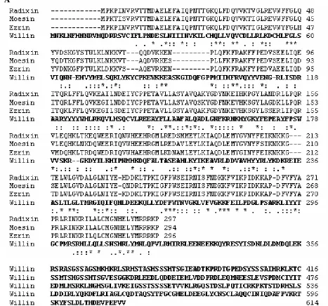
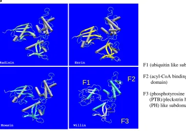
1.15 Sequence alignments of Willin, Ezrin Radixin and Moesin and predicted crystal structure of Willin FERM domain compared to
crystal structures of Ezrin, Radixin and Moesin FERM domains 33-34 1.16 Structural domains present in the Band 4.1 superfamily 34 1.17 Phospholipid blot overlay experiment comparing GST-Willin
and GST-Moesin phospholipid binding profiles and HEK-293
Figure
Page
1.18 Translocation of Willin-GFP in a PC12 cell stimulated with EGF 36 1.19 PC12 cells co-expressing Willin-GFP and RFP-ARNO treated
with wortmannin and EGF 37
Chapter 3
3.1 A Western blot of GST-Willin with Hen1 and APHen1 antibodies and pAP9143antibody control, gelatine block 65 3.2 A Western blot of GST-Willin with Hen2 and APHen2 antibodies
and pAP9143antibody control, gelatine block 65 3.3 A Western blot of GST-Willin with Hen2 and APHen2 antibodies
and pAP9143antibody control, BSA overnight block 66 3.4 A Western blot of GST-Willin with Hen1 and Hen2 antibodies
and pAP9143antibody control, 0.2% FSG block 67 3.5 A Western blot of GST-Willin with Hen1 antibody, 1% and 2%
FSG block 67
3.6 A Western blot of GST-Willin with Hen1 and Hen2 antibodies
and pAP9143antibody control, milk overnight block 68 3.7 A Western blot of GST-Willin with APHen1 and APHen2
antibodies and pAP9143antibody control, milk overnight block 69 3.8 A Western blot of COS-7 whole cell extract and BSA with
APHen2 antibody 70
3.9 A Western blot of COS-7 whole cell extract and BSA with
APHen2 antibody and two different secondary antibodies 70 3.10 A Western blot of GST-Willin with APHen2 antibody and
two different secondary antibodies 71 3.11 A Western blot of GST-Willin and rat sciatic nerve
with APHen2 antibody 72
3.12 A Western blot of HEK-293 whole cell extract and GST-Willin
Figure
Page
3.13 Peptide blocking experiment with APHen2 antibody 74 3.14 A Western blot of Willin2-GFP with APHen2 antibody 75 3.15 A Western blot of GST-Willin and BSA withWR1 and 2
antibodies 76
3.16 A Western blot of RIPA-extracted untransfected and Willin-GFP transfected COS-7 cells withWR1 and 2 and anti-GFP antibodies 77 3.17 A Western blot of RIPA-extracted untransfected and Willin-GFP
transfected HEK-293 cells withWR1 and anti-GFP antibodies 78 3.18 Peptide blocking experiment with GST-Willin andWR1 antibody 79 3.19 Peptide blocking experiment with Willin-GFP andWR1 antibody 79 3.20 A Western blot ofFRMD6 by Atlas Antibodies 80 3.21 A Western blot of GST-Willin withFRMD6 and pAP9143
antibodies 81
3.22 A Western blot of HEK-293 cells either untransfected or transfected with either Willin-GFP, M1GFP, GFP-Moesin or
GFP-Ezrin, withFRMD6 antibody 82
3.23 A Western blot of untransfected and Willin-GFP transfected
COS-7 cells withFRMD6 and anti-GFP antibodies 83 3.24 Immunocytochemistry ofFRMD6 on cells expressing
mito-ABAD-GFP 84
Chapter 4
4.1 Two-step cloning strategy for pWillin-FLAG 93 4.2 A Western blot of HEK-293 cell expression Willin-FLAG 94 4.3 Immunocytochemistry of a HEK-293 cell expressing
Willin-FLAG 95
Figure
Page
4.5 A COS-7 cell expressing pWillin-DsRed2 97 4.6 Bar graph showing percentage of apoptotic cells at two time
points post-transfection for Willin-GFP, Merlin1-GFP and
GFP-Moesin expressing cells 100
4.7 A Western blot of RIPA-extracted Schwann cells 101 4.8 A Western blot of a DRM subfractionation of COS-7 cells
expressing Willin-GFP 103
4.9 Western blots of DRM subfractionations of HEK-293 cells
expressing Willin-GFP or GFP and treated with cytochalasin D 104 4.10 A Western blot of a DRM subfractionation of PC12 cells
expressing Willin-GFP and treated with EGF 105 4.11 Western blots of Optiprep gradient subfractionations of HEK-293
cells expressing Willin-GFP or Merlin1-GFP 106 4.12 Cloning strategy for pMouse Willin2-GFP 107 4.13 A Western blot of COS-7 cells expressing Mouse Willin2-GFP 107 4.14 COS-7 cells expressing Mouse Willin2-GFP and Willin-GFP 108 4.15 Cloning strategy for truncated pGST-Willin constructs 112 4.16 A Western blot of BL21/DE3 E. coli expressing truncated
GST-Willin proteins 113
Chapter 5
5.1 The yeast two-hybrid system 118
5.2 Summary of yeast two-hybrid constructs used 121 5.3 The tandem affinity purification method 124 5.4 The initial cloning strategy for Willin-pIRESpuro2
CBP/TEV protein A 125-126
5.5 The second cloning strategy for Willin-pIRESpuro2
Figure
Page
5.6 The Stratagene tandem affinity purification method 128 5.7 The cloning strategy for pWillin-CTAP A 129-130 5.8 A Western blot of a COS-7 cell expressing Willin-CTAP A 130 5.9 A Western blot of the fractions of a Stratagene TAP experiment
with Willin-CTAP A 131
5.10 The cloning strategy for pWillin239-NTAP A 132 5.11 A Western blot of the fractions of a Stratagene TAP experiment
with Willin239-NTAP A 133
5.12 Western blots of the fractions of a pCMV/NTAP experiment with
CMV/NTAP neurofascinCT 134
5.13 Western blots of Willin-FLAG immunoprecipitation experiments
with neurofascin155 135
5.14 A Western blot of GST-Willin pulldown experiment with
neurofascin155 136
5.15 Western blots of Willin-FLAG immunoprecipitation experiments
with Merlin1 137
5.16 A Coomassie-stained gel and Western blot of the actin binding
biochem kit experiment 139
5.17 HEK-293 cells co-expressing Willin-GFP and neurofascin155 141 5.18 HEK-293 cells expressing GFP-Ezrin, neurofascin155 or both 142 5.19 HEK-293 cells expressing Merlin1-GFP and neurofascin155 144 5.20 A summary of results for putative Willin binding partners 150
Chapter 6
6.1 A proposed mechanism for FERM protein action in paranode
List of Tables
Table
Page
2.1 Reagent volumes for Lipofectamine transfection of
different sized dishes 46
2.2 Reagent volumes for GeneJammer transfection of
different sized dishes 47
2.3 Updated reagent volumes for GeneJammer transfection
of different sized dishes 47
2.4 Experimental setup for actin binding kit protocol 55 5.1 A summary of yeast two-hybrid results for rat N-terminal
Chapter 1: Introduction
1.1 The Band 4.1 Protein Superfamily
The Band 4.1 superfamily is a group of proteins characterised by a conserved domain known as the Four point one Ezrin Radixin Moesin (FERM) domain. Band 4.1, the prototype of the superfamily, is an erythrocyte membrane protein and a major component of the cortical cytoskeleton. Its N-terminal half was found to be well conserved throughout a variety of proteins, most of which have interactions with both the membrane and the cytoskeleton. This interesting ability has led to a high level of interest in these proteins, and a classification system for the band 4.1 proteins was proposed by Takeuchi (Takeuchi et al., 1994a) that divides them into five gene
families based on sequence analysis (though more may exist): the band 4.1 family; the ERM family, into which fall ezrin, radixin, moesin and merlin, along with novel band 4.1-like proteins 6 and 7 (NBL6 and NBL7); the talin family; the PTPH1 family, which includes PTPH1, PTPMEG, NBL1, NBL2 and NBL3; and the NBL4 family, which also includes NBL5. This section will focus on Band 4.1, the ERM family, Merlin and Willin.
1.1.1. Band 4.1
(Figure 1.1). Molecularly, the skeletal network and membrane structures become abnormally distributed (Yawata et al., 1997).
Figure 1.1. A) Scanning electron microscopy (EM) of normal red blood cells, with characteristic disc shape. B) Scanning EM of red blood cells from patient with
homozygous hereditary elliptocytosis. The cytoskeleton is improperly formed. Taken from Yawata et al., 1997.
Interest in 4.1 increased when it was discovered that it was able to bind membrane, cytoskeletal and membrane proteins, which, combined with the clincial evidence, suggested an important linking function (Hemming et al., 1994; Pasternack et al., 1985; Walensky et al., 1998). Three distinct binding domains have been observed in protein 4.1: a C-terminal domain, a spectrin-actin binding domain, and a ~30kDa N-terminal domain that mediates binding with the membrane and membrane proteins (Sun et al., 2002; see Figure 1.2.)
It was recognised that the N-terminal domain of 4.1 was conserved in a rapidly growing list of other proteins, and was termed the Four point one Ezrin Radixin Moesin (FERM) domain after the proteins initially discovered to have this domain (Chishti et al., 1998).
1.1.2. The FERM domain
The FERM domain is usually found in the N-terminus of proteins, though in myosinVIIA it is in the C-terminus (Chishti et al., 1998), and in at least one novel FERM-containing family it is centrally located (Ussar et al., 2006). Around 300 amino acids in length, it is hydrophobic, cysteine-rich (Conboy, 1986) and globular (Chishti et al., 1998). Studies of crystal structures have shown that there are three subdomains, F1/A, F2/B and F3/C, forming a ‘cloverleaf’ structure (Hamada et al., 2000; Pearson et al., 2000). F1/A has a ubiquitin-like structure, F2/B an acyl-CoA binding protein-like structure, and F3/C resembles a fold found in phosphotyrosine binding (PTB), pleckstrin homology (PH), and Enabled/VASP Homology 1 (EVH1) domains (Hamada et al., 2000; Pearson et al., 2000). The FERM domain of various proteins has been shown to bind such a diverse range of molecules as
Figure 1.3. Overlaid FERM domains of Ezrin (red), Radixin (cyan), and Moesin (active: green; dormant: violet). The dormant Moesin FERM domain is shown bound to the C-terminal domain (white). Ribbon figures prepared with Bobscript and
Raster3D structure programs. Image taken from Smith et al., 2003.
1.1.3. Ezrin, radixin and moesin
The ezrin/radixin/moesin (ERM) family consists of proteins that link the cell membrane with the actin cytoskeleton at the cell cortex (Bretscher et al., 2002). Regardless of the cell type in which they are found, the ERMs are generally localised at areas of rich actin activity, such as microvilli, filopodia, membrane ruffles and cell-cell contact sites (Turunen et al., 1998), where they are involved in the formation of those structures (Bonilha et al., 1999; Crepaldi et al., 1997; Martin et al., 1997; Takeuchi et al., 1994a; Yonemura and Tsukita, 1999); cell shape and motility (Lamb et al., 1997); cell-substrate and cell-cell adhesion (Kaul et al., 1996; Martin et al., 1995; Takeuchi et al., 1994a); and membrane trafficking (Cao et al., 1999; Defacque et al., 2000).
This structure is highly conserved: ezrin shows about 97% identity amongst the mammalian forms, and 99% identity in the FERM domain between mouse and human forms; ezrin, radixin and moesin have 73-81% sequence identity (Turunen et al., 1998). The FERM domain connects to the cell membrane either directly through phosphoinositols, especially phosphatidylinositol-4,5-bisphosphate (Niggli et al., 1995), or by binding to membrane proteins such as CD44 (Tsukita et al., 1994) CD43 and ICAM-2 (Yonemura et al., 1998). The C-terminal domain has a highly-conserved actin binding site in the last 34 amino-acids (Turunen et al., 1994). Recently, the central coiled-coil domain has been shown to participate in masking of the FERM domain region and regulation of FERM binding (Li et al., 2007).
Figure 1.4. The ERM proteins share an overall structure consisting of an N-terminal FERM domain, a central coiled-coil domain and a C-terminal domain that includes a highly-conserved actin-binding motif in the final 34 amino acids. Image taken from Sun et al., 2002.
1.1.3.1. The ERM Association Domains regulate ERM protein activation
The ERM family members form intra- and intermolecular associations via N and C-terminal areas known as ERM Association Domains (ERMAD). The
active form (Krieg and Hunter, 1992). It has also been observed that EGF stimulation of A431 cells not only activates ERMs to break the intramolecular association, but also stimulates ERM oligomer formation, suggesting the oligomeric form could be a transition form in the activation pathway (Bretscher et al., 2000; Gautreau et al., 2002). Deactivation of the ERMs by dephosphorylation is also important for cell dynamics, and has been observedin vivoto correlate with breakdown of microvilli in such situations as anoxia and apoptosis (Chen and Mandel, 1997).
1.1.3.2. Expression of ERM proteins
In vivo, ERM proteins are found in most tissues, with each family member showing unique expression in different tissues. Ezrin was initially purified from intestinal microvilli, but is also present in the placenta, stomach, lung and kidney at high levels, and in lower levels in the spleen. Subcellularly, its distribution is largely in actin-rich surface projections, and in the tissues in which it can be found, ezrin is mainly associated with the apical surface of epithelial cells (Berryman et al., 1993). In Schwann cells, Ezrin has been found to localise at the paranodal microvilli that project into the Node of Ranvier (Gatto et al., 2003).
Moesin was first isolated from bovine uterine cells and originally thought to be an extracellular heparin-binding protein (Lankes et al., 1988), but was soon shown to be intracellular and very similar to ezrin (Lankes and Furthmayr, 1991; Sato et al., 1992). Berryman et al. (1993) found it to be most abundant in lung and spleen, and to a lesser extent in kidney, while others have observed it in macrophages, lymphocytes, fibroblastic, endothelial, epithelial and neuronal cell lines as well (Amieva and Furthmayr, 1995). Like ezrin, its subcellular localisation is specific to certain areas, chiefly filopodia, microvilli, microspikes and retraction fibres (Amieva and
Radixin was initially purified from hepatic adherens junctions (Tsukita et al., 1989), but its subcellular localisation has been somewhat uncertain due to conflicting results with radixin antibody studies (Bretscher et al., 1997); different groups have shown it to localise to adherens junctions (Tsukita et al., 1989), microvilli (Amieva et al., 1994) contractile rings (Henry et al., 1995), focal contacts and cleavage furrows (Sato et al., 1991). Li and Crouch (2000) carried out experiments in chicken tissue and found high levels of radixin in kidney, liver, ovary and bone marrow; lower levels were detected in lung, thymus, colon and skin.
In contrast to the tissue-specific distributionin vivo, all three ERMs are usually co-expressed in cultured cells, perhaps due to the unique conditions of thein vitroenvironment (Franck et al., 1993; Sato et al., 1992). The tissue-specific
distribution pattern of the ERMs implies functional differences amongst the family members, but there is redundancy to the extent that deleting one or even two of the proteins via antisense oligonucleotide inhibition (Takeuchi et al., 1994b) or in transgenic knockout mice (Doi et al., 1999) produces no observable changes in phenotype.
1.1.3.3. ERM proteins and disease
interactions in which healthy cells were captured by the tumour. This seems to imply that ezrin plays a key role in the cell motility and adhesion involved in metastasis. Further evidence of this is seen in glial cell tumours, known as gliomas, in the brain, where ezrin appears to be involved in a hepatocyte growth factor signalling cascade that promotes tumour migration (Wick et al., 2001). It is also likely that Ezrin is able to activate signalling pathways for cell survival, such as MAPK and Akt (see section 1.1.3.4), allowing metastatic cells to survive in what might have otherwise been a hostile environment (Curto and McClatchey, 2004). In addition, increased expression of Ezrin in some cancer types correlated with both metastatic potential and poor prognosis (Curto and McClatchey, 2004), while cytoplasmic ezrin in head and neck cancers correlates with poor outcome (Madan et al., 2006).
1.1.3.4. ERM proteins are involved in cell signalling
(Morrison et al., 2007). Further regulation and activation of the Rho family by ERMs can also be accomplished through ERM binding of RhoGDI (Takahashi et al., 1997), which inhibits all Rho members, or Dbl, which stimulates all Rho members, and it appears that the interaction with these two regulators is mutually exclusive (Takahashi et al., 1998).
However, Rho-independent activation mechanisms also exist; it was observed that in the kidney-derived cell line MDCK ERM proteins appeared to be active in the absence of Rho, and that phosphatidylinositol-4,5-bisphosphate (PI(4,5)P2), a
membrane lipid, could regulate activation (Yonemura et al., 2002). The FERM domain of Ezrin is able to bind PI(4,5)P2(Niggli et al., 1995), which in turn has a
great deal of influence on the intracellular localisation of Ezrin (Barret et al., 2000). PI(4,5)P2may in fact mediate the interaction of ERMs with adhesion molecules such
as CD44 (Niggli et al., 1995).
ERM proteins have been implicated in additional pathways that may explain their potential role in cancer and tumour metastasis. They can activate cell survival signalling pathways through phosphatidylinositol 3-kinase (PI3K)/Akt signalling (Gautreau et al., 1999), such as in cases of apoptotic stress, where ERMs interact with NHE1 to mediate PI3K and Akt cell survival messages (Wu et al., 2004). Protein kinase C theta (PKC) and alpha (PCK) forms are able to phosphorylate moesin (Pietromonaco et al., 1998) and ezrin (Ng et al., 2001) respectively, leading to formation of membrane protrusions and migratory cell behaviour.
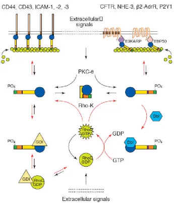
Figure 1.5 shows a model for ERM activation in the context of the above signalling pathways (Mangeat et al., 1999). Active, phosphorylated ERM proteins are shown to interact with the membrane either through PI(4,5)P2-mediated interaction
proteins indirectly via PDZ domain-containing binding partners such as Ezrin binding protein 50 (EBP50) and E3KARP. ERMs can be phosphorylated by Rho, and when active ERMs are then able to recruit positive and negative regulators of Rho, Dbl and RhoGDI respectively, to maintain activation of the pathway. Also, PKC is another potential activator of the ERM proteins through ERMAD phosphorylation, perhaps under the influence of different extracellular signals than those that affect Rho.
1.1.4. Merlin
It has long been known that mutations of merlin cause tumours in the nervous system. Merlin (moesin/ezrin/radixin-like protein), or schwannomin as it is also called, is the product of the brain tumour suppressor neurofibromatosis 2 (NF2) gene and shares 45% identity with the ERMs, including a FERM domain with over 60% idenitity between the human forms of merlin and ezrin (Turunen et al., 1998). Lallemand et al. (2003) observed that merlin appears to be required in the formation of adherens junctions and contact-dependent growth arrest.
1.1.4.1. Neurofibromatosis type 2 and the NF2 tumour suppressor gene
Neurofibromatosis type 2 (NF2) is an autosomal dominant syndrome characterised by first of all bilateral vestibular schwannomas, and usually also schwannomas of the cranial, spinal and cutaneous nerves; meningiomas and
ependymomas are also commonly reported in NF2 patients (Ahronowitz et al., 2007; Evans et al., 1992). These tumours are slow-growing, making them resistant to chemotherapy and treatable only through repeated, often deforming and debilitating, surgical resections (McClatchey and Giovannini, 2005). TheNF2gene is found on chromosome 22q12 (Trofatter et al., 1993), with 17 exons that can be alternatively spliced into isoform 1 (exons 1-17) or isoform 2 (exons 1-15 and 17), but only isoform 1 appears to have tumor suppressor function (Sherman et al., 1997).
which cells will survive (McLaughlin et al., 2007). It also inhibits Rac (Okada et al., 2005) and Pak1 (Kissil et al., 2003), which allows for contact inhibition to occur. Lallemand et al. (2003) found that loss of NF2 destabilised adherens junctions, thus allowing tumourigenesis. Cell cycle control is also a potential function for Merlin, as it has been found to interact with the cell cycle regulator HEI10 and affect its
targeting (Gronholm et al., 2006). Additionally, Merlin is shuttled to the nucleus in a cell cycle and density-dependent manner; once there it can inhibit activation of cell cycle promoter ERK2 (Muranen et al., 2005).
1.1.4.2. Merlin expression
Understandably, Merlin has mostly been studied within the context of its expression in Schwann cells of the peripheral nervous system, where it is
developmentally regulated and a component of the paranode and Schmidt-Lanterman incisures (Scherer and Gutmann, 1996). It may play a role in Schwann cell
differentiation from the pro-myelinating to the myelinating stage (Hung et al., 2002), and NF2 null mutants show abnormal myelination (Giovannini et al., 2000).
However, Merlin has been found in other cell types, such as fibroblasts (Shaw et al., 2001); and in neurons of the central nervous system it is also a component of
paranodes, though this time interacting with membrane glycoprotein Caspr on the axonal side (Denisenko-Nehrbass et al., 2003).
Subcellularly, Merlin shows strong cortical membrane localisation,
buoyant fraction (Figure 1.6), potentially containing different signalling molecules (Stickney et al., 2004); this is typical of raft-associated proteins, which are thought to have a dynamic relationship with rafts to allow for rapid adaptability to signals (Simons and Ehehalt, 2002). It is believed that this raft interaction is important in the function of Merlin, as many NF2 mutations make Merlin more soluble in Triton X-100 (Deguen et al., 1998; Stokowski and Cox, 2000), and as Schwann cells
differentiate, Merlin goes from being a soluble cytoplasmic component to an insoluble interactant of1 integrin (Obremski et al., 1998).
Figure 1.6. A model of how Merlin buoyancy within lipid rafts is affected by cell density-dependent activation. High cell density is associated with active Merlin and light raft localisation. Image taken from Stickney et al., 2004.
Like the ERMs, Merlin also shows a close association with the cytoskeleton, butin vivothis interaction may occur indirectly through the association of Merlin and II-spectrin (Gutmann, 2001). Certainly any interaction with actin is different in
1.1.4.3. ERM Association Domains and activation of Merlin
Merlin has ERMADs as well, but their interactions are weaker and more dynamic than those in ezrin, radixin and moesin (Nguyen et al., 2001).
Phosphorylation of a serine residue is required to break the association rather than the conserved threonine, but it has not yet been determined which form of merlin is active. It has been proposed that in fact the merlin oligomeric or ‘closed’ conformation is active, based on evidence that the ERMAD interaction and
dephosphorylation is a requisite step in merlin function (Shaw et al., 2001). Hetero-oligomers of ezrin and merlin have been detected using affinity binding assays of their domains (Nguyen et al., 2001), coimmunoprecipitation, yeast two-hybrid, blot overlay and affinity precipitation (Gronholm et al., 1999; Meng et al., 2000), and it has been observed that the merlin C-ERMAD has a much stronger affinity for the ezrin N-ERMAD than its own (Meng et al., 2000; Nguyen et al., 2001). It is possible that this interaction exists in a regulatory capacity to control the activity of ezrin and/or merlin (Gautreau et al., 2002). This seems to coincide with their opposing functions in growth regulation.
1.1.5. FERM-containing proteins in Drosophila melanogaster
1.1.5.1 DMoesin
Drosophilahave only one ERM protein homologue, DMoesin, which has 58% sequence homology with human moesin, including 26% identity in the C-terminal divergent region (McCartney and Fehon, 1996). This unique expression makes it possible to side-step the problem of redundant function found in vertebrate mutants (see section 1.1.3.2). In a molecular context, loss of DMoesin show that this protein is essential for cytoskeletal distribution, maintenance of apical-basal polarity, and epithelial integrity; this study also suggests that DMoesin acts antagonistically to the Rho pathway, contradicting the results discussed in section 1.1.3.4 (Speck et al., 2003), but furtherin vivostudies are required in vertebrates. Its subcellular
localisation was found to be primarily in apical membrane regions (McCartney and Fehon, 1996).
Physiologically, loss of DMoesin causes severe developmental problems, with the posterior structures missing completely from the fly embryos; in addition,
imaginal discs, adherens junctions and photoreceptors are all dependent on DMoesin for correct organisation and assembly (Polesello and Payre, 2004).
1.1.5.2. DMerlin and expanded
As with the human protein, DMerlin exerts a growth-suppressive function at the plasma membrane (LaJeunesse et al., 1998). In the developing eye, DMerlin appears to regulate normal apoptosis, with mutants exhibiting overgrowth due to a higher number of cells than the normal fly (Pellock et al., 2007), and wings show broadening and cross-vein disruption (McCartney et al., 2000).
Recent work has shown that DMerlin acts closely with another FERM-containing protein, expanded, to regulate growth, proliferation and differentiation in Drosophila tissues (McCartney et al., 2000). Like DMerlin mutants, expanded
mutants show overgrowth phenotypes in various tissues (Boedigheimer and Laughon, 1993); (Blaumueller, and Mlodzik 2000) due to overproliferation (Boedigheimer et al., 1997).
DMerlin and expanded co-localise in Drosophila cells, both in tissues and in culture (McCartney et al., 2000). The two act through the Hippo tumour suppressor signalling pathway, causing downstream activation of the Hippo/Salvador complex and Warts/Mats complex (Hamaratoglu et al., 2006), inhibiting the transcriptional coactivator Yorkie, which induces growth (Huang et al., 2005); this leads to further activation of theDMerlinandexpandedgenes, as well as repression ofcyclin Eand
Drosophila inhibitor of apoptosis protein 1(diap1) genes (Hamaratoglu et al., 2006). Cyclin E induces entry into S-phase from G1 (Richardson et al., 1995), so DMerlin and expanded are in this case preventing re-entry into the cell cycle; DIAP1, meanwhile, plays an essential role in cell survival through its inhibition of caspase-induced apoptosis (Wang et al., 1999), so the role of DMerlin and expanded here is to allow this apoptotic pathway to operate during differentiation.
al., 2005), and the Mats homologues (MOB proteins) also associate with NDR type kinases (Tamaskovic et al., 2003).
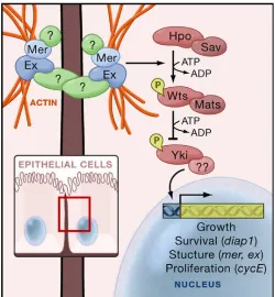
[image:39.595.173.423.304.574.2]Figure 1.7 shows a summary of this overall regulatory pathway as proposed by Edgar (2006); the receptor that initiates DMerlin and expanded signalling was not yet elucidated at the time, but has since been proposed to be Fat, a protocadherin already implicated in growth regulation in Drosophila development (Cho et al., 2006; Silva et al., 2006). This also has a vertebrate homologue, FAT4 (Silva et al., 2006), but the function of any pathway for this protein is not clear (Cho et al., 2006).
Figure 1.7. The Hippo pathway inDrosophilaepithelial cells. A membrane receptor, possibly Fat, activates DMerlin and expanded, which in turn regulate the Hippo signalling pathway. Hippo is modulated by Salvador and phosphorylates the Warts/Mats complex, leading to inhibition of Yorkie and continuing growth suppression and apoptosis in a feedback loop. Image taken from Edgar 2006.
1.1.5.3. Coracle
acids of the C-terminus, but lacks the spectrin-actin binding domain (Fehon et al., 1994). Coracle localises to the septate junctions of epithelial cells, where it is
required for correct septate junction structure, but unlike protein 4.1, not apical-basal polarity or structural integrity of epithelial cells (Lamb et al., 1998). This septate junction role is probably linked to correct proliferation in embryonic development, as coracle mutants show defects in proliferative aspects such as dorsal closure and cuticle thickness (Ward et al., 2001).
1.1.6. FERM-binding motifs
Several proteins have shown conserved motifs that recognise FERM domains for binding. One such site, MDWxxxxx(L/I)Fxx(L/F), is found in the C-terminus of Na+/H+exchanger regulatory factor (NHERF), an ERM binding partner that anchors ion channels and receptors, as a motif that binds to the F3/C lobe of the FERM domain (Terawaki et al., 2006). Another, and better-characterised, FERM-binding motif, (R/K/Q)xxT(Y/L)xx(A/G), is found in cell adhesion molecules at the
juxtamembrane region of their cytoplasmic tails (Dickson et al., 2002; Hamada et al., 2003; Terawaki et al., 2006) and been observed to bind ERMs. A similar motif is also found at the C-terminus of neurofascin (Gunn-Moore et al., 2006).
1.2 The L1 family of cell adhesion molecules
The L1 family of cell adhesion molecules share an overall structure of six IgG-like domains and three to five fibronectin III-IgG-like domains extracellularly, a single transmembrane domain, and a short (~110 amino acids) cytoplasmic C-terminus that is highly conserved (Hortsch, 2000). The high level of conservation of the
cytoplasmic C-terminus implies similar roles in intracellular signalling, and perhaps even shared binding partners. One known partner of L1 family members is the ankyrin family, a cytoskeletal molecule that binds to a highly conserved 36 amino acid domain found in all L1 proteins (Hortsch, 2000). This cytoskeletal link suggests a role in the regulation of cell morphology and structure for the L1 family.
Members of the family include the prototype, L1 (also known as Neuron-glia cell adhesion molecule or NgCAM), Close Homolog of L1 (CHL1), Neuron-glia-related cell adhesion molecule (NrCAM), and neurofascin in mammals, and neuroglian inDrosophila(Figure 1.8). The NrCAM and neurofascin genes are subject to extensive alternative splicing, a process which is tightly linked to
.
[image:42.595.153.445.65.349.2]L1/CHL1* neurofascin neurofascin NrCAM neuroglian 155 186
Figure 1.8. Overall structures of the L1 family of cell adhesion molecules. L1 and neuroglian each have 6 IgG-like and 5 fibronectin III-like repeats, while CHL1 has half of the last fibronectin repeat; neurofascin has either 4 fibronectin III domains (neurofascin155) or 3 fibronectin III domains and 1 mucin-like domain (neurofascin 186); NrCAM, like L1, has 6 IgG domains and 5 fibronectin domains.
1.2.1. L1
number of hippocampal neurons (Demyanenko et al., 1999). Schwann cell-axon interactions are also disrupted, leading to abnormal myelination (Itoh et al., 2005).
Mutations in L1, located on the X chromosome, cause a range of human syndromes known collectively as CRASH (corpus callosum hypoplasia, retardation, adducted thumbs, spastic paraplegia, hydrocephalus), and which show a great deal of diversity in both appearance and severity of symptoms depending on which mutation is present (Yamasaki et al., 1997).
1.2.2. CHL1
CHL1, like L1, promotes neuron survival and neurite outgrowth (Chen et al., 1999); it is believed to interact with integrins for regulation of cell migration, but with perhaps a differential preference for extracellular binding partners than L1 (Buhusi et al., 2003). Its expression during nervous system development is also distinct from L1, and in contrast to L1, the soluble form of CHL1 can promote neurite outgrowth (Hillenbrand et al., 1999).
1.2.3. NrCAM
In the nervous system, NrCAM is found in both glia and neurons, with most studies focusing on its neuronal functions. It is localised at the nodes of Ranvier and axon initial segment, where Na+channels cluster (Hillenbrand et al., 1999).
SAP102-NrCAM binding prevented neurite outgrowth (Davey et al., 2005), and it is required for axonal pathfinding in at least some neuronal systems, through mediation of growth cone-substrate interactions (Zelina et al., 2005).
NrCAM has been implicated in psychiatric disorders, particularly autism (Sakurai et al., 2006) and vulnerability to drug addiction (Ishiguro et al., 2006). Outside the nervous system, cancers such as melanomas (Reed et al., 2005), colon cancer (Conacci-Sorrell et al., 2002) and pancreatic cancer (Dhodapkar et al., 2001) are seen to upregulate NrCAM, and this may be a factor in tumour migratory behaviour (Conacci-Sorrell et al., 2002).
1.2.4. Neurofascin
Like NrCAM, neurofascin is extensively alternatively spliced (Volkmer et al., 1992); Hassel et al., 1997), with isoforms of 186kDa, 155kDa and 140kDa found in the brain (Davis et al., 1993). Alternative splicing strictly controls tissue localisation of neurofascin, with neurofascin186 localising to the Node of Ranvier in axons (Davis et al., 1996), while neurofascin155 localises to unmyelinated axons and the paranodal loops of glial cells in the CNS and PNS (Sherman et al., 2005; Tait et al., 2000). The 140kDa isoform is a minor component, and is present mainly in cerebellum (Davis et al., 1996).
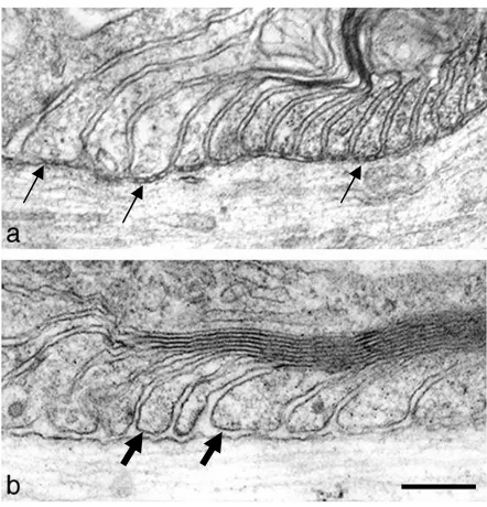
Figure 1.9. Electron micrographs of paranodes in wild type and neurofascin-null mice. In wild-type mice, paranodal loops connect to the axons with septate-like junctions as indicated by arrows (a), but these connections are disrupted and the gaps between loops and the axon (block arrows) are greater in neurofascin-null mice (b). Taken from Sherman et al., 2005.
In the PNS, neurofascin186 is anchored to the node by gliomedin, a component of Schwann cell microvilli, and can then act as a pioneer molecule for recruiting other binding partners, such as NrCAM, ankyrin and sodium channels (NaV). Neurofascin155, meanwhile, guides formation of the septate-like junctions of
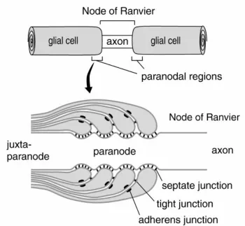
Figure 1.10. Construction of the node of Ranvier. Neurofascin186 and NrCAM in the axon and gliomedin in the Schwann cell microvilli establish the node, and neurofascin and NrCAM recruit additional binding partners to the node. In the paranodal loops, neurofascin 155 arrives independently of Caspr and contactin, and recruits them from the axonal side to form the paranodal complex. Taken form Sherman et al., 2005.
1.2.4.1. Neurofascin and multiple sclerosis
In multiple sclerosis (MS) demyelinating lesions, neurofascin155 expression is spread from its discreet paranodal localisation towards the juxtaparanode, and this leads to movement of juxtaparanode components, such as potassium channels (KV)
towards the node. After demylenation, neurofascin186 and NaVexpression is also
Figure 1.11. i) Neurofascin155 joins the paranodal loops to the axon, with
neurofascin186 and NaVchannels in the node and KVchannels in the juxtaparanode.
ii) Demyelination leads to neurofascin155 disruption and movement of KVchannels
towards the node. iii) Neurofascin155 is lost and the axon is demyelinated. iv) Nodes are disrupted, with neurofascin186 and NaVchannels distributed throughout the axon.
v) Abnormal triplicate neurofascin155 paranodal contacts occur during remyelination. vi) Binary nodes fuse, displacing the abnormal triplicate from the remyelination process. Taken from Howell et al., 2006.
1.2.4.2. Neurofascin localisation to lipid rafts is important in paranode formation
gene, disrupting production of two major glycolipids, show improper ion channel clustering, altered nodal length and diffuse distribution of Caspr along the internode (Ishibashi et al., 2002). Active MS lesions that show disrupted paranodes also have reduced neurofascin155 localisation to lipid rafts (Maier et al., 2007).
1.2.5. Neuroglian
Drosophilahave only one L1 family member, neuroglian. Like the other family members, neuroglian interacts with and may be necessary for membrane localisation of ankyrin (Dubreuil et al., 1996). In theDrosophilanervous system, neuroglian is important in neuronal pathfinding (Hall and Bieber, 1997) and axonal substrate choice (Garcia-Alonso et al., 2000); in other tissues, it is important for stabilisation of epithelial tissue at points of cell-to-cell contact (Wei et al., 2004), and indeed it has been found to localise to the ladder-like pleated septate junctions of the epithelium (Genova and Fehon, 2003) along with neurexin IV (Baumgartner et al., 1996) and contactin (Faivre-Sarrailh et al., 2004). These three adhesion molecules allow the formation of the Drosophila blood-brain barrier, which keeps the potassium-rich hemolymph separate from neurons. Septate junctions also form between
insulating glia around axon in a homologous structure to the vertebrate septate-like junctions of the paranodes (Banerjee et al., 2006).
1.2.6. The interaction of L1 family members and FERM-containing proteins
2005; Davis et al., 1993). Yeast two-hybrid screens for binding partners of the C-terminus of L1 family members have revealed multiple sites capable of bind ERM proteins in both L1 and neurofascin, but not NrCAM (Cheng et al., 2005; Dickson et al., 2002; Gunn-Moore et al., 2006).
1.2.6.1. L1 interaction with Ezrin is important for normal axonal morphogenesis
Dickson et al. (2002) first screened for cytoplasmic binding partners and found that the FERM domain of Ezrin bound to the neuronal isoform of L1 at a region encompassing the RSLE motif, a region previously shown to regulate sorting of L1 to the axonal growth cone (Kamiguchi and Lemmon, 1998). A second, juxtamembrane region also mediates ERM-binding, and both regions are involved in regulation of neurite outgrowth and branching (Dickson et al. 2002; Cheng et al. 2005). The L1-ERM interaction occurs in earlyin vitrodevelopment, with active, phosphorylated ERM expression peaking at 3 daysin vitro(DIV) and diminished by 21 DIV; between 21 and 28 DIV, mainly inactive ERMs are expressed, but after injury active ERMs are re-expressed for regeneration of neurites (Haas et al., 2004).
1.2.6.2. Neurofascin interacts with Ezrin in the microvilli of Schwann cells
Immunohistochemical staining of neurofascin155 and Ezrin in teased sciatic nerve fibre shows that the two proteins co-localise in the microvilli of Schwann cells that project from the paranode into the node (Figure 1.12); however, neurofascin155 expression is not required for localisation of Ezrin to these microvilli (Gunn-Moore et al., 2006).
Figure 1.12. Immunofluorescence localization of both neurofascin155 and Ezrin to the microvilli of Schwann cells. The axon is visualized using an antibody against the neurofilament M subunit (NFM). The location of the microvilli in this teased sciatic nerve fiber is shown by arrowheads and the paranodal axoglial junctions are identified by arrows. Scale bar 10μm. Images taken from Gunn-Moore et al. 2006.
1.2.6.3. Drosophila septate junctions are analogous to vertebrate paranode septate-like junctions
function (see section 1.2.5) in the insect blood-brain barrier (Tepass and Tanentzapf, 2001).
Figure 1.13. At the paranode, glia form septate-like junctions with the axon. Between paranodal loops are tight junctions and adherens junctions. Image from Tepass et al., 2001.
Several molecules have been identified in both junction types: Caspr is a vertebrate homologue of neurexin IV and localises to the paranodal septate-like junctions of myelinated axons (Einheber et al., 1997). It can also bind the neuronal isoform of protein 4.1, the homologue of coracle (Menegoz et al., 1997).
(Charles et al., 2002), an analogous set of structures becomes clear and is summarised in Figure 1.14.
Figure 1.14. The vertebrate paranodal septate-like junction (A) consists of
neurofascin155 on the glial side with its extracellular domains interacting with Caspr and contactin on the axonal side. The intracellular domain of Caspr interacts with the neuronal isoform of protein 4.1. The invertebrate septate junction (B) consist of neuroglian, the neurofascin homologue, neurexin IV, the Caspr homologue, and coracle, the 4.1 homologue. It is not known if theDrosophilacontactin homologue localises to septate junctions.
protein that interacts with neuroglian as Ezrin does with neurofascin155? As yet, all other FERM-containing proteins in Drosophila have been observed to localise to adherens, not septate, junctions (Boedigheimer et al., 1997; McCartney and Fehon, 1996), but as will be discussed in Chapter 6, this picture is not as clear cut as it may seem, and leaves some scope for further speculation.
1.2.6.4. A novel protein discovered from a yeast two-hybrid screen of neurofascin
During a yeast two-hybrid screen of the neurofascin C-terminus against a rat sciatic nerve library, a novel FERM-containing cDNA was identified and called 163ScII (accession number AF441249). This sequence was found to have 86% identity at the DNA level and 91% identity at the protein level to a full-length cDNA human clone (MGC:17921 image: 3941276) which has also been identified as Open Reading Frame 31 Chromosome 14 (accession number BC020521). The cDNA image clone, obtained from a human uterine leiomyosarcoma, was acquired from the MRC IMAGE consortium and termed Willin, after the founder of the Royal (Dick) School Veterinary College of the University of Edinburgh, William Dick, and is also known as FRMD6.
1.3. Willin
actin-binding motif nor the putative N-terminal actin-actin-binding regions of the ERMs and Merlin are conserved; to date, only the FERM domain is confirmed to be present (see Figure 1.16).
[image:54.595.84.551.204.640.2]B Radixin Ezrin Willin FERM DOMAINS F1 F2 F3
F1 (ubiquitin like subdomain)
F2 (acyl-CoA binding like sub-domain)
F3 (phosphotyrosine binding (PTB)/pleckstrin homology (PH) like subdomain)
[image:55.595.94.457.79.334.2]Moesin
Figure 1.15. A) Sequence alignments of Willin and the FERM domains of Ezrin, Radixin and Moesin. * = identical residue,:= conserved substitution,.= semi-conserved substitution. B) Predicted 3-dimensional structure of Willin FERM domain compared with Ezrin, Radixin and Moesin FERM domains from crystal structures. Structural prediction performed by V. Zaitsev.
Figure 1.16. Structural domains present in the Band 4.1 superfamily. The FERM domain is the only confirmed domain in Willin. Image taken from Diakowski et al., 2006.
ABD: Actin binding domain BB: blue box
CTD: carboxy terminal domain DHR:Drosophilahormone receptor PDZ: postsynaptic density-95 /discs large/zonula occludens-1 domain PH: pleckstrin homology domain PTP: protein tyrosine phosphatase domain
SABD: spectrin-actin-binding domain TKPD: tyrosine kinase phosphorylation domain
[image:55.595.92.375.438.684.2]Like the ERMs and Merlin, Willin has been observed in the cytoplasm and nucleus (Madan et al., 2006). We have also shown that Willin has a phosphoinositol binding profile comparable to that of Moesin (Figure 1.17), and Willin-GFP is often localised to the membrane; this is particularly true in adjoining cells.
A
B
Figure 1.17. A) A phospholipid blot overlay experiment using purified GST, GST-Moesin and GST-Willin. Image provided by Dr. Kanamarlapudi Venkateswarlu. B) A HEK-293 cell expressing Willin-GFP. Membrane localisation indicated by arrows. Image obtained by Dr. Frances Brannigan.
Figure 1.18) and nerve growth factor (NGF; data not shown), but the translocation is not blocked when the cells are treated with wortmannin (Figure 1.19). This indicates that phosphatidylinositol 3-kinase (PI3K) activity is not required to translocate Willin (Venkateswarlu et al., 1999). It is probable that, like Ezrin, EGF is causing tyrosine phosphorylation of Willin; EGF is a known activator of Ras (Marshall, 1995), Rho and Rac (Maddala et al., 2003), which in turn interact with ERM proteins (see section 1.1.3.4), so Rho family activation is a possible pathway for Willin translocation. These and other possibilities have yet to be studied.
Figure 1.19. PC12 cells co-expressing RFP-ARNO and Willin-GFP were treated with 100 ng/mL EGF alone (left) or pretreated with 100nM wortmannin 30 minutes before addition of 100 ng/mL EGF (right). ARNO, a PH-domain-containing protein, is dependent on PI3K activation for its transolaction, which is thus blocked by wortmannin, a PI3K inhibitor. Willin-GFP translocation is not blocked by wortmannin.
could be the mammalian homologue of theDrosophilatumour suppressor Expanded (Hamaratoglu et al., 2006).
Chapter 2: Materials and Methods
Unless otherwise stated, chemical reagents were obtained from Sigma. See appendix 4 for a list of suppliers.
2.1 Molecular biology and cloning
2.1.1 Polymerase Chain Reaction (PCR)
PCR was performed using PfuTURBO DNA Polymerase (Stratagene or Rovalab,Teltow, Germany) or Expand High Fidelity Taq Polymerase (Roche) according to manufacturer’s instructions in the buffer supplied and adding 200M dNTPs (Promega), 0.5M forward and reverse primers (Invitrogen) and 200-300ng
template. Thermal cycling was then done in a Biometra Tpersonal Combi thermal cycler (Biometra, Goettingen, Germany) as follows:
1. 94ºC for 5 minutes 2. 94ºC for 1 minute. 3. 54ºC for 1 minute.
4. 72ºC for 2minutes 30 seconds. (steps 2-4 repeated 34 times for a total of 35 cycles) 5. 72ºC for 10 minutes.
PCR products were run on a 1% or 2% agarose gel alongside a 1kb DNA ladder for size analysis.
2.1.2 Restriction enzyme digest
incubated at 37oC for 1-5 hours. They were then either heat inactivated at 65oC for 15 minutes for further treatment with alkaline phosphatase (see section 2.1.3) or run directly on an agarose gel for analysis or purification.
2.1.3 Alkaline phosphatase treatment of digested plasmids
To prevent religation of sticky ends in digested plasmids, phosphate groups were removed from their 5’ overhangs by the addition of 1 unit of calf intestinal alkaline phosphatase (CIAP) (Promega) incubated for 30 minutes at 37oC, followed by addition of another unit of CIAP and a further 30 minute incubation at 37oC. Blunt ends and 3’ overhangs were treated by addition of 1 unit CIAP for 15 minutes at 37oC and then 15 minutes at 56oC, repeated with a second unit of CIAP.
2.1.4 Klenow reaction
For cloning strategies where a blunt end was required for ligation, sticky ends were filled in using Klenow fragment and supplied buffer (Promega) according to manufacturer’s instructions.
2.1.5 Ligation reaction
Ligations were performed using T4 DNA ligase (Promega) according to manufacturer’s instructions.
2.1.6 Agarose gel electrophoresis
fragments > 1kb, and 2% (w/v) for fragments <1kb. The samples had 20% 6x agarose gel loading buffer (50% glycerol, 49.75% TBE, 0.25% bromophenol blue) added. For analytical gels, 1kb or 100bp DNA ladders were loaded into a lane in 1% and 2% gels respectively for sizing and quantification of DNA bands. Gels were run at 60-80V for 30-60 minutes. Analytical gels were visualised with a UV lamp and photographed by a digital camera (Mitsubishi 85mm lens, Thistle Scientific), while preparation gels were visualised with a low intensity UV lamp (230V-50Hz, Ultratec, Ltd.) and desired bands excised from the gel with a sterile scalpel blade.
2.1.7 Gel purification of digested DNA
Digested DNA was separated on a 1% agarose purification gel and purified with the Wizard SV Gel and PCR Cleanup Kit (Promega) according to manufacturer’s instructions.
2.1.8 Preparation of plasmid DNA
Small scale plasmid purification was done with the Qiagen Spin Miniprep Kit according to manufacturer’s instructions. Large scale purifications were done with the Qiagen Endofree Maxi Kit or the Promega PureYield Midi Prep kit according to manufacturer’s instructions.
2.1.9 Preparation of CaCl2-competent E. coli
A 5mL overnight culture of DH5or BL21/DE3E. coliwas grown shaking overnight at 37oC in LB without antibiotics. The next day, 50mL of LB without antibiotics were inoculated with 500L of preculture and grown shaking at 37o
C until A600was 0.3-0.4. Cells were then harvested by centrifugation at 3500gfor 10 minutes
at 4oC. The cell pellet was resuspended in 20mL of chilled sterile 100mM CaCl2and
3500gfor 5 minutes at 4oC and the pellet resuspended in 1mL 100mM CaCl2for an
additional 30 minute incubation on ice. Competent cells were then used for transformation or frozen at -80oC for later use.
2.1.10 Preparation of ‘super’-competent E. coli
DH5E. coliwere streaked from a glycerol stock onto an LB-Agar plate and grown overnight at 37oC. The following day, 10-12 colonies were picked to inoculate 250mL of SOB medium (2% (w/v) Bacto-tryptone, 0.5% (w/v) Bacto yeast extract, 10mM NaCl, 2.5mM KCl, 10mM MgCl2, 10mM MgSO4), which was the grown at
18oC with shaking until O.D.600reached 0.6. The cells were then incubated on ice for
10 minutes before being centrifuged at 2500gfor 10 minutes at 4oC. The pellet was gently resuspended in 80mL ice-cold TB (10mM PIPES, 15mM CaCl2, 250mM KCl,
pH 6.7, 55mM MnCl2, 0.45m filter sterilised), and aliquots snap frozen and stored in
liquid nitrogen. These cells were used for more difficult transformations.
2.1.11 Transformation of competent E. coli
The desired amount of plasmid or ligation was added to a 100-200L aliquot of competent cells in a 15mL tube, and the mixture was incubated on ice for 30 minutes. Cells were heat-shocked at 42oC for 45 seconds, incubated on ice for a further 2 minutes, then left to shake at 37oC in SOC medium (2% w/v tryptone, 0.5% bacto-yeast extract, 10mM NaCl, 2.5mM KCl, 10mM MgCl2, 10mM MgSO4, 20mM
2.2 Cell culture
2.2.1 Cell culture
All plasticware and glass coverslips were from Nunc/VWR. All cells were cultured in T-75 flasks (80cm3) at 37oC in the presence of 5% CO2. COS-7
(laboratory stocks) cells were routinely cultured in Dulbecco’s modified Eagle medium (DMEM) supplemented with 10% (v/v) foetal calf serum (FCS)
(Globepharm), 2mM L-glutamine, 100 units/mL penicillin (pen) and 0.1mg/mL streptomycin (strep). PC12 cells were routinely cultured in DMEM, 10% FCS, 10% horse serum (HS), 1mM L-glutamine, 100 units/mL pen and 0.1mg/mL strep. SK-UT-1 (Cell Lines Service, Eppelheim, Germany) cells were routinely cultured in minimum essential medium (MEM), 10% FCS, 2mM L-glutamate, 100 units/mL pen, 0.1mg/mL strep, 1% (v/v) non-essential amino acids (NEAA), and 1mM sodium pyruvate. Human embryonic kidney (HEK-293) cells (laboratory stocks) were routinely cultured in MEM, 10% FCS, 2mM L-glutamate, 100 units/mL pen, 0.1mg/mL strep and 1% NEAA. Stable cell lines were maintained in their normal medium supplemented with 1mg/mL G-418 sulfate (geneticin) (Melford
Laboratories).
2.2.2 Passage of cell lines
removing the trypsin-EDTA. SK-UT-1 cells were incubated for 2 minutes in the hood, then the trypsin-EDTA was removed and the flask incubated at 37oC for a further 8-10 minutes before being struck. Detached cells were harvested with 15mL medium by pipetting and seeded into flasks or Petri dishes at appropriate seeding densities as required.
2.2.3 Cryogenic storage of mammalian cell lines
Frozen stocks of cells were prepared by treating confluent cells as for passage, but upon harvesting the suspension was pelleted by centrifugation at 2000gfor 5 minutes at room temperature. The pellet was resuspended in 40% FCS, 50% normal growth medium, and 10% DMSO and 1.5mL aliquots pipetted into cryotubes. Cryotubes were slowly brought to -80oC by placing in a ‘Mr. Frosty’ cryo 1oC freezing container (Nalgene) in a -80oC freezer for 5-12 hours, then transferred to liquid nitrogen for long-term storage.
2.2.4 Rescue of frozen cell lines
Cell lines were rescued by rapid thawing of the cryotube at 37oC and seeding into a T-75 flask with 15mL usual medium. Medium was then changed 8-24 hours later to remove DMSO.
2.2.5 Transfection of mammalian cells with Lipofectamine Transfection Reagent
rinsed twice with Optimem to remove all serum, and a final volume of Optimem left on the dish. The contents of the tube were then added on top and the cells were left in usual growth conditions for 5-6 hours before the medium was replaced with normal growth medium. Table 2.1 shows the amounts of all reagents for the various sizes of dishes used in all experiments. Due to the high toxicity and low efficiency of
Lipofectamine transfection, its use was discontinued and replaced with GeneJammer.
Dish size (mm) g of Lipofectamine L of Optimem (each tube) g of DNA mL Optimem (dish) mL medium after 5-6 hrs
35 4 100 1 0.6 2
60 8 300 2 2.4 6
90/T75 16 800 4 6.4 10
Table 2.1. Reagent volumes for Lipofectamine transfection of different sized dishes.
2.2.6 Transfection of mammalian cells with GeneJammer Transfection Reagent