organic papers
Acta Cryst.(2006). E62, o1257–o1259 doi:10.1107/S1600536806007033 Wagner and Kubicki C
5H5N3O4
o1257
Acta Crystallographica Section E
Structure Reports Online
ISSN 1600-5368
3-Methyl-5-nitrouracil
Paweł Wagneraand Maciej Kubickib*
a
Nanomaterials Research Centre and MacDiarmid Institute for Advanced Materials, and Nanotechnology, Massey University, Private Bag 11 222, Palmerston North, New Zealand, andbDepartment of Chemistry, Adam Mickiewicz University, Grunwaldzka 6, 60-780 Poznan´, Poland
Correspondence e-mail: [email protected]
Key indicators
Single-crystal X-ray study
T= 295 K
Mean(C–C) = 0.002 A˚
Rfactor = 0.045
wRfactor = 0.128
Data-to-parameter ratio = 10.1
For details of how these key indicators were automatically derived from the article, see http://journals.iucr.org/e.
Received 23 February 2006 Accepted 27 February 2006
#2006 International Union of Crystallography All rights reserved
The molecules of the title compound, C5H5N3O4, are
approximately planar. The nitro group makes a dihedral angle
of 1.3 (4) with the plane of the six-membered ring. This
coplanar disposition is a reason for the changes in valence angles in the vicinity of the nitro group. Molecules are
connected into dimers by means of N—H O hydrogen
bonds, and these dimers make larger structures with the help
of relatively short C—H O hydrogen bonds.
Comment
Synthetic analogues of natural biopolymers, such as nucleic acids and peptides, are promising candidates as antisense and antigene drugs, and as diagnostic and biological tools. The improved hybridization properties exhibited by oligonucleo-tides containing LNAs (locked nucleic acids; Petersen &
Wengel, 2003) and PNAs (peptide nucleic acids; Larsenet al.,
1999) are particularly interesting for such purposes. The pyrimidine nucleosides and their analogues exhibit extremely diverse physiological activities. For example, 5-fluorouracil is an important anticancer agent widely used in oncology
(Longley et al., 2003),
1-[(2-hydroxyethoxy)methyl]-6-(phenylthio)thymine (HEPT) is applied as a non-nucleoside reverse transcriptase inhibitor in HIV-infection therapy
(Tanaka et al., 1992), and
5-nitro-1-[3-(5-nitro-2-furan-2-yl)acryloyl]uracil exhibits antitumour activity on leukaemia P388 cells (Trusuleet al., 1991).
5-Nitrouracil derivatives, besides their biological impor-tance, have also become more interesting for their application in non-linear optics (e.g.Puccetti et al., 1993). Knowledge of the factors influencing the crystal packing modes of these compounds is therefore very important and there is a need for more data regarding these modes. For 5-nitrouracil itself, the crystal structures of three polymorphic modifications have been reported, one monoclinic (P21/n; Kennedy et al., 1998)
and two orthorhombic [centrosymmetric Pbca reported by
Pierce & Wing (1986) and re-examined by Gopalan et al.
(2000), together with the non-centrosymmetricP212121form].
It should be noted, however, that for the orthorhombic structures there are no data in the Cambridge Structural Database (CSD; Version 5.27, January 2006 update; Allen, 2002). There are also two different solvates: a hydrate
(Craven, 1967) and a dimethyl sulfoxide solvate (Kennedyet
al., 1998). Interestingly, in the CSD there are many more
available, error-free). Therefore, new data on simple 3-methyluracils might be of interest. We report here the crystal structure of 3-methyl-5-nitrouracil, (I).
The pyrimidine ring is almost planar (Fig. 1), the largest
deviation from the least-squares plane being 0.024 (1) A˚ .
Carbonyl atom O2 lies in this plane [insignificant deviation of 0.007 (3) A˚ ], while the other three substituents deviate more significantly, although slightly, from the ring plane. Owing to the steric stress, the directions of deviations are opposite for
neighbouring atoms:0.019 (3) A˚ for C31, 0.092 (2) A˚ for O4
and 0.042 (3) A˚ for N5. Overall, the whole molecule is
almost planar; the dihedral angle between the ring plane and the nitro group is as small as 1.3 (4). Bond lengths and angles
are typical and similar to those of related compounds. There is an asymmetry in C—N—O angles caused by steric factors; the angle on the side of the C4/O4 group, C5—N5—O52, of 120.6 (2), is significantly larger than the angle on the other
side, C5—N5—O51 [117.8 (2)]. The same asymmetry is
observed for C—C—N angles; the C4—C5—N5 angle of
121.6 (2) is more than 3 larger than C6—C5—N5, of
117.3 (2). This proves that, in order to gain the advantage of
being coplanar with the-electron system and in the absence
of the steric hindrance, the nitro group has to accommodate the more flexible geometrical parameters, valence angles.
In the crystal structure the molecules are linked into centrosymmetric pairs by means of relatively strong and linear
N1—H1 O2 hydrogen bonds. Using graph-set notation
(Etter et al., 1990; Bernstein et al., 1995) the appropriate
symbol is R2
2(8). The tendency towards creating
hydrogen-bonded pairs in uracil derivatives is so strong that, even in the
case of 1,3-dimethyluracil, the C(methyl)—H O hydrogen
bonds can take the pattern-determining role (Banerjeeet al.,
1977). Only in the case of the non-centrosymmetric structure
of 5-nitrouracil (Gopalanet al., 2000) and, what is even more
surprising, in the centrosymmetric structure of 3-methyluracil
(Portaloneet al., 2002), are no dimers formed. Weaker C—
H O bonds are also involved in the determination of crystal
packing (cf. Table 2 and Fig. 2). These bonds connect
mol-ecules into C(5) chains which are interconnected by ring
organic papers
o1258
Wagner and Kubicki C [image:2.610.45.297.69.260.2] [image:2.610.315.562.74.294.2]5H5N3O4 Acta Cryst.(2006). E62, o1257–o1259
Figure 1
[image:2.610.49.292.317.510.2]A view of the molecular structure of (I) (Siemens, 1989). Displacement ellipsoids are drawn at the 50% probability level; H atoms are depicted as spheres of arbitrary radii.
Figure 2
A fragment of the hydrogen-bond pattern (Siemens, 1989); the ring graph symbols are shown. Hydrogen bonds are depicted as dashed lines.
Figure 3
[image:2.610.133.212.617.710.2]structures of R2
2(10); all these patterns make larger rings,
R4
3(15). These interactions, together with other weak hydrogen
bonds (cf. Table 2) and van der Waals interactions, create a
grid-like structure of molecules (Fig. 3).
Experimental
Direct alkylation by methyl iodide of 5-nitrouracil in dimethylform-amide in the presence of Bu4NOH gave 3-methyl-5-nitrouracil in
moderate yield (Blank & Fox, 1970). Crystals for X-ray data collec-tion were grown from a methanol solucollec-tion.
Crystal data
C5H5N3O4
Mr= 171.12
Monoclinic,P21=c a= 5.7510 (12) A˚
b= 10.176 (2) A˚
c= 11.775 (2) A˚
= 100.69 (3) V= 677.1 (2) A˚3
Z= 4
Dx= 1.679 Mg m 3 MoKradiation Cell parameters from 50
reflections
= 4–32 = 0.15 mm1
T= 295 (1) K Block, colourless 0.40.20.1 mm
Data collection
Kuma KM-4 diffractometer
!–2scans
Absorption correction: none 1256 measured reflections 1196 independent reflections 926 reflections withI> 2(I)
Rint= 0.066
max= 25.1
h=6!6
k= 0!12
l= 0!14
2 standard reflections every 100 reflections intensity decay: 0.3%
Refinement
Refinement onF2
R[F2> 2(F2)] = 0.045
wR(F2) = 0.128
S= 1.04 1196 reflections 119 parameters
H atoms treated by a mixture of independent and constrained refinement
w= 1/[2(F
o2) + (0.0911P)2 + 0.05P]
whereP= (Fo2+ 2Fc2)/3 (/)max< 0.001
max= 0.22 e A˚
3
min=0.27 e A˚
3
Extinction correction:SHELXL97
Extinction coefficient: 0.067 (11)
Table 1
Selected geometric parameters (A˚ ,).
N1—C6 1.329 (2) N1—C2 1.376 (2) C2—O2 1.220 (2) C2—N3 1.371 (2) N3—C4 1.404 (2) N3—C31 1.470 (2)
C4—O4 1.209 (2) C4—C5 1.449 (3) C5—C6 1.356 (2) C5—N5 1.440 (2) N5—O52 1.212 (2) N5—O51 1.216 (2)
C6—N1—C2 123.3 (2) N3—C2—N1 115.9 (2) C2—N3—C4 125.4 (2) N3—C4—C5 113.3 (1) C6—C5—N5 117.3 (2) C6—C5—C4 121.1 (2)
[image:3.610.312.568.111.183.2]N5—C5—C4 121.6 (2) O52—N5—O51 121.7 (2) O52—N5—C5 120.6 (2) O51—N5—C5 117.8 (2) N1—C6—C5 120.9 (2)
Table 2
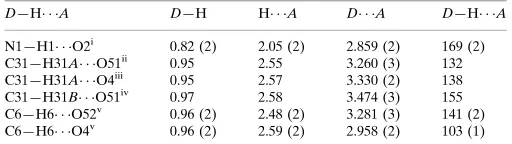
Hydrogen-bond geometry (A˚ ,).
D—H A D—H H A D A D—H A
N1—H1 O2i
0.82 (2) 2.05 (2) 2.859 (2) 169 (2) C31—H31A O51ii
0.95 2.55 3.260 (3) 132 C31—H31A O4iii
0.95 2.57 3.330 (2) 138 C31—H31B O51iv
0.97 2.58 3.474 (3) 155 C6—H6 O52v 0.96 (2) 2.48 (2) 3.281 (3) 141 (2) C6—H6 O4v
0.96 (2) 2.59 (2) 2.958 (2) 103 (1)
Symmetry codes: (i)xþ1;y;zþ1; (ii)x;y1 2;zþ
3
2; (iii)xþ1;y;zþ2;
(iv)xþ1;yþ1 2;zþ
1
2; (v)x;yþ 1 2;z
1 2.
The H atoms of the methyl group were found in a difference Fourier map and then refined as a rigid body (AFIX 3); theirUiso(H)
values were refined as a common FVAR.
Data collection: KM-4 Software (Kuma Diffraction, 1991); cell refinement: KM-4 Software; data reduction: KM-4 Software; program(s) used to solve structure: SHELXS97 (Sheldrick, 1990); program(s) used to refine structure:SHELXL97(Sheldrick, 1997); molecular graphics:Stereochemical Workstation Operation Manual (Siemens, 1989); software used to prepare material for publication: SHELXL97.
References
Allen, F. H. (2002).Acta Cryst.B58, 380–388.
Banerjee, A., Dattagupta, J. K., Saenger, W. & Rabczenko, A. (1977).Acta Cryst.B33, 90–94.
Bernstein, J., Davis, R. E., Shimoni, L. & Chang, N.-L. (1995).Angew. Chem. Int. Ed. Engl.34, 1555–1573.
Blank, H., U. & Fox, J. J. (1970).J. Heterocycl. Chem.7, 735–737. Craven, B. M. (1967).Acta Cryst.23, 376–383.
Etter, M. C., MacDonald, J. C. & Bernstein, J. (1990).Acta Cryst.B46, 256–262. Gopalan, R. S., Kulkarni, G. U. & Rao, C. N. R. (2000).ChemPhysChem,1,
127–135.
Kennedy, A. R., Okoth, M. O., Sheen, D. B., Sherwood, J. N. & Vrcelj, R. M. (1998).Acta Cryst.C54, 547–550.
Kuma Diffraction, (1991).KM-4 Software. User’s Guide, Version 5.0. Kuma Diffraction, Wrocław, Poland.
Larsen, H. J., Bentin, T. & Nielsen, P. E. (1999).Biochim. Biophys. Acta,1489, 159–166.
Longley, D. B., Harkin, D. P. & Johnston, P. G. (2003).Nat. Rev.3, 330–338. Petersen, M. & Wengel, J. (2003).Trends Biotechnol.21, 74–81.
Pierce, B. P. & Wing, R. M. (1986).Molecular and Polymeric Optoelectronic Materials: Fundamentals and Applications. Proc. Soc. Photo-Opt. Instrum. Eng.682, 27–35.
Portalone, G., Ballirano, P. & Maras, A. (2002).J. Mol. Struct.608, 35–39. Puccetti, G., Perigaud, A., Badan, J., Ledoux, I. & Zyss, J. (1993).J. Opt. Soc.
Am. B,10, 733–744.
Sheldrick, G. M. (1990).Acta Cryst.A46, 467–473.
Sheldrick, G. M. (1997).SHELXL97. University of Go¨ttingen, Germany. Siemens (1989).Stereochemical Workstation Operation Manual.Release 3.4.
Siemens Analytical X-ray Instruments Inc., Madison, Wisconsin, USA. Tanaka, H., Takashima, H., Ubasawa, M., Sekiya, K., Nitta, I., Baba, M.,
Shigeta, S., Walker, R. T., De Clercq, E. & Miyasaka, T. (1992).J. Med. Chem.35, 337–345.
Trusule, M., Kupce, E., Augustane, I., Verovskii, N. V., Lukevics, E., Baumane, L., Gavars, R. & Stradins, J. (1991).Khim. Geterotsikl. Soedin.12, 1687– 1694. (In Russian.)
organic papers
Acta Cryst.(2006). E62, o1257–o1259 Wagner and Kubicki C
supporting information
sup-1 Acta Cryst. (2006). E62, o1257–o1259
supporting information
Acta Cryst. (2006). E62, o1257–o1259 [https://doi.org/10.1107/S1600536806007033]
3-Methyl-5-nitrouracil
Pawe
ł
Wagner and Maciej Kubicki
3-Methyl-5-nitrouracil
Crystal data
C5H5N3O4 Mr = 171.12 Monoclinic, P21/c Hall symbol: -P 2ybc a = 5.7510 (12) Å b = 10.176 (2) Å c = 11.775 (2) Å β = 100.69 (3)° V = 677.1 (2) Å3 Z = 4
F(000) = 352 Dx = 1.679 Mg m−3
Mo Kα radiation, λ = 0.71073 Å Cell parameters from 50 reflections θ = 4–32°
µ = 0.15 mm−1 T = 295 K Block, colourless 0.4 × 0.2 × 0.1 mm
Data collection
Kuma KM-4 diffractometer
Radiation source: fine-focus sealed tube Graphite monochromator
ω–2θ scans
1256 measured reflections 1196 independent reflections 926 reflections with I > 2σ(I)
Rint = 0.066
θmax = 25.1°, θmin = 2.7° h = −6→6
k = 0→12 l = 0→14
2 standard reflections every 100 reflections intensity decay: 0.3%
Refinement
Refinement on F2 Least-squares matrix: full R[F2 > 2σ(F2)] = 0.045 wR(F2) = 0.128 S = 1.04 1196 reflections 119 parameters 0 restraints
Primary atom site location: structure-invariant direct methods
Secondary atom site location: difference Fourier map
Hydrogen site location: inferred from neighbouring sites
H atoms treated by a mixture of independent and constrained refinement
w = 1/[σ2(Fo2) + (0.0911P)2 + 0.05P] where P = (Fo2 + 2Fc2)/3
(Δ/σ)max < 0.001 Δρmax = 0.22 e Å−3 Δρmin = −0.27 e Å−3
supporting information
sup-2 Acta Cryst. (2006). E62, o1257–o1259
Special details
Geometry. All e.s.d.'s (except the e.s.d. in the dihedral angle between two l.s. planes) are estimated using the full covariance matrix. The cell e.s.d.'s are taken into account individually in the estimation of e.s.d.'s in distances, angles and torsion angles; correlations between e.s.d.'s in cell parameters are only used when they are defined by crystal symmetry. An approximate (isotropic) treatment of cell e.s.d.'s is used for estimating e.s.d.'s involving l.s. planes.
Refinement. Refinement of F2 against ALL reflections. The weighted R-factor wR and goodness of fit S are based on F2, conventional R-factors R are based on F, with F set to zero for negative F2. The threshold expression of F2 > σ(F2) is used only for calculating R-factors(gt) etc. and is not relevant to the choice of reflections for refinement. R-factors based on F2 are statistically about twice as large as those based on F, and R- factors based on ALL data will be even larger.
Fractional atomic coordinates and isotropic or equivalent isotropic displacement parameters (Å2)
x y z Uiso*/Ueq
N1 0.3041 (3) 0.10318 (16) 0.58178 (12) 0.0400 (5) H1 0.305 (4) 0.085 (2) 0.514 (2) 0.052 (6)* C2 0.4814 (3) 0.05458 (18) 0.66604 (14) 0.0374 (5) O2 0.6399 (3) −0.01339 (15) 0.64165 (11) 0.0545 (5) N3 0.4690 (3) 0.08839 (14) 0.77744 (11) 0.0339 (4) C31 0.6539 (3) 0.0368 (2) 0.86989 (15) 0.0439 (5)
H31A 0.6011 −0.0405 0.9018 0.082 (5)*
H31B 0.6728 0.0990 0.9328 0.082 (5)*
H31C 0.8032 0.0313 0.8459 0.082 (5)*
C4 0.2863 (3) 0.16141 (16) 0.81129 (14) 0.0347 (5) O4 0.2850 (3) 0.17753 (14) 0.91292 (10) 0.0532 (5) C5 0.1152 (3) 0.21061 (17) 0.71467 (14) 0.0357 (5) N5 −0.0782 (3) 0.29368 (16) 0.73154 (13) 0.0436 (5) O51 −0.2155 (3) 0.3304 (2) 0.64614 (14) 0.0964 (8) O52 −0.1018 (3) 0.32526 (15) 0.82795 (12) 0.0563 (5) C6 0.1302 (3) 0.17918 (18) 0.60439 (15) 0.0380 (5) H6 0.019 (4) 0.210 (2) 0.5393 (16) 0.044 (5)*
Atomic displacement parameters (Å2)
U11 U22 U33 U12 U13 U23
supporting information
sup-3 Acta Cryst. (2006). E62, o1257–o1259
Geometric parameters (Å, º)
N1—C6 1.329 (2) C31—H31C 0.95
N1—C2 1.376 (2) C4—O4 1.209 (2)
N1—H1 0.82 (2) C4—C5 1.449 (3)
C2—O2 1.220 (2) C5—C6 1.356 (2)
C2—N3 1.371 (2) C5—N5 1.440 (2)
N3—C4 1.404 (2) N5—O52 1.212 (2)
N3—C31 1.470 (2) N5—O51 1.216 (2)
C31—H31A 0.94 C6—H6 0.96 (2)
C31—H31B 0.96
C6—N1—C2 123.3 (2) H31B—C31—H31C 107
C6—N1—H1 118 (2) O4—C4—N3 119.7 (2)
C2—N1—H1 118.7 (2) O4—C4—C5 127.0 (2)
O2—C2—N3 122.8 (2) N3—C4—C5 113.3 (1)
O2—C2—N1 121.3 (2) C6—C5—N5 117.3 (2)
N3—C2—N1 115.9 (2) C6—C5—C4 121.1 (2)
C2—N3—C4 125.4 (2) N5—C5—C4 121.6 (2)
C2—N3—C31 117.4 (2) O52—N5—O51 121.7 (2)
C4—N3—C31 117.1 (1) O52—N5—C5 120.6 (2)
N3—C31—H31A 111 O51—N5—C5 117.8 (2)
N3—C31—H31B 107 N1—C6—C5 120.9 (2)
H31A—C31—H31B 104 N1—C6—H6 117 (1)
N3—C31—H31C 112 C5—C6—H6 123 (1)
H31A—C31—H31C 116
C6—N1—C2—O2 −178.9 (2) N3—C4—C5—C6 3.9 (3)
C6—N1—C2—N3 0.5 (3) O4—C4—C5—N5 3.2 (3)
O2—C2—N3—C4 −177.1 (2) N3—C4—C5—N5 −176.6 (2) N1—C2—N3—C4 3.5 (3) C6—C5—N5—O52 179.9 (2) O2—C2—N3—C31 −1.4 (3) C4—C5—N5—O52 0.4 (3) N1—C2—N3—C31 179.2 (2) C6—C5—N5—O51 0.0 (3) C2—N3—C4—O4 174.7 (2) C4—C5—N5—O51 −179.5 (2) C31—N3—C4—O4 −1.0 (3) C2—N1—C6—C5 −1.8 (3) C2—N3—C4—C5 −5.5 (3) N5—C5—C6—N1 179.8 (2) C31—N3—C4—C5 178.8 (2) C4—C5—C6—N1 −0.6 (3) O4—C4—C5—C6 −176.3 (2)
Hydrogen-bond geometry (Å, º)
D—H···A D—H H···A D···A D—H···A
supporting information
sup-4 Acta Cryst. (2006). E62, o1257–o1259
C6—H6···O52v 0.96 (2) 2.48 (2) 3.281 (3) 141 (2) C6—H6···O4v 0.96 (2) 2.59 (2) 2.958 (2) 103 (1)