organic papers
o2514
Tkachevet al. C12H9Cl2NO2S doi:10.1107/S160053680601943X Acta Cryst.(2006). E62, o2514–o2515
Acta Crystallographica Section E Structure Reports Online
ISSN 1600-5368
N
-(2,3-Dichlorophenyl)benzenesulfonamide
Valery V. Tkachev,a* Klaus-Ju¨rgen Schaper,b Nadezda N. Strakhovacand Vladimir P. Kazachenkoc
aLaboratory of Structural Chemistry, Institute of
Problems of Chemical Physics, Russian Academy of Sciences, 142432 Chernogolovka, Russian Federation,bResearch Center Borstel,
Leibniz Center for Medicine and Biosciences, D-23845 Borstel, Germany, andcLaboratory of
Computer-Aided Molecular Design, Institute of Physiologically Active Compounds, Russian Academy of Sciences, 142432 Chernogolovka, Russian Federation
Correspondence e-mail: [email protected]
Key indicators
Single-crystal X-ray study
T= 293 K
Mean(C–C) = 0.007 A˚
Rfactor = 0.049
wRfactor = 0.103
Data-to-parameter ratio = 12.1
For details of how these key indicators were automatically derived from the article, see http://journals.iucr.org/e.
Received 18 May 2006 Accepted 24 May 2006
#2006 International Union of Crystallography
All rights reserved
In the crystal structure of the title compound, C12H9Cl2NO2S, the dihedral angle between the two benzene rings is 54.8 (2).
Intermolecular N—H O hydrogen bonds lead to infinite helices along thebaxis.
Comment
Sulfanyls and sulfonamides are drugs used for the treatment of infections, some fungal and some protozoal. Other therapeutic applications are as diuretic and hypoglycaemic agents. Furthermore, the compounds are very interesting from a fundamental point of view, e.g.for studying the relationship between van der Waals interactions and hydrogen-bond topology in the formation of crystal structures.
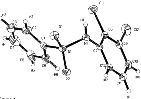
A view of the title compound, (I), with the atomic numbering is presented in Fig. 1. The conformation can be characterized in the following way. The torsion angle O1— S1—C1—C2, defining the orientation of the SO2group rela-tive to ring Ph1 (atoms C1–C6), is 7.4 (4). The benzene rings
are rotated relative to each other by 54.8 (2). The torsion
angle N1—S1—C1—C2 is 106.8 (3), whereas the torsion
[image:1.610.255.408.344.410.2] [image:1.610.211.459.552.726.2]angle S1—N1—C7—C12, defining the orientation of the SO2 group with respect to ring Ph2 (C7–C12), is63.8 (4).
Figure 1
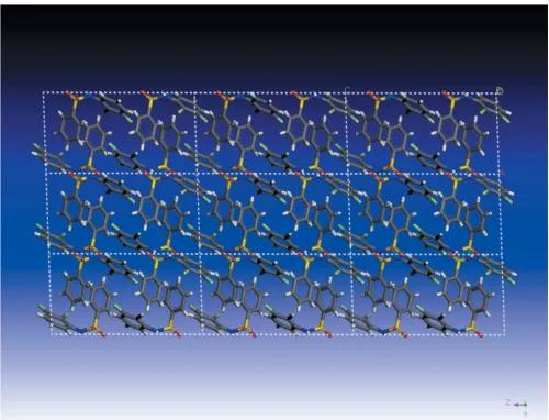
Intermolecular N—H O hydrogen bonds (dashed lines in Fig. 2) result in infinite helices along the b axis. Numeric details are given in Table 1. The hydrogen-bond network can be described by the graph-setC4 (Etter, 1990). The packing of molecules in the crystal structure is also illustrated in Fig. 3.
Experimental
The chemical synthesis of the compound has been performed by analogy with procedures described in papers by Crosleyet al.(1940), Andersonet al. (1942) and Gutscheet al. (1974), by reaction of a substituted aromatic amine (here 2,3-dichloroaniline) with benzene-sulfonyl chloride in dry pyridine, followed by precipitation of the end product by pouring the reaction mixture into water and by acid-ification to pH 5. Single crystals of the title compound were grown from a water–ethanol solution (20:1) by vapour diffusion. (Guillory, 1999).
Crystal data
C12H9Cl2NO2S
Mr= 302.16
Monoclinic,P21=c a= 8.466 (1) A˚
b= 9.805 (1) A˚
c= 15.876 (2) A˚ = 92.10 (1)
V= 1317.0 (3) A˚3
Z= 4
Dx= 1.524 Mg m3
MoKradiation = 0.64 mm1
T= 293 (2) K Prism, colourless 0.280.20.11 mm
Data collection
Bruker P4 diffractometer !–2scans
Absorption correction: none 2824 measured reflections 2022 independent reflections 1152 reflections withI> 2(I)
Rint= 0.042 max= 25.0 3 standard reflections
frequency: 120 min intensity decay: none
Refinement
Refinement onF2
R[F2> 2(F2)] = 0.049
wR(F2) = 0.103
S= 1.00 2022 reflections 167 parameters
H atoms treated by a mixture of independent and constrained refinement
w= 1/[2(F
o2) + (0.034P)2] whereP= (Fo2+ 2Fc2)/3 (/)max= 0.001
max= 0.24 e A˚3 min=0.21 e A˚3
Extinction correction:SHELXL97
Extinction coefficient: 0.0030 (9)
Table 1
Hydrogen-bond geometry (A˚ ,).
D—H A D—H H A D A D—H A
N1—H1 O2i
0.76 (4) 2.30 (4) 3.039 (4) 166 (4)
Symmetry code: (i)xþ2;y1 2;zþ
3 2.
C-bound H atoms were positioned geometrically and refined as riding, with C—H = 0.93 A˚ ; Uiso(H) values were set equal to
1.2Ueq(C). The coordinates of the N-bound H atom were determined
by an optimization procedure and refined freely [N—H = 0.76 (4) A˚ ] Data collection: CAD-4-PC Software (Enraf–Nonius, 1989); cell refinement: CELDIM in CAD-4-PC Software; data reduction:
XCAD4 (McArdle & Higgins, 1995); program(s) used to solve structure:SHELXS97(Sheldrick, 1997a); program(s) used to refine structure: SHELXL97 (Sheldrick, 1997a); molecular graphics:
SHELXTL(Sheldrick, 1997b); software used to prepare material for
This work was supported by ISTC (project No. 0888).
References
Anderson, G. W., Faith, H. E., Marson, H. W., Winnek, P. S. & Roblin, R. O. (1942).J. Am. Chem. Soc.64, 2902–2905.
Crosley, M. L., Northey, E. H. & Hultquist, M. E. (1940).J. Am. Chem. Soc.62, 372–374.
Enraf–Nonius (1989).CAD-4 Software. Version 5.0. Enraf–Nonius, Delft, The Netherlands.
Etter, M. C. (1990).Acc. Chem. Res.23, 120–126.
Guillory, J. K. (1999).Polymorphism in Pharmaceutical Solids, edited by H. G. Brittain. pp. 183–226. New York: Marcel Dekker Inc.
Gutsche, K., Schro¨der, E., Rufer, C. & Loge, O. (1974).Arzneim. Forsch. (Drug Res.),24, 1028–1039.
McArdle, P. & Higgins, T. (1995).XCAD. National University of Ireland, Galway, Ireland.
Sheldrick, G. M. (1997a). SHELXL97 and SHELXS97. University of Go¨ttingen, Germany.
Sheldrick, G. M. (1997b).SHELXTL. Bruker AXS Inc., Madison, Wisconsin,
Figure 2
[image:2.610.315.565.70.261.2]Projection of the molecular packing along theaaxis. Hydrogen bonds are indicated by dashed lines.
Figure 3
[image:2.610.314.565.305.494.2]supporting information
sup-1
Acta Cryst. (2006). E62, o2514–o2515
supporting information
Acta Cryst. (2006). E62, o2514–o2515 [https://doi.org/10.1107/S160053680601943X]
N
-(2,3-Dichlorophenyl)benzenesulfonamide
Valery V. Tkachev, Klaus-J
ü
rgen Schaper, Nadezda N. Strakhova and Vladimir P. Kazachenko
N-(2,3-Dichlorophenyl)benzenesulfonamide
Crystal data
C12H9Cl2NO2S Mr = 302.16 Monoclinic, P21/c a = 8.466 (1) Å
b = 9.805 (1) Å
c = 15.876 (2) Å
β = 92.10 (1)°
V = 1317.0 (3) Å3 Z = 4
F(000) = 616
Dx = 1.524 Mg m−3 Melting point: 387.2 K
Mo Kα radiation, λ = 0.71073 Å Cell parameters from 35 reflections
θ = 5–12°
µ = 0.64 mm−1 T = 293 K Prism, colourless 0.28 × 0.2 × 0.11 mm
Data collection
Bruker P4 diffractometer
Radiation source: fine-focus sealed tube Graphite monochromator
ω–2θ scans
2824 measured reflections 2022 independent reflections 1152 reflections with I > 2σ(I)
Rint = 0.042
θmax = 25.0°, θmin = 2.4° h = −10→1
k = −11→1
l = −18→18
3 standard reflections every 120 min intensity decay: none
Refinement
Refinement on F2 Least-squares matrix: full
R[F2 > 2σ(F2)] = 0.049 wR(F2) = 0.103 S = 1.00 2022 reflections 167 parameters 0 restraints
Primary atom site location: structure-invariant direct methods
Secondary atom site location: difference Fourier map
Hydrogen site location: inferred from neighbouring sites
H atoms treated by a mixture of independent and constrained refinement
w = 1/[σ2(F
o2) + (0.034P)2] where P = (Fo2 + 2Fc2)/3 (Δ/σ)max = 0.001
Δρmax = 0.24 e Å−3 Δρmin = −0.21 e Å−3
Special details
Geometry. All e.s.d.'s (except the e.s.d. in the dihedral angle between two l.s. planes) are estimated using the full covariance matrix. The cell e.s.d.'s are taken into account individually in the estimation of e.s.d.'s in distances, angles and torsion angles; correlations between e.s.d.'s in cell parameters are only used when they are defined by crystal symmetry. An approximate (isotropic) treatment of cell e.s.d.'s is used for estimating e.s.d.'s involving l.s. planes.
Refinement. Refinement of F2 against ALL reflections. The weighted R-factor wR and goodness of fit S are based on F2, conventional R-factors R are based on F, with F set to zero for negative F2. The threshold expression of F2 > σ(F2) is used only for calculating R-factors(gt) etc. and is not relevant to the choice of reflections for refinement. R-factors based on F2 are statistically about twice as large as those based on F, and R- factors based on ALL data will be even larger.
Fractional atomic coordinates and isotropic or equivalent isotropic displacement parameters (Å2)
x y z Uiso*/Ueq
S1 0.91244 (13) 0.08296 (11) 0.71431 (7) 0.0469 (3) Cl1 0.75548 (16) −0.18271 (12) 0.89767 (8) 0.0736 (5) Cl2 0.64362 (16) −0.07756 (15) 1.07167 (8) 0.0865 (5) O1 1.0071 (3) 0.0155 (3) 0.65482 (17) 0.0593 (9) O2 0.9226 (4) 0.2272 (3) 0.72520 (17) 0.0602 (9) N1 0.9593 (5) 0.0148 (3) 0.8059 (2) 0.0490 (10) H1 0.974 (5) −0.061 (4) 0.801 (2) 0.054* C1 0.7129 (5) 0.0437 (4) 0.6901 (2) 0.0440 (11) C2 0.6772 (6) −0.0515 (5) 0.6292 (3) 0.0705 (14) H2 0.7573 −0.0955 0.6013 0.085* C3 0.5197 (7) −0.0815 (5) 0.6097 (4) 0.0964 (19) H3 0.4938 −0.1437 0.5672 0.116* C4 0.4032 (7) −0.0197 (7) 0.6528 (4) 0.096 (2) H4 0.2981 −0.0428 0.6411 0.116* C5 0.4392 (7) 0.0756 (7) 0.7127 (3) 0.0825 (18) H5 0.3586 0.1188 0.7407 0.099* C6 0.5953 (6) 0.1086 (5) 0.7323 (3) 0.0666 (14) H6 0.6202 0.1737 0.7733 0.080* C7 0.8956 (5) 0.0629 (4) 0.8824 (2) 0.0401 (10) C8 0.8043 (5) −0.0214 (4) 0.9315 (3) 0.0458 (11) C9 0.7513 (5) 0.0268 (5) 1.0073 (3) 0.0553 (13) C10 0.7851 (6) 0.1582 (6) 1.0338 (3) 0.0667 (14) H10 0.7487 0.1898 1.0848 0.080* C11 0.8722 (6) 0.2411 (5) 0.9847 (3) 0.0665 (14) H11 0.8935 0.3302 1.0016 0.080* C12 0.9294 (5) 0.1931 (4) 0.9094 (3) 0.0535 (12) H12 0.9911 0.2496 0.8769 0.064*
Atomic displacement parameters (Å2)
U11 U22 U33 U12 U13 U23
supporting information
sup-3
Acta Cryst. (2006). E62, o2514–o2515
N1 0.053 (2) 0.037 (2) 0.056 (2) 0.006 (2) −0.005 (2) 0.001 (2) C1 0.045 (3) 0.044 (3) 0.043 (3) 0.007 (2) 0.002 (2) 0.011 (2) C2 0.052 (3) 0.060 (3) 0.099 (4) 0.007 (3) −0.007 (3) −0.014 (3) C3 0.067 (4) 0.071 (4) 0.149 (5) 0.001 (4) −0.033 (4) −0.018 (4) C4 0.047 (4) 0.109 (5) 0.133 (6) −0.001 (4) −0.003 (4) 0.036 (5) C5 0.057 (4) 0.124 (5) 0.067 (4) 0.035 (4) 0.013 (3) 0.024 (4) C6 0.061 (3) 0.090 (4) 0.049 (3) 0.022 (3) 0.006 (3) 0.007 (3) C7 0.045 (2) 0.032 (2) 0.043 (2) 0.003 (2) −0.007 (2) 0.000 (2) C8 0.047 (3) 0.043 (2) 0.047 (3) −0.002 (2) −0.009 (2) 0.001 (2) C9 0.056 (3) 0.063 (3) 0.047 (3) 0.000 (3) −0.004 (3) 0.006 (2) C10 0.076 (4) 0.077 (4) 0.046 (3) 0.013 (3) −0.010 (3) −0.016 (3) C11 0.092 (4) 0.046 (3) 0.060 (3) 0.003 (3) −0.009 (3) −0.009 (3) C12 0.057 (3) 0.044 (3) 0.059 (3) −0.004 (2) −0.010 (3) 0.005 (2)
Geometric parameters (Å, º)
S1—O1 1.424 (3) C4—C5 1.360 (7) S1—O2 1.427 (3) C4—H4 0.9300 S1—N1 1.636 (4) C5—C6 1.384 (6) S1—C1 1.761 (4) C5—H5 0.9300 Cl1—C8 1.716 (4) C6—H6 0.9300 Cl2—C9 1.729 (5) C7—C12 1.373 (5) N1—C7 1.427 (5) C7—C8 1.390 (5) N1—H1 0.76 (4) C8—C9 1.383 (5) C1—C2 1.369 (5) C9—C10 1.382 (6) C1—C6 1.377 (6) C10—C11 1.362 (6) C2—C3 1.389 (6) C10—H10 0.9300 C2—H2 0.9300 C11—C12 1.388 (6) C3—C4 1.363 (7) C11—H11 0.9300 C3—H3 0.9300 C12—H12 0.9300
C2—C3—H3 120.0 C10—C11—H11 119.9 C5—C4—C3 120.6 (6) C12—C11—H11 119.9 C5—C4—H4 119.7 C7—C12—C11 120.5 (4) C3—C4—H4 119.7 C7—C12—H12 119.7 C4—C5—C6 120.4 (5) C11—C12—H12 119.7 C4—C5—H5 119.8
O1—S1—N1—C7 175.8 (3) S1—N1—C7—C12 −63.8 (5) O2—S1—N1—C7 46.2 (4) S1—N1—C7—C8 118.4 (4) C1—S1—N1—C7 −68.6 (4) C12—C7—C8—C9 −1.0 (6) O1—S1—C1—C2 7.4 (4) N1—C7—C8—C9 176.8 (4) O2—S1—C1—C2 138.8 (3) C12—C7—C8—Cl1 178.0 (3) N1—S1—C1—C2 −106.8 (3) N1—C7—C8—Cl1 −4.2 (5) O1—S1—C1—C6 −172.6 (3) C7—C8—C9—C10 1.3 (6) O2—S1—C1—C6 −41.2 (4) Cl1—C8—C9—C10 −177.7 (3) N1—S1—C1—C6 73.2 (4) C7—C8—C9—Cl2 −177.6 (3) C6—C1—C2—C3 0.5 (7) Cl1—C8—C9—Cl2 3.4 (5) S1—C1—C2—C3 −179.4 (4) C8—C9—C10—C11 −0.2 (7) C1—C2—C3—C4 −2.1 (8) Cl2—C9—C10—C11 178.8 (4) C2—C3—C4—C5 2.7 (9) C9—C10—C11—C12 −1.3 (7) C3—C4—C5—C6 −1.6 (9) C8—C7—C12—C11 −0.5 (6) C2—C1—C6—C5 0.5 (6) N1—C7—C12—C11 −178.3 (4) S1—C1—C6—C5 −179.5 (3) C10—C11—C12—C7 1.7 (7) C4—C5—C6—C1 0.1 (8)
Hydrogen-bond geometry (Å, º)
D—H···A D—H H···A D···A D—H···A
N1—H1···O2i 0.76 (4) 2.30 (4) 3.039 (4) 166 (4)