Received 12 November 2019 Accepted 21 November 2019
Edited by A. J. Lough, University of Toronto, Canada
Keywords:crystal structure; benzimidazol-2-one; hydrogen bond; C—H (ring) inter-action;-stacking; Hirshfeld surface.
CCDC reference:1967468
Supporting information:this article has supporting information at journals.iucr.org/e
Crystal structure, Hirshfeld surface analysis and
interaction energy and DFT studies of
1-methyl-3-(prop-2-yn-1-yl)-2,3-dihydro-1
H
-1,3-benzodiazol-2-one
Asmaa Saber,aMohamed Srhir,a* Tuncer Ho¨kelek,bJoel T. Mague,cNoureddine Hamou Ahabchane,aNada Kheira Sebbard,aand El Mokhtar Essassia
aLaboratoire de Chimie Organique He´te´rocyclique URAC 21, Poˆle de Compe´tence Pharmacochimie, Av. Ibn Battouta, BP
1014, Faculte´ des Sciences, Universite´ Mohammed V, Rabat, Morocco,bDepartment of Physics, Hacettepe University,
06800 Beytepe, Ankara, Turkey,cDepartment of Chemistry, Tulane University, New Orleans, LA 70118, USA, and dLaboratoire de Chimie Applique´e et Environnement, Equipe de Chimie Bioorganique Applique´e, Faculte´ des Sciences,
Universite´ Ibn Zohr, Agadir, Morocco. *Correspondence e-mail: [email protected]
In the title molecule, C11H10N2O, the dihydrobenzimidazol-2-one moiety is essentially planar, with the prop-2-yn-1-yl substituent rotated well out of this plane. In the crystal, C—HMthy (ring) interactions and C—HProp ODhyr (Mthy = methyl, Prop = prop-2-yn-1-yl and Dhyr = dihydro) hydrogen bonds form corrugated layers parallel to (101), which are associated through additional C—HBnz ODhyr(Bnz = benzene) hydrogen bonds and head-to-tail, slipped, -stacking [centroid-to-centroid distance = 3.7712 (7) A˚ ] interactions between dihydrobenzimidazol-2-one moieties. The Hirshfeld surface analysis of the crystal structure indicates that the most important contributions to the crystal packing are from H H (44.1%), H C/C H (33.5%) and O H/H O (13.4%) interactions. Hydrogen-bonding and van der Waals interactions are the dominant interactions in the crystal packing. Computational chemistry calculations indicate that in the crystal, C—H O hydrogen-bond energies are 46.8 and 32.5 (for C—HProp ODhyr) and 20.2 (for C—HBnz ODhyr) kJ mol1. Density functional theory (DFT) optimized structures at the B3LYP/ 6–311 G(d,p) level are compared with the experimentally determined molecular structure in the solid state. The HOMO–LUMO behaviour was elucidated to determine the energy gap.
1. Chemical context
Benzimidazole is an aromatic heterocyclic organic compound that plays an important role in medicinal chemistry and pharmacology. The most prominent benzimidazole moiety present in nature is N-ribosyl-dimethylbenzimidazole and it serves as the axial ligand for cobalt in vitamin B12 (Waliaet al., 2011). Benzimidazole derivatives possess many biological activities such as anti-microbial, anti-fungal, anti-histaminic, anti-inflammatory, anti-viral, anti-oxidant, anti-cancer and anti-ulcerative (Farukh & Mubashira, 2009; Ayhan-Kılcıgil et al., 2007; Soderlindet al., 1999; Luo et al., 2011; Navarrete-Va´zquezet al., 2011). They are considered to be an important moiety for the development of molecules of pharmaceutical interest (Mondieig et al., 2013; Lakhrissi et al., 2008). As a continuation of our research on the development of N-substituted benzimidazole derivatives and the evaluation of their potential pharmacological activities (Saber et al., 2018a,b, 2020; Ouzidan et al., 2011), we have studied the
alkylation reaction of iodomethane with 1-(prop-2-ynyl)-1H-benzoimidazol-2(3H)-one in the presence of tetra-n-butyl-ammonium bromide as catalyst and potassium carbonate as base, to give the title compound, Iin good yield. We report herein on its synthesis, the molecular and crystal structures along with the Hirshfeld surface analysis and the inter-molecular interaction energies and the density functional theory (DFT) computational calculations carried out at the B3LYP/6–311 G(d,p) level for comparison with the experi-mentally determined molecular structure in the solid state.
2. Structural commentary
In the title compound, the dihydrobenzimidazol-2-one moiety is planar to within 0.0160 (8) A˚ (r.m.s. deviation = 0.0082) with atom C7 deviating the most from the mean plane and a prop-2-yn-1-yl substituent rotated well out of this plane as shown by the C1—N2—C9—C10 torsion angle of 62.16 (13)(Fig. 1).
3. Supramolecular features
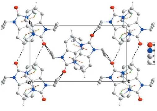
In the crystal, inversion dimers are formed by pairs of C— HMthy Cg1iinteractions [Mthy = methyl; symmetry code: (i)
x, 1y, 1z;Cg1 is the centroid of the benzene (A; C1– C6), ring]; which are connected along theb-axis direction by C—HBnz ODhyrhydrogen bonds (Bnz = benzene and Dhyr = dihydro) and along the a-axis direction atca 90 to this and
[image:2.610.93.247.197.288.2] [image:2.610.314.567.331.509.2]parallel to (101) by inversion-related C—HProp ODhyr hydrogen bonds (Table 1). The resulting corrugated layers are parallel to (101) and are connected in pairs by slipped, head-to-tail -stacking interactions between the dihydro-benzimidazol-2-one moieties, [Cg2 Cg1ii = 3.7712 (7) A˚ , dihedral angle = 0.96 (6); symmetry code: (ii) 1 –x, 1 –y, 1 –z; Cg1 andCg2 are the centroids of ringsAandB(N1/N2/C1/C6/ C7) and C—HProp ODhyr(Prop = prop-2-yn-1-yl) hydrogen bonds (Table 1, Figs. 2 and 3).
Figure 1
The molecular structure of the title compound with the atom-numbering scheme. Displacement ellipsoids are drawn at the 50% probability level.
Table 1
Hydrogen-bond geometry (A˚ ,).
Cg1 is the centroid of the C1–C6 benzene ring.
D—H A D—H H A D A D—H A
C3—H3 O1ix 1.005 (15) 2.566 (15) 3.4885 (15) 152.6 (11) C8—H8C Cg1v 1.004 (16) 2.626 (15) 3.5413 (13) 151.1 (12)
C9—H9B O1vi 0.978 (15) 2.347 (15) 3.3198 (14) 172.9 (12) C11—H11 O1vii 1.010 (15) 2.181 (15) 3.1569 (15) 162.1 (12)
Symmetry codes: (v) x;yþ1;zþ1; (vi) xþ1 2;y
1 2;zþ
3 2; (vii) xþ3
2;y 1 2;zþ
3
[image:2.610.62.267.526.722.2]2; (ix)x;y1;z.
Figure 3
A partial packing diagram viewed along the b-axis direction with intermolecular interactions depicted as in Fig. 2.
Figure 2
[image:2.610.318.567.560.711.2]4. Hirshfeld surface analysis
In order to visualize the intermolecular interactions in the crystal of the title compound, a Hirshfeld surface (HS) analysis (Hirshfeld, 1977; Spackman & Jayatilaka, 2009) was carried out usingCrystal Explorer 17.5(Turneret al., 2017). In
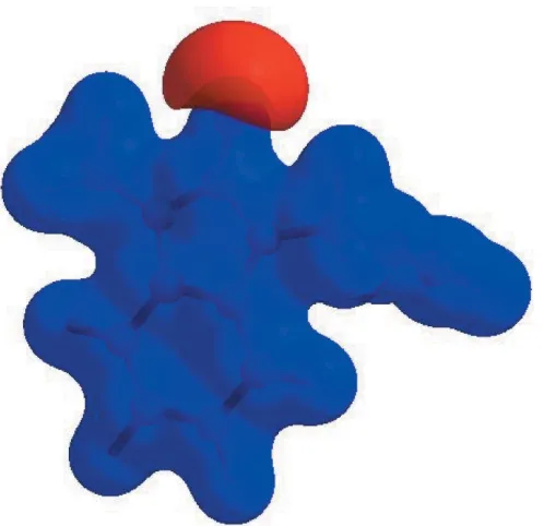
the HS plotted overdnorm(Fig. 4), the white surface indicates contacts with distances equal to the sum of van der Waals radii, and the red and blue colours indicate distances shorter (in close contact) or longer (distant contact) than the van der Waals radii, respectively (Venkatesanet al., 2016). The bright-red spots appearing near O1 and the hydrogen atom H11 indicate their roles as the donors and/or acceptors, respec-tively; they also appear as blue and red regions corresponding to positive and negative potentials on the HS mapped over electrostatic potential (Spackmanet al., 2008; Jayatilakaet al., 2005) as shown in Fig. 5. The blue regions indicate positive electrostatic potential (hydrogen-bond donors), while the red regions indicate negative electrostatic potential (hydrogen-bond acceptors). The shape-index of the HS is a tool to visualize
–stacking by the presence of adjacent red and blue trian-gles; if there are no adjacent red and/or blue triangles, then there are no–interactions. Fig. 6 clearly suggests that there are–interactions in (I).
Figure 4
[image:3.610.48.294.74.308.2] [image:3.610.313.563.78.206.2]View of the three-dimensional Hirshfeld surface of the title compound plotted overdnormin the range0.3997 to 1.3219 a.u.
Figure 5
View of the three-dimensional Hirshfeld surface of the title compound plotted over electrostatic potential energy in the range0.0500 to 0.0500 a.u. using the STO-3 G basis set at the Hartree–Fock level of theory. Hydrogen-bond donors and acceptors are shown as blue and red regions around the atoms corresponding to positive and negative potentials, respectively.
Table 2
Selected interatomic distances (A˚ ).
O1 H9A 2.491 (14) C11 O1vii 3.1569 (15)
O1 H3i 2.566 (15) C2 H8Aiv 2.82 (2)
O1 H8B 2.516 (19) C3 H8Cv 2.859 (15)
O1 H9Bii 2.346 (14) C3 H8Aiv 2.92 (2)
O1 H11iii 2.181 (15) C4 H8Cv 2.810 (15)
C2 C10 3.3889 (16) C5 H8Cv 2.935 (15)
C3 C8iv 3.5335 (17) C8 H5 2.983 (14)
C4 C8v 3.4947 (17) C9 H2 2.975 (14)
C4 C7iv 3.5437 (16) C10 H4viii 2.976 (15)
C5 C8v 3.5884 (17) C11 H5iv 2.865 (15)
C6 C6iv 3.5349 (14) C11 H4viii 2.705 (15)
C9 O1vi 3.3198 (14)
Symmetry codes: (i)x;yþ1;z; (ii)xþ1 2;yþ
1 2;zþ
3 2; (iii)xþ
3 2;yþ
1 2;zþ
3 2; (iv) xþ1;yþ1;zþ1; (v) x;yþ1;zþ1; (vi) xþ1
2;y 1 2;zþ
3 2; (vii) xþ3
2;y 1 2;zþ
3 2; (viii)xþ
1 2;yþ
1 2;zþ
[image:3.610.51.300.441.682.2]1 2.
Figure 6
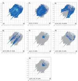
[image:3.610.319.562.506.730.2]The overall two-dimensional fingerprint plot, Fig. 7a, and those delineated into H H, H C/C H, H O/O H, C C, H N/N H and N C/C N contacts (McKinnonet al., 2007) are illustrated in Fig. 7b–g, respectively, together with their relative contributions to the Hirshfeld surface. The most important interaction is H H contributing 44.1% to the overall crystal packing, which is reflected in Fig. 7bas widely scattered points of high density due to the large hydrogen content of the molecule with the tip at de=di= 1.22 A˚ . The presence of C—H interactions gives rise to pairs of characteristic wings in the fingerprint plot delineated into H C/C H contacts, Fig. 7c., contributing 33.5% to the HS (Table 2); these are viewed as pairs of spikes with the tips atde +di= 2.56 A˚ . The pair of wings in Fig. 7dhas a symmetrical distribution of points with the edges atde+di= 2.09 A˚ arising from the H O/O H contacts (13.4% contribution). The C C contacts, Fig. 7e, have an arrow-shaped distribution of points with the tip at de = di = 1.75 A˚ . The H N/N N contacts, contributing 2.9% to the overall crystal packing, are depicted in Fig. 7f as widely scattered points. Finally, the N C/C N interactions, contributing 2.4% to the overall crystal packing, are shown in Fig. 7g as tiny characteristic wings with the tips atde+di= 3.45 A˚ .
The Hirshfeld surface representations with the function dnorm plotted onto the surface are shown for the H H, H C/C H and H O/O H interactions in Fig. 8a–c, respectively.
The Hirshfeld surface analysis confirms the importance of H-atom contacts in establishing the packing. The large number of H H, H C/C H and H O/O H interactions suggest that van der Waals interactions and hydrogen bonding play the major roles in the crystal packing (Hathwaret al., 2015).
5. Interaction energy calculations
The intermolecular interaction energies were calculated using the CE–B3LYP/6–31G(d,p) energy model available in Crys-talExplorer17.5(Turneret al., 2017), where a cluster of mol-ecules is generated by applying crystallographic symmetry operations with respect to a selected central molecule within the default radius of 3.8 A˚ (Turner et al., 2014). The total intermolecular energy (Etot) is the sum of electrostatic (Eele), polarization (Epol), dispersion (Edis) and exchange-repulsion (Erep) energies (Turneret al., 2015) with scale factors of 1.057, 0.740, 0.871 and 0.618, respectively (Mackenzieet al., 2017). Hydrogen-bonding interaction energies (in kJ mol1) were calculated to be17.4 (Eele), 3.5 (Epol),62.6 (Edis), 46.5 (Erep) and46.8 (Etot) for C11—H11 O1,12.4 (Eele),1.9 (Epol), 41.6 (Edis), 29.6 (Erep) and 32.5 (Etot) for C9— H9B O1 and 13.7 (Eele), 3.7 (Epol), 15.5 (Edis), 17.0 (Erep) and20.2 (Etot) for C3—H3 O1.
6. DFT calculations
[image:4.610.45.298.66.326.2]The optimized structure of the title compound in the gas phase was generated theoretically via density functional theory (DFT) using the standard B3LYP functional and 6–311 G(d,p) basis-set calculations (Becke, 1993) as implemented in GAUSSIAN 09 (Frisch et al., 2009). The theoretical and Figure 7
The full two-dimensional fingerprint plots for the title compound, showing (a) all interactions, and delineated into (b) H H, (c) H C/ C H, (d) H O/O H, (e) C C, (f) H N/N H and (g) N C/ C N interactions. The di and de values are the closest internal and
external distances (in A˚ ) from given points on the Hirshfeld surface contacts.
Figure 8
The Hirshfeld surface representations with the functiondnormplotted
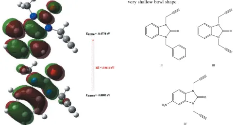
[image:4.610.309.563.74.305.2]experimental results are in good agreement (Table 3). The highest-occupied molecular orbital (HOMO), acting as an electron donor, and the lowest-unoccupied molecular orbital (LUMO), acting as an electron acceptor, are very important parameters for quantum chemistry. When the energy gap is small, the molecule is highly polarizable and has high chemical reactivity. The DFT calculations provide some important information on the reactivity and site selectivity of the mol-ecular framework. EHOMO andELUMOclarify the inevitable charge-exchange collaboration inside the studied material and are given in Table 4 along with the electronegativity (), hardness (), potential (), electrophilicity (!) and softness (). The significance of and is for the evaluation of both the reactivity and stability. The electron transition from the HOMO to the LUMO energy level is shown in Fig. 9. The
HOMO and LUMO are localized in the plane extending from the whole 1-methyl-3-(prop-2-yn-1-yl)-2,3-dihydro-1H-1,3-benzodiazol-2-one ring. The energy band gap [E=ELUMO EHOMO] of the molecule is about 5.4115 eV, and the frontier molecular orbital energies, EHOMO and ELUMO are 5.8885 and0.4770 eV, respectively.
7. Database survey
The syntheses of several N-substituted benzimidazol-2-one analogues have been reported (Saber et al., 2018a,b; 2020; Belazizet al., 2012; Bouayadet al., 2015; Belazizet al., 2013). In a search of the Cambridge Crystallographic Database (CSD; Version 5.40, update of September 2019; Groomet al., 2016) using benzimidazol-2-one with an exocyclic carbon atom bound to each nitrogen generated 94 hits. In these, the bicyclic ring system is either planar, has a slight twist end-to-end, or, in the cases where the exocyclic substituents form a ring, has a very shallow bowl shape.
[image:5.610.314.565.91.224.2]The closest examples to the title compound, I, are II (HISFUN; Saberet al., 2018b),III(URAQAG; Ouzidanet al., Figure 9
[image:5.610.44.296.92.240.2]The energy band gap of the title compound. Table 3
Comparison of the selected (X-ray and DFT) geometric data (A˚ ,).
Bonds/angles X-ray B3LYP/6–311 G(d,p)
O1—C7 1.2281 (13) 1.24660
N1—C7 1.3735 (14) 1.39764
N1—C6 1.3874 (15) 1.40100
N1—C8 1.4526 (14) 1.45375
N2—C7 1.3807 (14) 1.40268
N2—C1 1.3910 (13) 1.40222
N2—C9 1.4545 (14) 1.46036
C7—N1—C6 110.19 (9) 110.10303
C7—N1—C8 124.14 (10) 122.94288
C6—N1—C8 125.66 (10) 126.95366
C7—N2—C1 110.16 (9) 110.18664
C7—N2—C9 123.55 (9) 122.02491
C1—N2—C9 126.00 (9) 126.78733
C2—C1—N2 131.64 (10) 132.00719
Table 4
Calculated energies for the title compound.
Molecular Energy (a.u.) (eV)
Total EnergyTE(eV) 16594.1662
EHOMO(eV) 5.8885
ELUMO(eV) 0.4770
Energy gap,E(eV) 5.4115
Dipole moment,(Debye) 2.8313
Ionization potential,I(eV) 5.8885
Electron affinity,A 2.6040
Electro negativity, 0.31828
Hardness, 2.7058
Electrophilicity index,! 1.8719
Softness, 0.3696
[image:5.610.51.535.465.723.2]2011a) andIV (AGAXOX; Kandri Rodiet al., 2013). In the title compound, the C—N bonds to the exocyclic groups are 1.4526 (14) and 1.4545 (19) A˚ while in II–IV the corre-sponding distances range from 1.445 (3) to 1.4632 (11) A˚ , and so are quite comparable. The exocyclic groups inI are in an anti-arrangement with the prop-2-yn-1-yl group rotated by 62.16 (13) out of the plane of the bicyclic moiety (as measured by the C1—N2—C9—C10 torsion angle). In the other three, these substituents are also anti and in II the corresponding torsion angle is 73.46 (18)while in
IIIthey are 82.58 (15) and 74.31 (14). In
IV the torsion angles are 106.0 (3) and 113.4 (3) indicating a rotation in the opposite direction from the first three.
8. Synthesis and crystallization
To a mixture of 1-(prop-2-ynyl)-1H-benzimidazol-2(3H)-one (3.61 mmol), iodomethane (6.73 mmol) and potassium carbonate (6.24 mmol) in DMF (15 ml) was added a catalytic amount of tetra-n-butylammonium bromide (0.37 mmol). The mixture was stirred for 24 h. The solid material was removed by filtration and the solvent evaporated under vacuum. The solid product was purified by recrystallization from ethanol to afford colorless crystals (yield: in 82%).
9. Refinement
Crystal data, data collection and structure refinement details are summarized in Table 5. Hydrogen atoms were located in a difference Fourier map and refined freely.
Funding information
The support of NSF–MRI grant No. 1228232 for the purchase of the diffractometer and Tulane University for support of the Tulane Crystallography Laboratory are gratefully acknowl-edged. TH is grateful to Hacettepe University Scientific Research Project Unit (grant No. 013 D04 602 004).
References
Ayhan-Kılcıgil, G., Kus, G., O¨ zdamar, E. D., Can-Eke, B. & Iscan, M. (2007).Arch. Pharm. Chem. Life Sci.340, 607–611.
Becke, A. D. (1993).J. Chem. Phys.98, 5648–5652.
Belaziz, D., Kandri Rodi, Y., Essassi, E. M. & El Ammari, L. (2012). Acta Cryst.E68, o1276.
Belaziz, D., Kandri Rodi, Y., Ouazzani Chahdi, F., Essassi, E. M., Saadi, M. & El Ammari, L. (2013).Acta Cryst.E69, o122. Bouayad, K., Kandri Rodi, Y., Ouzidan, Y., Essassi, E. M., Saadi, M.
& El Ammari, L. (2015).Acta Cryst.E71, o735–o736.
Brandenburg, K. & Putz, H. (2012). DIAMOND. Crystal Impact GbR, Bonn, Germany.
Bruker (2016). APEX3,SAINT and SADABS. Bruker AXS, Inc., Madison, Wisconsin, USA.
Farukh, A. & Mubashira, A. (2009).Eur. J. Med. Chem.44, 834–844. Frisch, M. J.,et al.(2009).GAUSSIAN09. Gaussian Inc., Wallingford,
CT, US
Groom, C. R., Bruno, I. J., Lightfoot, M. P. & Ward, S. C. (2016).Acta Cryst.B72, 171–179.
Hathwar, V. R., Sist, M., Jørgensen, M. R. V., Mamakhel, A. H., Wang, X., Hoffmann, C. M., Sugimoto, K., Overgaard, J. & Iversen, B. B. (2015).IUCrJ,2, 563–574.
Hirshfeld, H. L. (1977).Theor. Chim. Acta,44, 129–138.
Jayatilaka, D., Grimwood, D. J., Lee, A., Lemay, A., Russel, A. J., Taylor, C., Wolff, S. K., Cassam-Chenai, P. & Whitton, A. (2005). TONTO – A System for Computational Chemistry.Available at: http://hirshfeldsurface.net/
Kandri Rodi, Y., Misbahi, K., El-Ghayoury, A., Zorina, L., Essassi, E. M. & El Ammari, L. (2013).Acta Cryst.E69, o1159.
Krause, L., Herbst-Irmer, R., Sheldrick, G. M. & Stalke, D. (2015).J. Appl. Cryst.48, 3–10.
Lakhrissi, B., Benksim, A., Massoui, M., Essassi, E. M., Lequart, V., Joly, N., Beaupe`re, D., Wadouachi, A. & Martin, P. (2008). Carbohydr. Res.343, 421–433.
Luo, Y., Yao, J. P., Yang, L., Feng, C. L., Tang, W., Wang, G. F., Zuo, J. P. & Lu, W. (2011).Arch. Pharm. Pharm. Med. Chem.344, 78– 83.
Mackenzie, C. F., Spackman, P. R., Jayatilaka, D. & Spackman, M. A. (2017).IUCrJ,4, 575–587.
McKinnon, J. J., Jayatilaka, D. & Spackman, M. A. (2007).Chem. Commun.pp. 3814–3816.
Mondieig, D., Lakhrissi, L., El Assyry, A., Lakhrissi, B., Negrier, P., Essassi, E. M., Massoui, M., Michel Leger, J. & Benali, B. (2013).J. Mar. Chim. Heterocycl.12, 51–61.
Navarrete-Va´zquez, G., Cedillo, R., Herna´ndez-Campos, A., Ye´pez, L., Herna´ndez-Luis, F., Valdez, J., Morales, R., Corte´s, R., Herna´ndez, M. & Castillo, R. (2011). Bioorg. Med. Chem. 11, 187–190.
Ouzidan, Y., Kandri Rodi, Y., Fronczek, F. R., Venkatraman, R., El Ammari, L. & Essassi, E. M. (2011). Acta Cryst. E67, o362– o363.
[image:6.610.312.565.90.366.2]Ouzidan, Y., Kandri Rodi, Y., Jasinski, J. P., Butcher, R. J., Golen, J. A. & El Ammari, L. (2011a).Acta Cryst.E67, o1091.
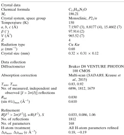
Table 5
Experimental details.
Crystal data
Chemical formula C11H10N2O
Mr 186.21
Crystal system, space group Monoclinic,P21/n
Temperature (K) 150
a,b,c(A˚ ) 7.1507 (3), 8.8177 (4), 15.4602 (7)
() 97.914 (2)
V(A˚3) 965.52 (7)
Z 4
Radiation type CuK
(mm1) 0.68
Crystal size (mm) 0.320.310.12
Data collection
Diffractometer Bruker D8 VENTURE PHOTON
100 CMOS
Absorption correction Multi-scan (SADABS; Krauseet al., 2015)
Tmin,Tmax 0.83, 0.92
No. of measured, independent and observed [I> 2(I)] reflections
6896, 1812, 1679
Rint 0.030
(sin /)max(A˚ 1
) 0.610
Refinement
R[F2> 2(F2)],wR(F2),S 0.033, 0.086, 1.06
No. of reflections 1812
No. of parameters 168
H-atom treatment All H-atom parameters refined
max,min(e A˚ 3
) 0.18,0.19
Saber, A., Sebbar, N. K., Ho¨kelek, T., El hafi, M., Mague, J. T. & Essassi, E. M. (2018b).Acta Cryst.E74, 1842–1846.
Saber, A., Sebbar, N. K., Ho¨kelek, T., Hni, B., Mague, J. T. & Essassi, E. M. (2018a).Acta Cryst.E74, 1746–1750.
Saber, A., Sebbar, N. K., Sert, Y., Alzaqri, N., Ho¨kelek, T., El Ghayati, L., Talbaoui, A., Mague, J. T., Filali Baba, Y., Urrutigoıˆty, M. & Essassi, E. M. (2020).J. Mol. Struct.1200, 127174.
Sheldrick, G. M. (2008).Acta Cryst.A64, 112–122. Sheldrick, G. M. (2015a).Acta Cryst.A71, 3–8. Sheldrick, G. M. (2015b).Acta Cryst.C71, 3–8.
Soderlind, K. J., Gorodetsky, B., Singh, A. K., Bachur, N., Miller, G. G. & Lown, J. W. (1999).Anticancer Drug. Des.14, 19–36.
Spackman, M. A. & Jayatilaka, D. (2009).CrystEngComm,11, 19–32.
Spackman, M. A., McKinnon, J. J. & Jayatilaka, D. (2008). CrystEngComm,10, 377–388.
Turner, M. J., Grabowsky, S., Jayatilaka, D. & Spackman, M. A. (2014).J. Phys. Chem. Lett.5, 4249–4255.
Turner, M. J., McKinnon, J. J., Wolff, S. K., Grimwood, D. J., Spackman, P. R., Jayatilaka, D. & Spackman, M. A. (2017). CrystalExplorer17. The University of Western Australia.
Turner, M. J., Thomas, S. P., Shi, M. W., Jayatilaka, D. & Spackman, M. A. (2015).Chem. Commun.51, 3735–3738.
Venkatesan, P., Thamotharan, S., Ilangovan, A., Liang, H. & Sundius, T. (2016).Spectrochim. Acta Part A,153, 625–636.
sup-1
Acta Cryst. (2019). E75, 1940-1946
supporting information
Acta Cryst. (2019). E75, 1940-1946 [https://doi.org/10.1107/S2056989019015779]
Crystal structure, Hirshfeld surface analysis and interaction energy and DFT
studies of 1-methyl-3-(prop-2-yn-1-yl)-2,3-dihydro-1
H
-1,3-benzodiazol-2-one
Asmaa Saber, Mohamed Srhir, Tuncer H
ö
kelek, Joel T. Mague, Noureddine Hamou Ahabchane,
Nada Kheira Sebbar and El Mokhtar Essassi
Computing details
Data collection: APEX3 (Bruker, 2016); cell refinement: SAINT (Bruker, 2016); data reduction: SAINT (Bruker, 2016);
program(s) used to solve structure: SHELXT (Sheldrick, 2015a); program(s) used to refine structure: SHELXL2018
(Sheldrick, 2015b); molecular graphics: DIAMOND (Brandenburg & Putz, 2012); software used to prepare material for
publication: SHELXTL (Sheldrick, 2008).
1-Methyl-3-(prop-2-yn-1-yl)-2,3-dihydro-1H-1,3-benzodiazol-2-one
Crystal data
C11H10N2O
Mr = 186.21
Monoclinic, P21/n
a = 7.1507 (3) Å
b = 8.8177 (4) Å
c = 15.4602 (7) Å
β = 97.914 (2)°
V = 965.52 (7) Å3
Z = 4
F(000) = 392
Dx = 1.281 Mg m−3
Cu Kα radiation, λ = 1.54178 Å Cell parameters from 5848 reflections
θ = 5.8–70.1°
µ = 0.68 mm−1
T = 150 K Plate, colourless 0.32 × 0.31 × 0.12 mm
Data collection
Bruker D8 VENTURE PHOTON 100 CMOS diffractometer
Radiation source: INCOATEC IµS micro-focus source
Mirror monochromator
Detector resolution: 10.4167 pixels mm-1
ω scans
Absorption correction: multi-scan (SADABS; Krause et al., 2015)
Tmin = 0.83, Tmax = 0.92
6896 measured reflections 1812 independent reflections 1679 reflections with I > 2σ(I)
Rint = 0.030
θmax = 70.1°, θmin = 5.8°
h = −8→8
k = −10→9
l = −18→18
Refinement
Refinement on F2
Least-squares matrix: full
R[F2 > 2σ(F2)] = 0.033
wR(F2) = 0.086
S = 1.06 1812 reflections 168 parameters 0 restraints
Primary atom site location: dual
Secondary atom site location: difference Fourier map
Hydrogen site location: difference Fourier map All H-atom parameters refined
w = 1/[σ2(F
o2) + (0.0402P)2 + 0.2239P]
where P = (Fo2 + 2Fc2)/3
sup-2
Acta Cryst. (2019). E75, 1940-1946
Δρmax = 0.18 e Å−3
Δρmin = −0.19 e Å−3
Extinction correction: SHELXL2018 (Sheldrick, 2015b), Fc*=kFc[1+0.001xFc2λ3/sin(2θ)]-1/4
Extinction coefficient: 0.0100 (12)
Special details
Geometry. All esds (except the esd in the dihedral angle between two l.s. planes) are estimated using the full covariance matrix. The cell esds are taken into account individually in the estimation of esds in distances, angles and torsion angles; correlations between esds in cell parameters are only used when they are defined by crystal symmetry. An approximate (isotropic) treatment of cell esds is used for estimating esds involving l.s. planes.
Refinement. Refinement of F2 against ALL reflections. The weighted R-factor wR and goodness of fit S are based on F2,
conventional R-factors R are based on F, with F set to zero for negative F2. The threshold expression of F2 > 2sigma(F2) is
used only for calculating R-factors(gt) etc. and is not relevant to the choice of reflections for refinement. R-factors based on F2 are statistically about twice as large as those based on F, and R- factors based on ALL data will be even larger.
Fractional atomic coordinates and isotropic or equivalent isotropic displacement parameters (Å2)
x y z Uiso*/Ueq
O1 0.31019 (11) 0.82725 (9) 0.64517 (6) 0.0345 (2) N1 0.24854 (12) 0.64929 (11) 0.53316 (6) 0.0280 (2) N2 0.36075 (12) 0.56773 (10) 0.66470 (6) 0.0250 (2) C1 0.33940 (14) 0.44082 (11) 0.61080 (7) 0.0235 (2) C2 0.37638 (15) 0.28918 (12) 0.62754 (8) 0.0289 (3) H2 0.426 (2) 0.2543 (16) 0.6872 (10) 0.039 (4)* C3 0.34025 (16) 0.18941 (14) 0.55731 (8) 0.0353 (3) H3 0.364 (2) 0.0783 (17) 0.5684 (10) 0.043 (4)* C4 0.27117 (17) 0.24106 (15) 0.47421 (8) 0.0378 (3) H4 0.246 (2) 0.1678 (17) 0.4255 (10) 0.046 (4)* C5 0.23359 (16) 0.39392 (15) 0.45751 (7) 0.0339 (3) H5 0.190 (2) 0.4305 (16) 0.3992 (10) 0.042 (4)* C6 0.26803 (14) 0.49324 (12) 0.52720 (7) 0.0255 (3) C7 0.30715 (14) 0.69684 (12) 0.61712 (7) 0.0260 (2) C8 0.17860 (17) 0.75002 (16) 0.46162 (8) 0.0381 (3) H8A 0.255 (3) 0.747 (2) 0.4146 (13) 0.076 (6)* H8B 0.176 (3) 0.854 (2) 0.4867 (13) 0.072 (5)* H8C 0.044 (2) 0.7264 (17) 0.4370 (10) 0.047 (4)* C9 0.44506 (16) 0.56993 (13) 0.75585 (7) 0.0283 (3) H9A 0.4344 (19) 0.6753 (16) 0.7764 (9) 0.033 (3)* H9B 0.376 (2) 0.5012 (16) 0.7898 (9) 0.038 (3)* C10 0.64427 (15) 0.52362 (12) 0.76752 (7) 0.0281 (3) C11 0.80385 (17) 0.48197 (14) 0.77883 (8) 0.0342 (3) H11 0.938 (2) 0.4443 (17) 0.7926 (10) 0.050 (4)*
Atomic displacement parameters (Å2)
U11 U22 U33 U12 U13 U23
sup-3
Acta Cryst. (2019). E75, 1940-1946
C2 0.0249 (5) 0.0281 (6) 0.0337 (6) −0.0008 (4) 0.0041 (4) 0.0011 (4) C3 0.0298 (6) 0.0300 (6) 0.0469 (7) −0.0021 (4) 0.0088 (5) −0.0072 (5) C4 0.0339 (6) 0.0427 (7) 0.0384 (6) −0.0083 (5) 0.0107 (5) −0.0158 (5) C5 0.0286 (6) 0.0486 (7) 0.0248 (6) −0.0089 (5) 0.0048 (4) −0.0032 (5) C6 0.0207 (5) 0.0307 (6) 0.0254 (5) −0.0046 (4) 0.0037 (4) 0.0014 (4) C7 0.0189 (5) 0.0261 (5) 0.0328 (6) −0.0007 (4) 0.0025 (4) 0.0025 (4) C8 0.0294 (6) 0.0450 (7) 0.0380 (7) −0.0013 (5) −0.0019 (5) 0.0189 (6) C9 0.0296 (6) 0.0332 (6) 0.0218 (5) 0.0004 (4) 0.0019 (4) −0.0014 (4) C10 0.0333 (6) 0.0293 (5) 0.0206 (5) −0.0018 (4) −0.0004 (4) 0.0014 (4) C11 0.0330 (6) 0.0381 (6) 0.0298 (6) 0.0022 (5) −0.0016 (4) 0.0030 (5)
Geometric parameters (Å, º)
O1—C7 1.2281 (13) C4—C5 1.3915 (19)
N1—C7 1.3735 (14) C4—H4 0.989 (16)
N1—C6 1.3874 (15) C5—C6 1.3839 (16)
N1—C8 1.4526 (14) C5—H5 0.967 (15)
N2—C7 1.3807 (14) C8—H8A 0.97 (2)
N2—C1 1.3910 (13) C8—H8B 0.99 (2)
N2—C9 1.4545 (14) C8—H8C 1.004 (16)
C1—C2 1.3805 (15) C9—C10 1.4689 (16)
C1—C6 1.4011 (14) C9—H9A 0.988 (14)
C2—C3 1.3937 (17) C9—H9B 0.978 (15)
C2—H2 0.991 (15) C10—C11 1.1885 (17)
C3—C4 1.3883 (19) C11—H11 1.009 (16)
C3—H3 1.005 (15)
O1···H9A 2.491 (14) C11···O1vii 3.1569 (15)
O1···H3i 2.566 (15) C2···H8Aiv 2.82 (2)
O1···H8B 2.516 (19) C3···H8Cv 2.859 (15)
O1···H9Bii 2.346 (14) C3···H8Aiv 2.92 (2)
O1···H11iii 2.181 (15) C4···H8Cv 2.810 (15)
C2···C10 3.3889 (16) C5···H8Cv 2.935 (15)
C3···C8iv 3.5335 (17) C8···H5 2.983 (14)
C4···C8v 3.4947 (17) C9···H2 2.975 (14)
C4···C7iv 3.5437 (16) C10···H4viii 2.976 (15)
C5···C8v 3.5884 (17) C11···H5iv 2.865 (15)
C6···C6iv 3.5349 (14) C11···H4viii 2.705 (15)
C9···O1vi 3.3198 (14)
C7—N1—C6 110.19 (9) C5—C6—N1 132.12 (10)
C7—N1—C8 124.14 (10) C5—C6—C1 120.83 (11)
C6—N1—C8 125.66 (10) N1—C6—C1 107.04 (9)
C7—N2—C1 110.16 (9) O1—C7—N1 127.43 (10)
C7—N2—C9 123.55 (9) O1—C7—N2 126.43 (10)
C1—N2—C9 126.00 (9) N1—C7—N2 106.14 (9)
C2—C1—N2 131.64 (10) N1—C8—H8A 112.7 (12)
sup-4
Acta Cryst. (2019). E75, 1940-1946
N2—C1—C6 106.45 (9) H8A—C8—H8B 111.1 (16)
C1—C2—C3 117.07 (11) N1—C8—H8C 111.9 (9)
C1—C2—H2 120.7 (8) H8A—C8—H8C 108.5 (15)
C3—C2—H2 122.3 (8) H8B—C8—H8C 105.7 (13)
C4—C3—C2 121.20 (11) N2—C9—C10 112.38 (9)
C4—C3—H3 120.5 (9) N2—C9—H9A 106.5 (8)
C2—C3—H3 118.3 (9) C10—C9—H9A 109.8 (8)
C3—C4—C5 121.63 (11) N2—C9—H9B 109.9 (8)
C3—C4—H4 119.6 (9) C10—C9—H9B 108.2 (8)
C5—C4—H4 118.8 (9) H9A—C9—H9B 110.1 (11)
C6—C5—C4 117.35 (11) C11—C10—C9 177.63 (12)
C6—C5—H5 120.9 (8) C10—C11—H11 176.1 (9)
C4—C5—H5 121.7 (8)
C7—N2—C1—C2 178.91 (11) C2—C1—C6—C5 −0.69 (15)
C9—N2—C1—C2 4.89 (17) N2—C1—C6—C5 178.96 (9)
C7—N2—C1—C6 −0.69 (11) C2—C1—C6—N1 −179.67 (9)
C9—N2—C1—C6 −174.72 (9) N2—C1—C6—N1 −0.02 (11)
N2—C1—C2—C3 −179.33 (10) C6—N1—C7—O1 179.51 (10)
C6—C1—C2—C3 0.23 (15) C8—N1—C7—O1 0.13 (17)
C1—C2—C3—C4 0.37 (16) C6—N1—C7—N2 −1.13 (11)
C2—C3—C4—C5 −0.54 (18) C8—N1—C7—N2 179.49 (9)
C3—C4—C5—C6 0.08 (17) C1—N2—C7—O1 −179.51 (10)
C4—C5—C6—N1 179.20 (11) C9—N2—C7—O1 −5.31 (16)
C4—C5—C6—C1 0.52 (15) C1—N2—C7—N1 1.12 (11)
C7—N1—C6—C5 −178.10 (11) C9—N2—C7—N1 175.32 (9)
C8—N1—C6—C5 1.27 (18) C7—N2—C9—C10 −111.11 (11)
C7—N1—C6—C1 0.72 (11) C1—N2—C9—C10 62.16 (13)
C8—N1—C6—C1 −179.91 (9)
Symmetry codes: (i) x, y+1, z; (ii) −x+1/2, y+1/2, −z+3/2; (iii) −x+3/2, y+1/2, −z+3/2; (iv) −x+1, −y+1, −z+1; (v) −x, −y+1, −z+1; (vi) −x+1/2, y−1/2, −z+3/2; (vii) −x+3/2, y−1/2, −z+3/2; (viii) x+1/2, −y+1/2, z+1/2.
Hydrogen-bond geometry (Å, º)
Cg1 is the centroid of the C1–C6 benzene ring.
D—H···A D—H H···A D···A D—H···A
C3—H3···O1ix 1.005 (15) 2.566 (15) 3.4885 (15) 152.6 (11)
C8—H8C···Cg1v 1.004 (16) 2.626 (15) 3.5413 (13) 151.1 (12)
C9—H9B···O1vi 0.978 (15) 2.347 (15) 3.3198 (14) 172.9 (12)
C11—H11···O1vii 1.010 (15) 2.181 (15) 3.1569 (15) 162.1 (12)
![Crystal structure, Hirshfeld surface analysis and DFT studies of 6 [(E) 2 (thiophen 2 yl)ethenyl] 4,5 dihydropyridazin 3(2H) one](data:image/gif;base64,R0lGODlhAQABAIAAAP///wAAACH5BAEAAAAALAAAAAABAAEAAAICRAEAOw==)