Received 17 September 2016 Accepted 6 October 2016
Edited by H. Stoeckli-Evans, University of Neuchaˆtel, Switzerland
Keywords:crystal structure; isatin; C—H O hydrogen bonds.
CCDC reference:1508554
Supporting information:this article has supporting information at journals.iucr.org/e
Crystal and geometry-optimized structure, and
Hirshfeld surface analysis of
1-(2-bromoethyl)-indoline-2,3-dione
N. Sharmila,aT. V. Sundar,a* G. Sathishband P. Venkatesanc
a
Postgraduate and Research Department of Physics, National College (Autonomous), Tiruchirappalli 620 001, Tamilnadu, India,bSchool of Chemistry, Bharathidasan University, Tiruchirappalli 620 024, Tamilnadu, India, andcLaboratorio de Polı´imeros, Centro de Quı´mica Instituto de Ciencias, Beneme´rita Universidad Auto´noma de Puebla (BUAP), Complejo de Ciencias, ICUAP, Edif. 103H, 22 Sur y San Claudio, C.P. 72570 Puebla, Puebla, Mexico. *Correspondence e-mail: [email protected]
In the title compound, C10H8BrNO2, the isatin (1H-indole-2,3-dione) moiety is nearly planar (r.m.s. deviation = 0.026 A˚ ). In the crystal, molecules are linked by C—H O hydrogen bonds, forming layers parallel to the ab plane, and enclosingR44(24) loops. There are a low percentage (19.3%) of intermolecular H H contacts in the structure, as estimated by the analysis of Hirshfeld surfaces. This could be due to the presence of the Br atom, present in the bromoethylene group, which makesca18.7% Br H contacts.
1. Chemical context
Isatin (1H-indole-2,3-dione) is an endogenous compound that has been identified in humans and possesses a wide range of biological activities, such as anxiogenic and sedative activities. It serves as a synthetically useful substrate which can be used to prepare a broad range of heterocyclic compounds, including molecules of pharmacological significance (Bekircan & Bektas, 2008). A variety of biological activities are associated with isatin, including central nervous system (CNS) activities (Raj, 2012). As part of our interest in the identification of bioactive compounds, we report herein on the synthesis, the crystal structure, and the geometry optimization and Hirshfeld surface analysis of the title isatin derivative, (I).
2. Structural commentary
The molecular structure of the title isatin derivative, (I), is illustrated in Fig. 1. It crystallized in the orthorhombic space group P212121 with an absolute structure parameter of 0.015 (8). The bond lengths and angles of the isatin moiety are comparable with those reported for similar N-substituted isatin derivatives (Qachchachiet al., 2016a,b).
In compound (I), the isatin ring system is almost planar, with an r.m.s. deviation of the fitted atoms C1–C8/N1/O1/O2 of 0.026 A˚ . The sum of the bond angles around atom N1 isca 360, indicating little evidence for the presence of ansp3
lone pair.
3. Supramolecular features
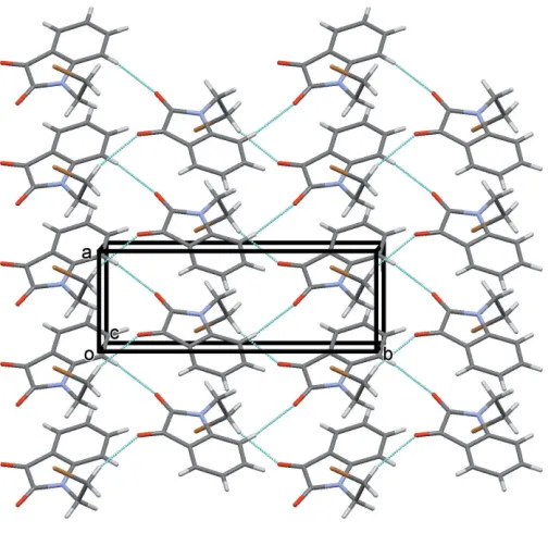
In the crystal of (I), molecules are linked by C—H O hydrogen bonds, viz C2—H2 O1 and C10—H10A O2 (Table 1), which individually form C(6) and C(7) chains, respectively. Together they form layers parallel to theabplane and encloseR44(24) loops (Table 1 and Fig. 2). An analysis of the crystal packing of (I) indicated that no further significant intermolecular interactions were present (PLATON; Spek, 2009).
4. Database survey
A search of the Cambridge Structural Database (Version 5.37, update May 2016; Groomet al., 2016) forN-substituted isatin derivatives yielded 58 hits. These include five reports of the structure of isatin itself and four reports of the structure of N-methylisatin. 13 of the structures involve an alkyl chain of two or more C atoms. The compound most similar to the title compound is 1-(3-bromopropyl)-1H-indole-2,3-dione (AKO-BIN), whose structure was published very recently
(Qach-chachiet al., 2016a). A view of the structural overlap of this compound with that of compound (I) is shown in Fig. 3.
5. Geometry optimization
[image:2.610.315.567.69.314.2]The geometry optimization of compound (I) was performed using the density functional theory (DFT) method with a 6-311++G** basis set. The crystal structure in the solid state was used as the starting structure for the calculations. The DFT calculations are performed with the GAUSSIAN09 program package (Frischet al., 2013). The resulting geome-trical parameters are compared with those obtained from an X-ray crystallography study. A superimposed analysis of (I) with its optimized structure gives an r.m.s. deviation of 0.068 A˚
Figure 1
[image:2.610.43.295.93.133.2]The molecular structure of compound (I), showing the atom labelling. Displacement ellipsoids are drawn at the 30% probability level.
Table 1
Hydrogen-bond geometry (A˚ ,).
D—H A D—H H A D A D—H A
C2—H2 O1i 0.93 2.41 3.286 (6) 156
C10—H10A O2ii 0.97 2.42 3.309 (6) 151
Symmetry codes: (i)xþ1;yþ1 2;zþ
1
2; (ii)xþ2;yþ 1 2;zþ
1 2.
Figure 2
A view along thec axis of the crystal packing of compound (I). The hydrogen bonds are shown as dashed lines (see Table 1) and, for clarity, only H atoms H2 and H10Ahave been included.
Figure 3
[image:2.610.322.551.560.709.2](Fig. 4). This indicates a twist leading to further separation between the isatin moiety and the benzene ring. Also, this suggests that the crystal packing could be influenced by the collective effect of the intermolecular interactions. To probe further, structure-based theoretical parameters, viz. HOMO and LUMO energy levels, total energy and dipole moment, were calculated and found to be 6.860 eV, 3.091 eV,
86134.81 eV and 7.2176 Debye, respectively. As a further structure-based test, semi-empirical molecular orbital calcu-lations are carried out using the PM7 method inMOPAC2012 (Stewart, 2012; Maiaet al., 2012). The PM7 method gave the HOMO and LUMO energy levels, total energy and dipole moment as 9.276 eV, 1.271 eV, 2334.96 eV and 5.8952 Debye, respectively. Also, the superimposed analysis of the X-ray structure with the isolated molecule in the gas phase by the PM7 method gave an r.m.s. deviation of 0.211 A˚ . Further, the N1—C8 and N1—C1 (X-ray: 1.367 A˚ ; DFT: 1.392 A˚; PM7: 1.424 A˚ ) bond lengths increased, while the bond angles O2— C7—C6 (X-ray: 131.3; DFT: 130.8; PM7: 131.2) and O1— C8—N1 (X-ray: 127.4; DFT: 126.8; PM7: 123.8) decreased. These confirm the influence of the packing interactions in the solid state of the molecule. The relative conformation about the bond joining the isatin and bromoethylene moieties of (I) is defined by the N1—C9—C10—Br1 torsion angle of
62.0 (5). This indicates that the conformation of the molecule is (+)-synclinal.
6. Hirshfeld surface analysis
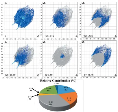
[image:3.610.97.244.73.241.2]A detailed Hirshfeld surface analysis is useful for identifing the various intermolecular interactions and intermolecular contacts present in crystal structures, with the aid of decom-posed two-dimensional fingerprint plots. The Hirshfeld surface (HS) and the two-dimensional fingerprint plots were generated based on the di and de distances using Crystal Explorer(Wolffet al., 2012);diis the distance from the nearest atom inside the surface, whiledeis the distance from the HS to the nearest atom outside the surface. This analysis identified the various intermolecular contacts (O—H, H—H, C—H, C—C and H—Br) and their relative contributions in the crystal structure. The bond lengths (C—H = 1.083 A˚ , N—H = 1.009 A˚ and O—H = 0.983 A˚) were adjusted to typical neutron diffraction values before the HS calculation (Venkatesanet al., 2015, 2016a,b). In Hirshfeld surface diagrams, the contacts with distances shorter than the sum of the van der Waals radii are indicated as red and the contacts with distances longer than the van der Waals radii are represented as blue, whereas the contacts with distances equal to the sum of the van der Waals radii are indicated as white. The HS area of compound Figure 5
Views of the Hirshfeld surfaces mapped with dnorm in two different
[image:3.610.313.561.96.395.2]orientations for compound (I). The represented interactions are labelled (see Table 1).
Figure 4
[image:3.610.47.294.597.705.2]Superimposed fit of the molecule of compound (I) in the crystalline state (red) and after energy minimization (blue).
Table 2
Experimental details.
Crystal data
Chemical formula C10H8BrNO2
Mr 254.08
Crystal system, space group Orthorhombic,P212121
Temperature (K) 293
a,b,c(A˚ ) 4.6834 (2), 12.9567 (7), 16.1130 (8)
V(A˚3) 977.76 (8)
Z 4
Radiation type MoK
(mm1) 4.18
Crystal size (mm) 0.250.200.20
Data collection
Diffractometer Bruker Kappa APEXII CCD
Absorption correction Multi-scan (SADABS; Bruker, 2004)
Tmin,Tmax 0.419, 0.498
No. of measured, independent and observed [I> 2(I)] reflections
8226, 3150, 1663
Rint 0.037
(sin/)max(A˚ 1
) 0.762
Refinement
R[F2> 2(F2)],wR(F2),S 0.055, 0.084, 1.02
No. of reflections 3150
No. of parameters 127
H-atom treatment H-atom parameters constrained
max,min(e A˚ 3
) 0.70,0.59
Absolute structure Flackxdetermined using 503 quotients
[(I+)(I
)]/[(I+)+(I )] (Parsonset al., 2013) Absolute structure parameter 0.015 (8)
(I) is shown in Fig. 5, and the respective points of inter-molecular interactions are labelled.
Two-dimensional fingerprint plots are used to quantify and visualize the intermolecular interactions present in the crystal structure and the same for the title compound is shown in Fig. 6. The result suggests that the share of intermolecular H H contacts in (I) is about 19.3%. The low percentage could be attributed to the presence of the Br atom in the bromoethylene group, which makesca18.7% contacts with H atoms (Br H). The next significant intermolecular contacts observed in the structure,i.e.O H, C H and C C, have relative contributions of 30.6, 18.8 and 3.1%, respectively.
7. Synthesis and crystallization
To a solution of 1-{2-[(2-bromoethyl)amino]phenyl}ethanone (1 equivalent) in DMSO were added I2(0.1 equivalents) and TBHP (1 equivalent, 70% in H2O) at ambient temperature, and the mixture was heated to 353 K. The progress of the reaction was monitored by thin-layer chromatography. Upon completion, the reaction mixture was allowed to cool to ambient temperature and was quenched with aqueous sodium
thiosulfate and ethyl acetate. The organic phase was sepa-rated, dried over Na2SO4, filtered and concentrated. The crude product was purified by silica-gel column chromato-graphy using hexane–ethyl acetate (9:1v/v) as eluent. The title compound was obtained as a red solid (yield: 71%, 74.5 mg; m.p. 404–406 K). It was dissolved in a mixture of hexane–ethyl acetate (9:1 v/v) and left to slowly evaporate at room temperature, yielding brown block-like crystals after a period of 3 d.
8. Refinement
Crystal data, data collection and structure refinement details are summarized in Table 2. C-bound H atoms were included in calculated positions and treated as riding, with C—H = 0.93– 0.97 A˚ andUiso(H) = 1.2Ueq(C).
Acknowledgements
[image:4.610.101.509.71.457.2]NS thanks the Sophisticated Analytical Instrumentation Facility (SAIF), Indian Institute of Technology Madras, India, for help with the data collection and Professor A. Ilangovan, Figure 6
School of Chemistry, Bharathidasan University, India, for fruitful discussions.
References
Bekircan, O. & Bektas, H. (2008).Molecules,13, 2126–2135. Bruker (2004).APEX2,SAINT,XPREPandSADABS. Bruker AXS
Inc., Madison, Wisconsin, USA.
Farrugia, L. J. (2012).J. Appl. Cryst.45, 849–854.
Frisch, M. J., Trucks, G. W., Schlegel, H. B., Scuseria, G. E., Robb, M. A., Cheeseman, J. R.,et al.(2013).GAUSSIAN09. Gaussian, Inc., Wallingford, CT, USA.
Gans, J. & Shalloway, D. (2001).J. Mol. Graph. Model.19, 557–559. Groom, C. R., Bruno, I. J., Lightfoot, M. P. & Ward, S. C. (2016).Acta
Cryst.B72, 171–179.
Macrae, C. F., Bruno, I. J., Chisholm, J. A., Edgington, P. R., McCabe, P., Pidcock, E., Rodriguez-Monge, L., Taylor, R., van de Streek, J. & Wood, P. A. (2008).J. Appl. Cryst.41, 466–470.
Maia, J. D. C., Carvalho, G. A. U., Mangueira, C. P. Jr, Santana, S. R., Cabral, L. A. F. & Rocha, G. B. (2012).J. Chem. Theory Comput.8, 3072–3081.
Parsons, S., Flack, H. D. & Wagner, T. (2013).Acta Cryst.B69, 249– 259.
Qachchachi, F. Z., Kandri Rodi, Y., Haoudi, H., Essassi, E. M., Capet, F. & Zouihri, H. (2016a).IUCrData,1, x160593.
Qachchachi, F. Z., Kandri Rodi, Y., Haoudi, H., Essassi, E. M., Capet, F. & Zouihri, H. (2016b).IUCrData,1, x160609.
Raj, V. (2012).Int. J. Curr. Pharm. Res.4, 1–9. Sheldrick, G. M. (2008).Acta Cryst.A64, 112–122. Sheldrick, G. M. (2015).Acta Cryst.C71, 3–8. Spek, A. L. (2009).Acta Cryst.D65, 148–155.
Stewart, J. J. P. (2012).MOPAC2012. http://OpenMOPAC.net. Venkatesan, P., Rajakannan, V., Venkataramanan, N. S., Ilangovan,
A., Sundius, T. & Thamotharan, S. (2016a).J. Mol. Struct. 1119, 259–268.
Venkatesan, P., Thamotharan, S., Ilangovan, A., Liang, H. & Sundius, T. (2016b).Spectrochim. Acta A Mol. Biomol. Spectrosc.153, 625– 636.
Venkatesan, P., Thamotharan, S., Kumara, R. G. & Ilangovan, A. (2015).CrystEngComm,17, 904–915.
sup-1 Acta Cryst. (2016). E72, 1569-1573
supporting information
Acta Cryst. (2016). E72, 1569-1573 [https://doi.org/10.1107/S2056989016015760]
Crystal and geometry-optimized structure, and Hirshfeld surface analysis of
1-(2-bromoethyl)indoline-2,3-dione
N. Sharmila, T. V. Sundar, G. Sathish and P. Venkatesan
Computing details
Data collection: APEX2 (Bruker, 2004); cell refinement: APEX2 (Bruker, 2004) and SAINT (Bruker, 2004); data
reduction: SAINT (Bruker, 2004) and XPREP (Bruker, 2004); program(s) used to solve structure: SHELXS97 (Sheldrick,
2008); program(s) used to refine structure: SHELXL2014 (Sheldrick, 2015); molecular graphics: QMOL (Gans &
Shalloway, 2001) and Mercury (Macrae et al., 2008).; software used to prepare material for publication: WinGX
(Farrugia, 2012) and PLATON (Spek, 2009).
1-(2-Bromoethyl)indoline-2,3-dione
Crystal data
C10H8BrNO2 Mr = 254.08
Orthorhombic, P212121 a = 4.6834 (2) Å
b = 12.9567 (7) Å
c = 16.1130 (8) Å
V = 977.76 (8) Å3 Z = 4
F(000) = 504
Dx = 1.726 Mg m−3 Melting point: 406 K
Mo Kα radiation, λ = 0.71073 Å Cell parameters from 2844 reflections
θ = 2.5–26.7°
µ = 4.18 mm−1 T = 293 K Block, brown
0.25 × 0.20 × 0.20 mm
Data collection
Bruker Kappa APEXII CCD diffractometer
Radiation source: fine-focus sealed tube
ω and φ scan
Absorption correction: multi-scan (SADABS; Bruker, 2004)
Tmin = 0.419, Tmax = 0.498 8226 measured reflections
3150 independent reflections 1663 reflections with I > 2σ(I)
Rint = 0.037
θmax = 32.8°, θmin = 2.0°
h = −7→6
k = −19→17
l = −22→18
Refinement
Refinement on F2 Least-squares matrix: full
R[F2 > 2σ(F2)] = 0.055 wR(F2) = 0.084 S = 1.02 3150 reflections 127 parameters 0 restraints
Primary atom site location: structure-invariant direct methods
Secondary atom site location: difference Fourier map
Hydrogen site location: inferred from neighbouring sites
H-atom parameters constrained
w = 1/[σ2(F
sup-2 Acta Cryst. (2016). E72, 1569-1573
(Δ/σ)max < 0.001 Δρmax = 0.70 e Å−3 Δρmin = −0.59 e Å−3
Absolute structure: Flack x determined using 503 quotients [(I+)-(I-)]/[(I+)+(I-)] (Parsons et al., 2013)
Absolute structure parameter: 0.015 (8)
Special details
Geometry. All esds (except the esd in the dihedral angle between two l.s. planes) are estimated using the full covariance
matrix. The cell esds are taken into account individually in the estimation of esds in distances, angles and torsion angles; correlations between esds in cell parameters are only used when they are defined by crystal symmetry. An approximate (isotropic) treatment of cell esds is used for estimating esds involving l.s. planes.
Fractional atomic coordinates and isotropic or equivalent isotropic displacement parameters (Å2)
x y z Uiso*/Ueq
Br1 0.83217 (12) 0.83821 (4) 0.06227 (4) 0.0609 (2)
O1 0.4527 (9) 0.7005 (3) 0.2349 (3) 0.0719 (13)
O2 0.8447 (9) 0.6409 (2) 0.3684 (2) 0.0688 (10)
N1 0.6516 (8) 0.8607 (2) 0.2584 (2) 0.0378 (8)
C1 0.8490 (10) 0.9005 (3) 0.3156 (3) 0.0326 (9)
C2 0.9288 (9) 1.0020 (3) 0.3268 (3) 0.0429 (12)
H2 0.8520 1.0548 0.2947 0.051*
C3 1.1289 (12) 1.0218 (4) 0.3883 (3) 0.0538 (14)
H3 1.1896 1.0894 0.3966 0.065*
C4 1.2414 (10) 0.9446 (4) 0.4377 (4) 0.0572 (14)
H4 1.3755 0.9606 0.4783 0.069*
C5 1.1551 (10) 0.8445 (4) 0.4267 (3) 0.0503 (11)
H5 1.2276 0.7920 0.4599 0.060*
C6 0.9592 (9) 0.8233 (4) 0.3656 (3) 0.0362 (11)
C7 0.8222 (12) 0.7276 (3) 0.3414 (3) 0.0447 (12)
C8 0.6167 (11) 0.7569 (4) 0.2703 (3) 0.0458 (13)
C9 0.4985 (10) 0.9198 (4) 0.1964 (3) 0.0464 (12)
H9A 0.4179 0.9805 0.2227 0.056*
H9B 0.3412 0.8784 0.1757 0.056*
C10 0.6765 (12) 0.9533 (3) 0.1248 (3) 0.0499 (12)
H10A 0.8325 0.9956 0.1451 0.060*
H10B 0.5612 0.9955 0.0881 0.060*
Atomic displacement parameters (Å2)
U11 U22 U33 U12 U13 U23
Br1 0.0714 (3) 0.0570 (3) 0.0543 (3) −0.0008 (3) 0.0059 (3) −0.0069 (3)
O1 0.090 (3) 0.055 (2) 0.071 (3) −0.036 (2) −0.003 (2) −0.012 (2)
O2 0.102 (3) 0.0323 (18) 0.072 (3) 0.005 (2) 0.013 (3) 0.0138 (17)
N1 0.042 (2) 0.035 (2) 0.036 (2) −0.0055 (19) −0.001 (2) 0.0022 (16)
C1 0.038 (2) 0.032 (2) 0.028 (2) −0.001 (2) 0.007 (2) −0.0028 (18)
C2 0.057 (3) 0.029 (2) 0.043 (3) −0.001 (2) 0.006 (2) 0.000 (2)
C3 0.069 (4) 0.045 (3) 0.047 (3) −0.016 (3) 0.011 (3) −0.015 (3)
C4 0.060 (3) 0.078 (4) 0.034 (3) −0.011 (2) 0.003 (3) −0.007 (3)
sup-3 Acta Cryst. (2016). E72, 1569-1573
C6 0.045 (2) 0.038 (3) 0.026 (3) 0.002 (2) 0.006 (2) −0.001 (2)
C7 0.060 (3) 0.032 (2) 0.043 (3) 0.003 (3) 0.017 (3) 0.002 (2)
C8 0.058 (3) 0.038 (3) 0.041 (3) −0.011 (3) 0.010 (3) −0.005 (2)
C9 0.041 (3) 0.053 (3) 0.045 (3) 0.004 (2) −0.004 (3) 0.000 (3)
C10 0.061 (3) 0.040 (2) 0.048 (3) 0.005 (3) −0.005 (3) 0.005 (2)
Geometric parameters (Å, º)
Br1—C10 1.942 (5) C4—C5 1.370 (7)
O1—C8 1.204 (6) C4—H4 0.9300
O2—C7 1.210 (5) C5—C6 1.373 (6)
N1—C8 1.367 (5) C5—H5 0.9300
N1—C1 1.404 (6) C6—C7 1.449 (6)
N1—C9 1.449 (6) C7—C8 1.544 (7)
C1—C2 1.380 (6) C9—C10 1.488 (7)
C1—C6 1.384 (6) C9—H9A 0.9700
C2—C3 1.388 (7) C9—H9B 0.9700
C2—H2 0.9300 C10—H10A 0.9700
C3—C4 1.382 (7) C10—H10B 0.9700
C3—H3 0.9300
C8—N1—C1 110.4 (4) C1—C6—C7 107.2 (4)
C8—N1—C9 123.9 (4) O2—C7—C6 131.3 (5)
C1—N1—C9 125.7 (4) O2—C7—C8 123.3 (5)
C2—C1—C6 120.7 (4) C6—C7—C8 105.4 (4)
C2—C1—N1 128.0 (4) O1—C8—N1 127.4 (5)
C6—C1—N1 111.2 (4) O1—C8—C7 126.9 (5)
C1—C2—C3 116.9 (4) N1—C8—C7 105.7 (4)
C1—C2—H2 121.5 N1—C9—C10 114.3 (4)
C3—C2—H2 121.5 N1—C9—H9A 108.7
C4—C3—C2 122.3 (5) C10—C9—H9A 108.7
C4—C3—H3 118.8 N1—C9—H9B 108.7
C2—C3—H3 118.8 C10—C9—H9B 108.7
C5—C4—C3 119.9 (5) H9A—C9—H9B 107.6
C5—C4—H4 120.1 C9—C10—Br1 112.9 (3)
C3—C4—H4 120.1 C9—C10—H10A 109.0
C4—C5—C6 118.6 (5) Br1—C10—H10A 109.0
C4—C5—H5 120.7 C9—C10—H10B 109.0
C6—C5—H5 120.7 Br1—C10—H10B 109.0
C5—C6—C1 121.5 (4) H10A—C10—H10B 107.8
C5—C6—C7 131.3 (5)
C8—N1—C1—C2 −175.9 (5) C5—C6—C7—O2 −0.3 (9)
C9—N1—C1—C2 2.7 (7) C1—C6—C7—O2 −178.2 (6)
C8—N1—C1—C6 2.1 (5) C5—C6—C7—C8 178.1 (5)
C9—N1—C1—C6 −179.4 (4) C1—C6—C7—C8 0.2 (5)
C6—C1—C2—C3 1.9 (7) C1—N1—C8—O1 175.8 (5)
sup-4 Acta Cryst. (2016). E72, 1569-1573
C1—C2—C3—C4 −1.2 (7) C1—N1—C8—C7 −1.8 (5)
C2—C3—C4—C5 −0.1 (8) C9—N1—C8—C7 179.6 (4)
C3—C4—C5—C6 0.7 (8) O2—C7—C8—O1 2.0 (8)
C4—C5—C6—C1 0.0 (7) C6—C7—C8—O1 −176.6 (5)
C4—C5—C6—C7 −177.6 (5) O2—C7—C8—N1 179.6 (5)
C2—C1—C6—C5 −1.4 (7) C6—C7—C8—N1 1.0 (5)
N1—C1—C6—C5 −179.5 (4) C8—N1—C9—C10 −107.9 (5)
C2—C1—C6—C7 176.8 (4) C1—N1—C9—C10 73.7 (6)
N1—C1—C6—C7 −1.3 (5) N1—C9—C10—Br1 62.0 (5)
Hydrogen-bond geometry (Å, º)
D—H···A D—H H···A D···A D—H···A
C2—H2···O1i 0.93 2.41 3.286 (6) 156
C10—H10A···O2ii 0.97 2.42 3.309 (6) 151