Methyl (
E
)-3-(2-formylphenoxy)acrylate
S. Karthikeyan,aK. Sethusankar,a* R. Selvakumarband M. Bakthadossb
a
Department of Physics, RKM Vivekananda College (Autonomous), Chennai 600 004, India, andbDepartment of Organic Chemistry, University of Madras, Maraimalai Campus, Chennai 600 025, India
Correspondence e-mail: [email protected]
Received 2 May 2014; accepted 9 May 2014
Key indicators: single-crystal X-ray study;T= 293 K; mean(C–C) = 0.002 A˚; Rfactor = 0.045;wRfactor = 0.155; data-to-parameter ratio = 14.7.
In the title compound, C11H10O4, the methyl acrylate
substituent adopts an extended E conformation with all torsion angles close to 180. The conformation of the keto group with respect to the olefinic double bond is typically S-trans. In the crystal, molecules are linked via pairs of C— H O hydrogen bonds, forming inversion dimers with an R2
2
(8) graph-set motif. The dimers are further linkedviaC— H O hydrogen bonds, forming chains along [001], which enclose R3
2
(16) graph-set ring motifs. The keto group O atomaccepts two C—H O interactions.
Related literature
For applications of acrylate derivatives, see: Xiaoet al.(2008); Deet al.(2011); Sharma (2011). For related crystal structures, see: Karthikeyan et al. (2012). For E-conformation aspects, see: Dunitz & Schweizer (1982). For resonance effects of acrylate, see: Merlino (1971); Vargheseet al.(1986). For graph-set motif notation, see: Bernsteinet al.(1995).
Experimental
Crystal data
C11H10O4 Mr= 206.19 Monoclinic,P21=c a= 17.7458 (8) A˚
b= 4.0629 (2) A˚
c= 14.5745 (7) A˚ = 107.868 (3)
V= 1000.13 (8) A˚3
Z= 4
MoKradiation = 0.11 mm1
T= 293 K
0.200.150.10 mm
Data collection
Bruker SMART APEXII CCD diffractometer
13052 measured reflections
2015 independent reflections 1523 reflections withI> 2(I)
Rint= 0.027
Refinement
R[F2> 2(F2)] = 0.045
wR(F2) = 0.155 S= 1.06 2015 reflections
137 parameters
H-atom parameters constrained
max= 0.23 e A˚
3 min=0.17 e A˚
3
Table 1
Hydrogen-bond geometry (A˚ ,).
D—H A D—H H A D A D—H A
C9—H9 O2i 0.93 2.54 3.440 (2) 164 C8—H8 O4ii
0.93 2.61 3.529 (2) 171 C11—H11C O2iii
0.96 2.63 3.578 (2) 168 Symmetry codes: (i) xþ1;yþ2;zþ2; (ii) x;yþ3
2;z 1 2; (iii) xþ1;y1
2;zþ 3 2.
Data collection:APEX2(Bruker, 2008); cell refinement:SAINT (Bruker, 2008); data reduction:SAINT; program(s) used to solve structure:SHELXS97(Sheldrick, 2008); program(s) used to refine structure: SHELXL97 (Sheldrick, 2008); molecular graphics: ORTEP-3 for Windows(Farrugia, 2012) andMercury(Macraeet al., 2008); software used to prepare material for publication:SHELXL97 andPLATON(Spek, 2009).
SK and KS thank Dr D. Velmurugan, CAS in Crystal-lography and Biophysics, University of Madras, Maraimalai Campus, Chennai, India, for the X-ray intensity data collec-tion.
Supporting information for this paper is available from the IUCr electronic archives (Reference: SU2732).
References
Bernstein, J., Davis, R. E., Shimoni, L. & Chang, N.-L. (1995).Angew. Chem. Int. Ed. Engl.34, 1555–1573.
Bruker (2008).APEX2,SAINTandSADABS. Bruker AXS Inc., Madison, Wisconsin, USA.
De, P., Baltas, M. & Bedos-Belvan, F. (2011).Curr. Med. Chem.18, 1672–1703. Dunitz, J. D. & Schweizer, B. W. (1982).Helv. Chim. Acta,65, 1547–1554. Farrugia, L. J. (2012).J. Appl. Cryst.45, 849–854.
Karthikeyan, S., Sethusankar, K., Devaraj, A. & Bakthadoss, M. (2012).Acta Cryst.E68, o1273.
Macrae, C. F., Bruno, I. J., Chisholm, J. A., Edgington, P. R., McCabe, P., Pidcock, E., Rodriguez-Monge, L., Taylor, R., van de Streek, J. & Wood, P. A. (2008).J. Appl. Cryst.41, 466–470.
Merlino, S. (1971).Acta Cryst.B27, 2491–2492. Sharma, P. (2011).J. Chem. Pharm. Res.3, 403–423. Sheldrick, G. M. (2008).Acta Cryst.A64, 112–122. Spek, A. L. (2009).Acta Cryst.D65, 148–155.
Varghese, B., Srinivasan, S., Padmanabhan, P. V. & Ramadas, S. R. (1986).Acta Cryst.C42, 1544–1546.
Xiao, Z.-P., Fang, R.-Q., Li, H.-Q., Xue, J.-Y., Zheng, Y. & Zhu, H.-L. (2008).
Eur. J. Med. Chem.43, 1828–1836. Acta Crystallographica Section E
Structure Reports Online
supporting information
Acta Cryst. (2014). E70, o709 [doi:10.1107/S1600536814010617]
Methyl (E)-3-(2-formylphenoxy)acrylate
S. Karthikeyan, K. Sethusankar, R. Selvakumar and M. Bakthadoss
S1. Comment
Cinnamic acid derivatives have received attention in medicinal research as traditional as well as recent synthetic
antitumor agents (De et al., 2011). They also posses significant antibacterial activities against staphylococcus aureus
(Xiao et al., 2008). Different substitutions on the basic moiety lead to various pharmacological activities, such as
antioxidant, hepatoprotective, anxiolytic, insect repellent, antidiabetic, and anticholesterolemic (Sharma, 2011).
In the title molecule, Fig. 1, the methyl acrylate group is essentially planar, with a maximum deviation of 0.0264 (19) Å
for atom C9. Its mean plane forms a dihedral angle of 31.74 (6)° with the benzene ring (C2—C7). The molecular
dimensions are in excellent agreement with the those reported for a closely related compound (Karthikeyan et al., 2012).
The configuration of the keto group with respect to the olefinic double bond is typically S-trans, with the O2═C10—
C9═C8 torsion angle = 178.78 (19)°. The methyl acrylate group adopts an extended E conformation with torsion angles
C8═C9—C10═O2 = 178.78 (19)°, C8═ C9—C10—O1 = -1.2 (3)°, C9—C10—O1—C11 = -178.82 (16)° and O2═ C10
—O1—C11 = 1.2 (3)°. The extended conformation is supported by the fact that the bond angles involving carbonyl O
atoms are invariably enlarged (Dunitz & Schweizer, 1982).
The significant difference in the bond lengths C10—O1 = 1.342 (2) Å and C11—O1 = 1.438 (2) Å is attributed to a
partial contribution from the O-—C═O+—C resonance structure of the O2═C10—O1—C11 group (Merlino, 1971). This
feature, commonly observed for the carboxylic ester group of substituents in various compounds gives average values of
1.340 Å and 1.447 Å, respectively (Varghese et al., 1986).
The crystal packing (Fig. 2 and Table 1) is stabilized by C—H···O intermolecular interactions. The molecules are linked
into inversion dimers via C9—H9···O2 interactions resulting in an R2
2(8) graph-set motif (Bernstein et al., 1995). The
dimers are further consolidated by R2
3(16) graph-set ring motifs via C8—H8···O4 and C11—H11C···O2 interactions
resulting in chains of molecules running parallel to the c axis; the keto group O atom (O2) is involved in bifurcated
hydrogen bonding.
S2. Experimental
Salicylaldehyde (1 mmol) was dissolved in an aqueous solution of K2CO3 (1 mmol) and methyl propiolate (1 mmol) was
added. The reaction mixture was stirred vigorously at room temperature. A turbid solution was formed by consumption
of salicylaldehyde (monitored by TLC) in 5 min, the reaction mixture then became clear. The title compound was
precipitated as a solid in water. The product was isolated by filtration without further purification [Yield 75%]. Block-like
colourless crystals were obtained by slow evaporation of a solution in ethylacetate.
S3. Refinement
The H atoms could all be located in difference electron-density maps. In the final cycles of refinement they were treated
1.2Ueq(C) for other H atoms.
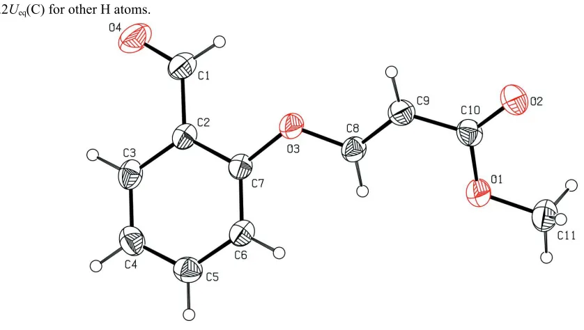
Figure 1
The molecular structure of the title molecule, with atom labelling. Displacement ellipsoids are drawn at the 30%
[image:3.610.128.489.362.449.2]probability level.
Figure 2
The crystal packing of the title compound viewed along the b axis, showing the formation of the R2
2(8) graph-set motif.
The dimers are further consolidated by R2
3(16) graph-set ring motifs. Hydrogen bonds are shown as dashed lines (see
Table 1 for details; H atoms not involved in these interactions have been omitted for clarity).)
Methyl (E)-3-(2-formylphenoxy)acrylate
Crystal data
C11H10O4
Mr = 206.19 Monoclinic, P21/c Hall symbol: -p 2ybc
a = 17.7458 (8) Å
b = 4.0629 (2) Å
c = 14.5745 (7) Å
β = 107.868 (3)°
V = 1000.13 (8) Å3
Z = 4
F(000) = 432
Dx = 1.369 Mg m−3
Mo Kα radiation, λ = 0.71073 Å Cell parameters from 2015 reflections
θ = 1.2–26.3°
µ = 0.11 mm−1
Data collection
Bruker SMART APEXII CCD diffractometer
Radiation source: fine-focus sealed tube Graphite monochromator
ω scans
13052 measured reflections 2015 independent reflections
1523 reflections with I > 2σ(I)
Rint = 0.027
θmax = 26.3°, θmin = 1.2°
h = −22→21
k = −5→5
l = −18→18
Refinement
Refinement on F2 Least-squares matrix: full
R[F2 > 2σ(F2)] = 0.045
wR(F2) = 0.155
S = 1.06 2015 reflections 137 parameters 0 restraints
Primary atom site location: structure-invariant direct methods
Secondary atom site location: difference Fourier map
Hydrogen site location: inferred from neighbouring sites
H-atom parameters constrained
w = 1/[σ2(F
o2) + (0.1001P)2 + 0.0957P] where P = (Fo2 + 2Fc2)/3
(Δ/σ)max < 0.001 Δρmax = 0.23 e Å−3 Δρmin = −0.17 e Å−3
Special details
Geometry. All e.s.d.'s (except the e.s.d. in the dihedral angle between two l.s. planes) are estimated using the full covariance matrix. The cell e.s.d.'s are taken into account individually in the estimation of e.s.d.'s in distances, angles and torsion angles; correlations between e.s.d.'s in cell parameters are only used when they are defined by crystal symmetry. An approximate (isotropic) treatment of cell e.s.d.'s is used for estimating e.s.d.'s involving l.s. planes.
Refinement. Refinement of F2 against ALL reflections. The weighted R-factor wR and goodness of fit S are based on F2, conventional R-factors R are based on F, with F set to zero for negative F2. The threshold expression of F2 > σ(F2) is used only for calculating R-factors(gt) etc. and is not relevant to the choice of reflections for refinement. R-factors based on F2 are statistically about twice as large as those based on F, and R- factors based on ALL data will be even larger.
Fractional atomic coordinates and isotropic or equivalent isotropic displacement parameters (Å2)
x y z Uiso*/Ueq
C1 0.19462 (11) 0.6483 (5) 1.09554 (11) 0.0570 (5)
H1 0.2426 0.7588 1.1079 0.068*
C2 0.16126 (8) 0.4928 (4) 1.00055 (10) 0.0437 (4)
C3 0.08802 (9) 0.3378 (4) 0.97865 (12) 0.0518 (5)
H3 0.0621 0.3250 1.0252 0.062*
C4 0.05341 (10) 0.2034 (5) 0.88901 (13) 0.0574 (5)
H4 0.0040 0.1033 0.8746 0.069*
C5 0.09250 (10) 0.2182 (5) 0.82056 (12) 0.0545 (5)
H5 0.0692 0.1265 0.7600 0.065*
C6 0.16554 (9) 0.3671 (4) 0.84083 (11) 0.0484 (4)
H6 0.1918 0.3734 0.7946 0.058*
C7 0.19943 (8) 0.5068 (4) 0.93033 (10) 0.0425 (4)
C8 0.30241 (9) 0.7868 (4) 0.88985 (11) 0.0463 (4)
H8 0.2706 0.8014 0.8260 0.056*
C9 0.37579 (9) 0.8937 (5) 0.91462 (12) 0.0542 (5)
H9 0.4075 0.8649 0.9780 0.065*
C11 0.39059 (12) 1.2299 (6) 0.68764 (14) 0.0655 (5)
H11A 0.4103 1.4452 0.7097 0.098*
H11B 0.3494 1.2484 0.6272 0.098*
H11C 0.4329 1.0976 0.6794 0.098*
O1 0.35946 (7) 1.0781 (3) 0.75754 (8) 0.0598 (4)
O2 0.47632 (7) 1.1596 (4) 0.86851 (10) 0.0723 (5)
O3 0.27287 (6) 0.6544 (3) 0.95771 (7) 0.0541 (4)
O4 0.16367 (9) 0.6413 (5) 1.15808 (9) 0.0821 (5)
Atomic displacement parameters (Å2)
U11 U22 U33 U12 U13 U23
C1 0.0536 (10) 0.0738 (13) 0.0448 (8) 0.0075 (8) 0.0171 (7) 0.0039 (8)
C2 0.0403 (8) 0.0498 (10) 0.0419 (8) 0.0095 (6) 0.0142 (6) 0.0084 (7)
C3 0.0432 (9) 0.0604 (11) 0.0562 (9) 0.0071 (7) 0.0220 (7) 0.0119 (8)
C4 0.0442 (9) 0.0590 (11) 0.0665 (11) −0.0046 (8) 0.0134 (8) 0.0077 (8)
C5 0.0531 (10) 0.0542 (10) 0.0504 (9) 0.0009 (8) 0.0074 (7) 0.0007 (8)
C6 0.0485 (9) 0.0547 (10) 0.0443 (8) 0.0048 (7) 0.0175 (7) 0.0049 (7)
C7 0.0357 (7) 0.0485 (10) 0.0429 (7) 0.0054 (6) 0.0115 (6) 0.0082 (6)
C8 0.0411 (8) 0.0565 (10) 0.0435 (8) 0.0025 (7) 0.0162 (6) 0.0013 (7)
C9 0.0425 (8) 0.0710 (12) 0.0490 (9) −0.0019 (8) 0.0137 (7) −0.0027 (8)
C10 0.0383 (8) 0.0609 (11) 0.0580 (9) −0.0018 (7) 0.0184 (7) −0.0063 (8)
C11 0.0639 (11) 0.0721 (13) 0.0669 (11) −0.0039 (10) 0.0297 (9) 0.0095 (10)
O1 0.0487 (7) 0.0767 (9) 0.0553 (7) −0.0123 (6) 0.0181 (5) 0.0033 (6)
O2 0.0431 (7) 0.1021 (13) 0.0731 (8) −0.0180 (7) 0.0197 (6) −0.0050 (7)
O3 0.0403 (6) 0.0800 (9) 0.0427 (6) −0.0071 (5) 0.0139 (5) 0.0042 (5)
O4 0.0787 (10) 0.1250 (15) 0.0509 (7) −0.0008 (9) 0.0322 (7) −0.0084 (7)
Geometric parameters (Å, º)
C1—O4 1.200 (2) C7—O3 1.3778 (18)
C1—C2 1.471 (2) C8—C9 1.314 (2)
C1—H1 0.9300 C8—O3 1.3640 (18)
C2—C3 1.390 (2) C8—H8 0.9300
C2—C7 1.3910 (19) C9—C10 1.454 (2)
C3—C4 1.375 (3) C9—H9 0.9300
C3—H3 0.9300 C10—O2 1.2035 (19)
C4—C5 1.380 (2) C10—O1 1.342 (2)
C4—H4 0.9300 C11—O1 1.438 (2)
C5—C6 1.378 (2) C11—H11A 0.9600
C5—H5 0.9300 C11—H11B 0.9600
C6—C7 1.380 (2) C11—H11C 0.9600
C6—H6 0.9300
O4—C1—C2 123.98 (17) O3—C7—C2 115.83 (13)
O4—C1—H1 118.0 C6—C7—C2 120.61 (14)
C2—C1—H1 118.0 C9—C8—O3 120.00 (14)
C3—C2—C1 119.24 (14) O3—C8—H8 120.0
C7—C2—C1 121.91 (15) C8—C9—C10 122.98 (15)
C4—C3—C2 120.77 (15) C8—C9—H9 118.5
C4—C3—H3 119.6 C10—C9—H9 118.5
C2—C3—H3 119.6 O2—C10—O1 122.10 (15)
C3—C4—C5 119.52 (16) O2—C10—C9 124.32 (16)
C3—C4—H4 120.2 O1—C10—C9 113.58 (14)
C5—C4—H4 120.2 O1—C11—H11A 109.5
C6—C5—C4 120.83 (15) O1—C11—H11B 109.5
C6—C5—H5 119.6 H11A—C11—H11B 109.5
C4—C5—H5 119.6 O1—C11—H11C 109.5
C5—C6—C7 119.46 (15) H11A—C11—H11C 109.5
C5—C6—H6 120.3 H11B—C11—H11C 109.5
C7—C6—H6 120.3 C10—O1—C11 115.85 (13)
O3—C7—C6 123.51 (13) C8—O3—C7 120.10 (12)
O4—C1—C2—C3 2.7 (3) C3—C2—C7—C6 −0.5 (2)
O4—C1—C2—C7 −179.85 (17) C1—C2—C7—C6 −178.03 (15)
C7—C2—C3—C4 −0.6 (2) O3—C8—C9—C10 −176.28 (16)
C1—C2—C3—C4 176.94 (17) C8—C9—C10—O2 178.78 (19)
C2—C3—C4—C5 1.0 (3) C8—C9—C10—O1 −1.2 (3)
C3—C4—C5—C6 −0.3 (3) O2—C10—O1—C11 1.2 (3)
C4—C5—C6—C7 −0.9 (3) C9—C10—O1—C11 −178.82 (16)
C5—C6—C7—O3 178.55 (15) C9—C8—O3—C7 −172.25 (16)
C5—C6—C7—C2 1.3 (2) C6—C7—O3—C8 26.2 (2)
C3—C2—C7—O3 −178.02 (14) C2—C7—O3—C8 −156.37 (14)
C1—C2—C7—O3 4.5 (2)
Hydrogen-bond geometry (Å, º)
D—H···A D—H H···A D···A D—H···A
C9—H9···O2i 0.93 2.54 3.440 (2) 164
C8—H8···O4ii 0.93 2.61 3.529 (2) 171
C11—H11C···O2iii 0.96 2.63 3.578 (2) 168