
research communications
Acta Cryst.(2014).E70, 207–209 doi:10.1107/S1600536814020418
207
Received 30 August 2014 Accepted 11 September 2014
Edited by M. Weil, Vienna University of Technology, Austria
†
Keywords:crystal structure; anhydrous bis(sarcosinato)copper(II); non-proteinogenic amino acid
CCDC reference:961026
Supporting information:this article has supporting information at journals.iucr.org/e
Crystal structure of anhydrous poly[bis(
l
2-sarcosi-nato-
j
3O
,
N
:
O
000)copper(II)]
Ray J. Butcher,a* Greg Brewerband Matthew Zembab
aDepartment of Chemistry, Howard University, 525 College Street NW, Washington, DC 20059, USA, andbDepartment
of Chemistry, The Catholic University of America, 620 Michigan Ave., N.E., Washington, DC 20064, USA. *Correspondence e-mail: [email protected]
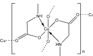
The title compound, [Cu(C3H6NO2)2]n, is a bis-complex of the anion of sarcosine (N-methylglycine). The asymmetric unit consists of a copper(II) ion, located on a center of inversion, and one molecule of the uninegative sarcosinate anion. The copper(II) ion exhibits a typical Jahn–Teller distorted [4 + 2] coordination geometry. The four shorter equatorial bonds are to the nitrogen and carboxylate O atoms of two sarcosinate anions, and the longer axial bonds are to carboxylate O atoms of neighboring complexes. The overall structure is made up from two chains formed by these longer axial Cu—O bonds, one extending parallel to [011] and the other parallel to [011]. Each one-dimensional array is connected by the equatorial bridging moieties to the chains on either side, creating an extended two-dimensional framework parallel to (100). There is a single intermolecular hydrogen-bonding interaction within the sheets between the amino NH group and an O atom of an adjacent molecule.
1. Chemical context
The-amino acids are essential for life as they are the building blocks of all proteins and enzymes and a great deal is known about their structures and complexes.N-Methyl amino acids, such as sarcosine, are non-proteinogenic and hence differ from the proteinogenic amino acids used in living systems in that the amino N atom is achiral in the free molecule but chiral,R
or S, when bound to a metal. Examples of complexes of sarcosine that exhibit chirality due to coordination of the amino N atom have been reported (Blountet al., 1967; Larsen
et al., 1968; Proutet al., 1972). This is similar to the chirality that is observed on the binding of reduced tripodal Schiff base complexes of metals (Brewer et al., 2014; Al-Obaidi et al., 1996). In these cases, the binding of three achiral (due to rapid inversion) amine N atoms of the free ligand to the same metal resulted in the observation of a single enantiomeric pair (RRR
andSSS) or a single enantiomer (RRRorSSS) if the molecule crystallized in one of the Sohncke space groups. In these cases, the binding of an organic ligand containing three achiral N atoms to a metal resulted in a preference for chirality corre-lation of the N atoms, RRR or SSS, resulting in homochiral complexes. Similarly, the reduced Schiff base complexes of the condensate of amino acids with salicylaldehyde have an energetic preference for the stereoisomer in which the chirality of the-C atom and the amine N atom are correlated (Kohet al., 1996).
This preference for homochirality is not always observed: the copper complex of a Schiff base condensate of tyrosine is heterochiral as is the bis-adduct of cobalt(III) with histidine (Pradeepet al., 2006; Zieet al., 2007). The present complex was investigated to determine if there was a preference for homo-(RRor SS) versus heterochirality (RS) in a M(sarcosinato)2
complex (M= divalent transition metal). Heterochirality,RS, was observed in this complex. Future work will focus on related complexes such as M0(sarcosinato)3 (M0 = trivalent
transition metal) to determine if the presence of three chiral ligands bound to a single metal favors homochirality, which can serve as a method of enantiomeric separation.
2. Structural commentary
The title compound, [Cu(C3H6NO2)2]n, is a bis-complex of the sarcosinate anion with copper(II). The central metal cation is located on a center of inversion. It is six-coordinate and has a distorted octahedral [4 + 2] coordination sphere characteristic for Jahn–Teller systems. The four shorter equatorial bonds are to the amino N atom and carboxylate O atom of two sarco-sinate anions (Fig. 1). The N and O atoms are transto one another. The related [Cu(sarcosinato)2]2H2O structure
(Krishnakumar et al., 1994) is much simpler in that the two longer axial bonds are to water molecules so that there is no extended bonding to neighboring complexes. In both struc-tures, the equatorial Cu—O and Cu—N bond lengths are very similar [Cu—O = 1.9758 (8) A˚ and Cu—N = 2.0046 (9) A˚ in the title compound, and 1.970 and 2.007 A˚ in the dihydrate], but the axial Cu—O distances are significantly different at 2.5451 (10) and 2.461 A˚ .
3. Supramolecular features
In the title compound, the individual coordination polyhedra are linked by longer axial Cu—O bonds into two chains, one extending parallel to [011] and the other parallel to [011]. The one-dimensional array is linked by equatorial bridging bonds to the chains on either side, creating an extended two-dimensional framework (Fig. 2) parallel to (100). There is a single intermolecular hydrogen-bonding interaction within the sheets between the amino NH and an carboxylate O atom of an adjacent molecule (Table 1).
4. Database survey
The structure of the zwitterionic form of sarcosine has been reported by Rodrigues et al. (2005). The structure of the copper(II) and nickel(II) complexes of this same ligand have been reported as their dihydrates by Krishnakumar et al.
(1994) and Guha (1973), respectively.
5. Synthesis and crystallization
Sarcosine (N-methylglycine) was purchased from Aldrich Chemical. Sarcosine (1.87 mmol, 0.166 g) was dissolved in 0.10
Maqueous potassium hydroxide (18.7 ml, 1.87 mmol). Copper chloride dihydrate (0.468 mmol, 0.078 g) was added to the above solution and crystals of the title compound were grown by slow evaporation.
208
Butcheret al. [Cu(C3H6NO2)2] Acta Cryst.(2014).E70, 207–209
[image:2.610.89.251.81.179.2]research communications
Figure 1
Part of a chain in the title compound, with the atom-numbering scheme and atomic displacement parameters drawn at the 30% probability level. Hydrogen bonding is shown by dashed lines. [Symmetry codes: (A) 1x, 1y, 1z; (B) 1x,y1
2, 1
2z; (C)x, 3 2y,
[image:2.610.312.564.84.124.2] [image:2.610.315.566.534.715.2]1 2+z.]
Table 1
Hydrogen-bond geometry (A˚ ,).
D—H A D—H H A D A D—H A
N1—H1A O1i 0.93 2.13 2.9729 (13) 150
Symmetry code: (i)xþ1;y1 2;zþ
1 2.
Figure 2
[image:2.610.44.298.583.699.2]6. Refinement
Crystal data, data collection and structure refinement details are summarized in Table 2. H atoms were positioned geom-etrically and refined using a riding model, with C—H distances of 0.93–0.99 A˚ , and withUiso(H) = 1.2Ueq(C) or 1.5Ueq(C) for
methyl H atoms. The H attached to N was located in a difference Fourier map and refined using a riding model, with an N—H distance of 0.93 A˚ andUiso(H) = 1.2Ueq(N).
Acknowledgements
RJB wishes to acknowledge the NSF–MRI program (grant No. CHE-0619278) for funds to purchase the diffractometer. GB wishes to acknowledge support of this work from NASA (NNX10AK71A)
References
Agilent (2012). CrysAlis PRO. Agilent Technologies, Yarnton, England.
Al-Obaidi, A. H. R., Jensen, K. B., McGarvey, J. J., Toftlund, H., Jensen, B., Bell, S. E. J. & Carroll, J. G. (1996).Inorg. Chem.35, 5055–5060.
Altomare, A., Burla, M. C., Camalli, M., Carrozzini, B., Cascarano, G. L., Giacovazzo, C., Guagliardi, A., Moliterni, A. G. G., Polidori, G. & Rizzi, R. (1999).J. Appl. Cryst.32, 339–340.
Blount, J. F., Freeman, H. C., Sargensen, A. M. & Tumbull, K. R. (1967).Chem. Commun.pp. 324–326.
Brewer, G., Brewer, C. T., Butcher, R. J., Robichaux, G. T. & Viragh, C. (2014).Inorg. Chim. Acta,410, 171–177.
Guha, S. (1973).Acta Cryst.B29, 2167–2170.
Koh, L. L., Ranford, J. O., Robinson, W. T., Svensson, J. O., Tan, A. L. C. & Wu, D. (1996).Inorg. Chem.35, 6466–6472.
Krishnakumar, R. V., Natarajan, S., Bahadur, S. A. & Cameron, T. S. (1994).Z. Kristallogr.209, 443–445.
Larsen, S., Watson, K. J., Sargenson, A. M. & Turmbull, K. R. (1968). Chem. Commun.pp. 847–848.
Pradeep, C. P., Supriya, S., Zacharias, P. S. & Das, S. K. (2006). Polyhedron,25, 3588–3592.
Prout, C. K., Allison, G. B., Delbaere, L. T. J. & Gore, E. (1972).Acta Cryst.B28, 3043–3056.
Rodrigues, V. H., Ramos Silva, M., Matos Beja, A., Paixa˜o, J. A. & Costa, M. M. R. R. (2005).Acta Cryst.E61, o1631–o1633. Sheldrick, G. M. (2008).Acta Cryst.A64, 112–122.
Zie, Y., Wu, H. H., Yong, G. P., Wang, Z. Y., Fan, R., Li, R. P., Pan, G. Q., Tian, Y. C., Sheng, L. S., Pan, L. & Li, J. (2007).J. Mol. Struct.
833, 88–91.
research communications
Acta Cryst.(2014).E70, 207–209 Butcheret al. [Cu(C
[image:3.610.313.558.92.358.2]3H6NO2)2]
209
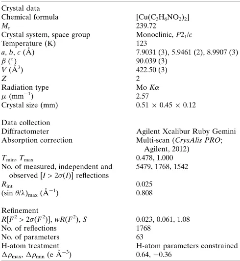
Table 2
Experimental details.
Crystal data
Chemical formula [Cu(C3H6NO2)2]
Mr 239.72
Crystal system, space group Monoclinic,P21/c
Temperature (K) 123
a,b,c(A˚ ) 7.9031 (3), 5.9461 (2), 8.9907 (3)
() 90.039 (3)
V(A˚3) 422.50 (3)
Z 2
Radiation type MoK
(mm1) 2.57
Crystal size (mm) 0.510.450.12
Data collection
Diffractometer Agilent Xcalibur Ruby Gemini
Absorption correction Multi-scan (CrysAlis PRO; Agilent, 2012)
Tmin,Tmax 0.478, 1.000
No. of measured, independent and observed [I> 2(I)] reflections
5479, 1768, 1542
Rint 0.025
(sin/)max(A˚
1) 0.808
Refinement
R[F2> 2(F2)],wR(F2),S 0.023, 0.061, 1.08
No. of reflections 1768
No. of parameters 63
H-atom treatment H-atom parameters constrained
max,min(e A˚
3) 0.64,0.36
Computer programs:CrysAlis PRO (Agilent, 2012),SIR97 (Altomareet al., 1999),
supporting information
sup-1
Acta Cryst. (2014). E70, 207-209
supporting information
Acta Cryst. (2014). E70, 207-209 [doi:10.1107/S1600536814020418]
Crystal structure of anhydrous poly[bis(
µ
2-sarcosinato-
κ
3O
,
N
:
O
′
)copper(II)]
Ray J. Butcher, Greg Brewer and Matthew Zemba
Computing details
Data collection: CrysAlis PRO (Agilent, 2012); cell refinement: CrysAlis PRO (Agilent, 2012); data reduction: CrysAlis
PRO (Agilent, 2012); program(s) used to solve structure: SIR97 (Altomare et al., 1999); program(s) used to refine
structure: SHELXL97 (Sheldrick, 2008); molecular graphics: SHELXTL (Sheldrick, 2008); software used to prepare
material for publication: SHELXTL (Sheldrick, 2008).
Poly[bis(µ2-sarcosinato-κ3O,N:O′)copper(II)]
Crystal data
[Cu(C3H6NO2)2] Mr = 239.72 Monoclinic, P21/c Hall symbol: -P 2ybc
a = 7.9031 (3) Å
b = 5.9461 (2) Å
c = 8.9907 (3) Å
β = 90.039 (3)°
V = 422.50 (3) Å3 Z = 2
F(000) = 246
Dx = 1.884 Mg m−3
Mo Kα radiation, λ = 0.71073 Å Cell parameters from 2393 reflections
θ = 3.4–35.0°
µ = 2.57 mm−1 T = 123 K Plate, dark blue 0.51 × 0.45 × 0.12 mm
Data collection
Agilent Xcalibur Ruby Gemini diffractometer
Radiation source: Enhance (Mo) X-ray Source Graphite monochromator
Detector resolution: 10.5081 pixels mm-1 ω scans
Absorption correction: multi-scan (CrysAlis PRO; Agilent, 2012)
Tmin = 0.478, Tmax = 1.000
5479 measured reflections 1768 independent reflections 1542 reflections with I > 2σ(I)
Rint = 0.025
θmax = 35.0°, θmin = 4.1° h = −12→11
k = −9→8
l = −14→14
Refinement
Refinement on F2 Least-squares matrix: full
R[F2 > 2σ(F2)] = 0.023 wR(F2) = 0.061 S = 1.08 1768 reflections 63 parameters 0 restraints
Primary atom site location: structure-invariant direct methods
Secondary atom site location: difference Fourier map
Hydrogen site location: inferred from neighbouring sites
H-atom parameters constrained
w = 1/[σ2(F
o2) + (0.0285P)2 + 0.0835P] where P = (Fo2 + 2Fc2)/3
supporting information
sup-2
Acta Cryst. (2014). E70, 207-209
Extinction correction: SHELXTL (Sheldrick, 2008), Fc*=kFc[1+0.001xFc2λ3/sin(2θ)]-1/4
Extinction coefficient: 0.014 (3)
Special details
Geometry. All e.s.d.'s (except the e.s.d. in the dihedral angle between two l.s. planes) are estimated using the full covariance matrix. The cell e.s.d.'s are taken into account individually in the estimation of e.s.d.'s in distances, angles and torsion angles; correlations between e.s.d.'s in cell parameters are only used when they are defined by crystal symmetry. An approximate (isotropic) treatment of cell e.s.d.'s is used for estimating e.s.d.'s involving l.s. planes.
Refinement. Refinement of F2 against ALL reflections. The weighted R-factor wR and goodness of fit S are based on F2, conventional R-factors R are based on F, with F set to zero for negative F2. The threshold expression of F2 > σ(F2) is used only for calculating R-factors(gt) etc. and is not relevant to the choice of reflections for refinement. R-factors based on F2 are statistically about twice as large as those based on F, and R- factors based on ALL data will be even larger.
Fractional atomic coordinates and isotropic or equivalent isotropic displacement parameters (Å2)
x y z Uiso*/Ueq
Cu 0.5000 0.5000 0.5000 0.01076 (7)
O1 0.53710 (10) 0.74236 (14) 0.35326 (9) 0.01390 (16) O2 0.72233 (12) 0.85036 (16) 0.17943 (10) 0.0221 (2) N1 0.69539 (12) 0.35314 (16) 0.39628 (10) 0.01218 (17)
H1A 0.6524 0.2775 0.3140 0.015*
C1 0.67866 (14) 0.72698 (19) 0.28370 (12) 0.01373 (19) C2 0.79685 (14) 0.5444 (2) 0.34046 (13) 0.0135 (2)
H2A 0.8718 0.4933 0.2590 0.016*
H2B 0.8685 0.6048 0.4215 0.016*
C3 0.79798 (15) 0.1919 (2) 0.48149 (14) 0.0171 (2)
H3A 0.8941 0.1425 0.4209 0.026*
H3B 0.7284 0.0615 0.5080 0.026*
H3C 0.8398 0.2642 0.5723 0.026*
Atomic displacement parameters (Å2)
U11 U22 U33 U12 U13 U23
Cu 0.00955 (10) 0.01254 (10) 0.01019 (10) 0.00153 (6) 0.00211 (6) 0.00253 (6) O1 0.0127 (3) 0.0158 (4) 0.0132 (3) 0.0014 (3) 0.0018 (3) 0.0034 (3) O2 0.0215 (4) 0.0249 (5) 0.0199 (4) 0.0014 (4) 0.0060 (3) 0.0105 (4) N1 0.0111 (4) 0.0142 (4) 0.0112 (4) −0.0001 (3) 0.0001 (3) 0.0001 (3) C1 0.0132 (4) 0.0152 (5) 0.0128 (4) −0.0016 (4) 0.0005 (4) 0.0008 (4) C2 0.0107 (4) 0.0165 (5) 0.0132 (5) −0.0008 (4) 0.0011 (4) 0.0012 (4) C3 0.0161 (5) 0.0168 (5) 0.0183 (5) 0.0040 (4) 0.0003 (4) 0.0020 (4)
Geometric parameters (Å, º)
Cu—O1i 1.9758 (8) N1—C2 1.4796 (15)
Cu—O1 1.9758 (8) N1—H1A 0.9300
Cu—N1i 2.0046 (9) C1—C2 1.5200 (17)
Cu—N1 2.0046 (9) C2—H2A 0.9900
Cu—O2ii 2.5451 (10) C2—H2B 0.9900
supporting information
sup-3
Acta Cryst. (2014). E70, 207-209
O1—C1 1.2853 (13) C3—H3B 0.9800
O2—C1 1.2396 (14) C3—H3C 0.9800
N1—C3 1.4705 (15)
O1i—Cu—O1 180.0 C3—N1—H1A 107.4
O1i—Cu—N1i 83.82 (3) C2—N1—H1A 107.4
O1—Cu—N1i 96.18 (3) Cu—N1—H1A 107.4
O1i—Cu—N1 96.18 (3) O2—C1—O1 124.66 (11)
O1—Cu—N1 83.82 (3) O2—C1—C2 120.34 (10)
N1i—Cu—N1 180.0 O1—C1—C2 114.98 (10)
O1i—Cu—O2ii 86.24 (3) N1—C2—C1 109.26 (9)
O1—Cu—O2ii 93.76 (3) N1—C2—H2A 109.8
N1i—Cu—O2ii 94.85 (3) C1—C2—H2A 109.8
N1—Cu—O2ii 85.15 (3) N1—C2—H2B 109.8
O1i—Cu—O2iii 93.76 (3) C1—C2—H2B 109.8
O1—Cu—O2iii 86.24 (3) H2A—C2—H2B 108.3
N1i—Cu—O2iii 85.15 (3) N1—C3—H3A 109.5
N1—Cu—O2iii 94.85 (3) N1—C3—H3B 109.5
O2ii—Cu—O2iii 180.0 H3A—C3—H3B 109.5
C1—O1—Cu 113.74 (7) N1—C3—H3C 109.5
C3—N1—C2 112.28 (9) H3A—C3—H3C 109.5
C3—N1—Cu 117.79 (7) H3B—C3—H3C 109.5
C2—N1—Cu 103.93 (7)
O1i—Cu—O1—C1 −64 (100) O1—Cu—N1—C2 29.24 (7)
N1i—Cu—O1—C1 166.53 (8) N1i—Cu—N1—C2 80 (100)
N1—Cu—O1—C1 −13.47 (8) O2ii—Cu—N1—C2 −65.08 (7)
O2ii—Cu—O1—C1 71.23 (8) O2iii—Cu—N1—C2 114.92 (7)
O2iii—Cu—O1—C1 −108.77 (8) Cu—O1—C1—O2 174.29 (10)
O1i—Cu—N1—C3 −25.85 (8) Cu—O1—C1—C2 −6.96 (12)
O1—Cu—N1—C3 154.15 (8) C3—N1—C2—C1 −167.81 (9)
N1i—Cu—N1—C3 −155 (100) Cu—N1—C2—C1 −39.44 (10)
O2ii—Cu—N1—C3 59.83 (8) O2—C1—C2—N1 −148.69 (11)
O2iii—Cu—N1—C3 −120.17 (8) O1—C1—C2—N1 32.50 (13)
O1i—Cu—N1—C2 −150.76 (7)
Symmetry codes: (i) −x+1, −y+1, −z+1; (ii) x, −y+3/2, z+1/2; (iii) −x+1, y−1/2, −z+1/2.
Hydrogen-bond geometry (Å, º)
D—H···A D—H H···A D···A D—H···A
N1—H1A···O1iii 0.93 2.13 2.9729 (13) 150
![Crystal structure of catena poly[[copper(II) μ2 salicylato [diaquacopper(II)] μ2 salicylato] dihydrate]](data:image/gif;base64,R0lGODlhAQABAIAAAP///wAAACH5BAEAAAAALAAAAAABAAEAAAICRAEAOw==)