R E S E A R C H
Open Access
Ventriculoarterial decoupling in human septic
shock
Fabio Guarracino
1*, Baldassare Ferro
1, Andrea Morelli
2, Pietro Bertini
1, Rubia Baldassarri
1and Michael R Pinsky
3Abstract
Introduction:Septic shock is the most severe manifestation of sepsis. It is characterized as a hypotensive cardiovascular state associated with multiorgan dysfunction and metabolic disturbances. Management of septic shock is targeted at preserving adequate organ perfusion pressure without precipitating pulmonary edema or massive volume overload. Cardiac dysfunction often occurs in septic shock patients and can significantly affect outcomes. One physiologic approach to detect the interaction between the heart and the circulation when both are affected is to examine ventriculoarterial coupling, which is defined by the ratio of arterial elastance (Ea) to left ventricular end-systolic elastance (Ees). In this study, we analyzed ventriculoarterial coupling in a cohort of patients admitted to ICUs who presented with vs without septic shock.
Methods:In this retrospective cross-sectional opportunity study, we measured routine hemodynamics using indwelling arterial and pulmonary arterial catheters and transthoracic echocardiograms in 25 septic patients
(group S) and 25 non–septic shock patients (group C) upon ICU admission. Ees was measured by echocardiography using a single-beat (EesSB) method. Ea was calculated as 0.9 systolic arterial pressure/stroke volume, and then the
Ea/EesSBratio was calculated (normal value <1.36).
Results:In group S, 21 patients had an Ea/EesSBratio >1.36 (uncoupled). The four patients with Ea/EesSB
ratios≤1.36 had higher EesSBvalues than patients with Ea/EesSBratios >1.36 (P= 0.007), although Ea measurements
were similar in both groups (P= 0.4). In group C, five patients had uncoupled Ea/EesSBratios. No correlation was
found between EesSBand left ventricular ejection fraction and between Ea/EesSBratio and mixed venous oxygen
saturation in septic shock patients.
Conclusions:Upon admission to the ICU, patients in septic shock often display significant ventriculoarterial decoupling that is associated with impaired left ventricular performance. Because Ea/Ees decoupling alters cardiovascular efficiency and cardiac energetic requirements independently of Ea or Ees, we speculate that septic patients with ventriculoarterial uncoupling may benefit from therapy aimed at normalizing the Ea/Ees ratio.
Introduction
Septic shock is characterized in the resuscitated patient as a hyperdynamic, hypotensive cardiovascular state as-sociated with multiorgan dysfunction and metabolic dis-turbances consistent with tissue dysoxia [1]. Importantly, peripheral vasodilation (relative hypovolemia and low systemic vascular resistance) can mask coexistent car-diac dysfunction [2,3]. Carcar-diac dysfunction in septic shock includes left ventricular (LV) diastolic and systolic
dysfunction secondary to primary myocardial injury or right ventricular dysfunction due to pulmonary hyper-tension [4]. The combined loss of peripheral vasomotor tone and cardiac contractile impairment may potentially result in ventriculoarterial decoupling, the consequences of which can be worsening cardiac energetics and performance.
Suga and Sagawa [5] and Sunagawa et al. [6] analyzed the circuit from the standpoint of LV ejection fraction (LVEF). In this model, LV contractility is described by the end-systolic pressure–volume relationship (ESPVR). The slope of the ESPVR, called end-systolic elastance (Ees), is a load-independent measure of cardiac contract-ility. End-systolic pressure is also a function of both LV
* Correspondence:[email protected]
1Department of Anesthesia and Critical Care Medicine, University Hospital of
Pisa, Via Paradisa 2, Pisa 56124, Italy
Full list of author information is available at the end of the article
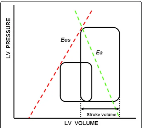
stroke volume (SV) and a characteristic of the arterial outflow tract. The greater the SV for a given vascular tone, the greater the systolic arterial pressure. Similarly, increases in arterial systolic pressure for a constant pre-load and Ees will decrease SV and increase end-systolic volume. The slope representing the relationship between SV and systolic arterial pressure as SV is varied is called arterial elastance (Ea) (Figure 1). Thus, SV is both limited and defines end-systolic pressure through arterioventricu-lar coupling [6]. Maximal myocardial efficiency, defined as the amount of external work performed for myocardial oxygen consumed, occurs when Ea is approximately one-half Ees [7] and has stronger dependence on Ea than on Ees [8]. Accordingly, the Ea/Ees ratio is a sensitive and independent estimate of cardiovascular efficiency [5]. A normally coupled human cardiovascular system has an Ea/Ees ratio = 1 ± 0.36 (median ± IQR), with normal values being 2.2 ± 0.8 mmHg/ml for Ea and 2.3 ± 1 mmHg/ml for Ees [9].
Uncoupling, defined as an Ea/Ees ratio >1.36, can re-sult from changes in Ea, Ees or both. Uncoupling reflects a reduction in LV ejection efficiency, which can promote LV energetic failure. Although septic shock is associated with hypotension, which unloads LV ejection, it may, if unbalanced, result in Ea/Ees decoupling. We recently documented a primary decrease in peripheral impedance and an increase in peripheral compliance associated with a decrease in central aortic compliance in a porcine model of early endotoxic shock [10]. We hypothesized that similar Ea/Ees decoupling may also be present in
septic patients and that this uncoupling may contribute to the observed impaired LV ejection effectiveness.
Ea is a lumped parameter of arterial tone which is influenced by arterial resistance and compliance, aortic impedance and systolic and diastolic time intervals. It reflects the net arterial load on LV ejection. Because end-systolic pressure follows maximal ejection pressure and systole occurs as LV relaxation starts, end-systolic pressure can be approximated as 90% of end-systolic arterial pressure and SV [5,6]. Similarly, Ees is defined as the slope of the LV ESPVR derived from a series of LV end-systolic pressure–volume points created by rapidly varying preload or afterload such that intrinsic contract-ility remains constant [6]. We previously showed that end-inspiratory hold maneuvers rapidly decrease pre-load, thus allowing the measurement of Ees [11]. Still, the need to rapidly alter either preload or afterload and also use invasive LV catheterization to measure LV volume has kept the bedside measurement of Ees out of the realm of routine bedside practice.
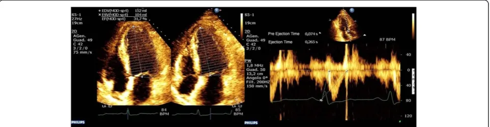
The demonstration that Ees can be estimated in a single beat (EesSB) [12], and the subsequent validation of a noninvasive echocardiographic method to measure EesSB[13] through the measure of LVEF, SV, pre-ejection time and systolic time interval when coupled with systolic and diastolic arterial pressure (Figure 2), made the clinical bedside measure possible. Thus, using these bedside esti-mates of Ea and Ees, we measured in patients presenting with vs without septic shock to analyze the presence of native ventriculoarterial decoupling.
Methods
After we obtained approval from the ethical committees for human biomedical research at both the University Hospital of Pisa and the University of Pittsburgh, we conducted a retrospective cross-sectional study between July 2011 and February 2013 to elaborate hemodynamic data obtained from (1) patients presenting with septic shock as defined by the international consensus confer-ence definition and treated them following the Surviving Sepsis Campaign guidelines [14] and (2) patients admit-ted to the ICU without presumed septic shock. Patients with a history of cardiac disease and preoperative cardiac surgery patients were excluded. Informed consent was obtained from all patients, and all were studied immedi-ately after initial fluid resuscitation in accordance with the Surviving Sepsis Campaign guidelines, but prior to starting therapy with any vasoactive pharmacological agents.
[image:2.595.56.291.467.679.2]We analyzed a series of measured hemodynamic vari-ables and calculated parameters at the time of diagnosis of septic shock in our septic shock cohort (8 women and 17 men, age 69 ± 8 years (median ± IQR)) and at the time of ICU admission in our non–septic shock cohort Figure 1Pressure–volume relationship in a cardiac cycle.
with neurological problems but otherwise hemody-namically stable (11 women and 14 men, age 60 ± 11 years). These data included cardiac index (CI), heart rate and mean arterial pressure (MAP) invasively measured at a radial arterial site, as well as pulmonary artery occlusion pressure (Ppao) and mixed venous oxygen satur-ation (SvO2) measured using a continuous cardiac output pulmonary artery catheter equipped with fiber optics (Swan-Ganz oximetry thermodilution catheter; Edwards Lifesciences, Irvine, CA, USA).
In nonseptic patients in whom a pulmonary artery catheter was not inserted at the discretion of the at-tending intensivist, the CI was measured using a pulse contour method (Vigileo Monitor/FloTrac Sensor System; Edwards Lifesciences), and central venous oxygen satur-ation (ScvO2) was measured instead of SvO2. None of the nonseptic patients were receiving vasoactive drug therapy at the time they were examined.
All patients in both groups had a transthoracic echo-cardiographic examination upon admission, all of which were performed by the same operator. Echocardiography was performed with a CX50 ultrasound system and an S5-1 Sector Array Transducer (Koninklijke Philips Elec-tronics NV, Eindhoven, the Netherlands).
Parameters calculated using data gathered from the echocardiographic examination included LVEF, SV, pree-jection time and systolic time. EesSB was estimated by using the method of Chen et al. [13]. Ea was calculated as 0.9 × (systolic arterial pressure/SV), and the Ea/EesSB ratio was then calculated.
Statistical data are expressed as median ± IQR. Con-sidering the small number of patients in our study, a nonparametric unpaired Mann–WhitneyUtest was used to compare groups. Spearman’s coefficient of rank correl-ation (ρ) was used to correlate variables. Fisher’s exact test was used in the analysis of contingency tables.
Results
The data derived from 50 patients were analyzed. The 25 patients with a diagnosis of septic shock presented in a hyperdynamic (median CI = 2.77 L/min/m2(IQR = 2.4 to 3.8) and HR = 115 beats/min (IQR = 109 to 124), hypotensive (MAP 58 IR 53–60) mmHg) state with a median Ppao of 13 mmHg (IQR = 10 to 14.2), median LVEF of 40% (IQR = 32.25 to 51.5), SvO2of 60% (IQR = 58 to 69) and an Ea/EesSB ratio of 1.81 (IQR = 1.49 to 2.02) following initial fluid resuscitation. Only four pa-tients had an Ea/EesSBratio≤1.36 at the time of diagno-sis of septic shock, whereas the remaining twenty-one patients had an Ea/EesSBratio >1.36 (Figure 3). The four patients with Ea/EesSB ratios ≤1.36 had higher EesSB compared with twenty-one patients with Ea/EesSB ra-tios >1.36 (P = 0.007), though Ea was similar in both the coupled and uncoupled septic shock patients (P= 0.4). We found no correlation between EesSBand LVEF (ρ=−0.0809,
P = 0.7007) or between Ea/EesSB ratio and SvO2in septic shock patients (ρ=−0.293,P= 0.15).
[image:3.595.58.540.90.215.2]The 25 patients admitted in a non–septic shock state presented with a normal systemic flow state (CI = 2.8 L/ min/m2(IQR = 2.6 to 3)); HR = 80 beats/min (IQR = 71 Figure 2Left ventricular end-systolic elastance was calculated by using the single-beat method.These echocardiographic scans display the evaluation of ejection fraction (left image) and preejection and ejection time (right image) using aortic Doppler waveforms. Normalized ventricular elastance at arterial end-diastole (End) was measured according to the following formula:
Ees sbð Þ¼ ½Pd−ðENd estð ÞPs0:9Þ=½SVENd estð Þ ENd avgð Þ¼ΣaitNd
i¼0
ENd estð Þ¼0:0275–0:165EFþ0:3656ðPd=PesÞþ0:515ENd avgð Þ
whereaivalues are 0.35695,−7.2266, 74.249,−307.39, 684.54.−856.92, 571.95 and−159.1 fori= 1 toi= 7, respectively. The valuetNdvalue was
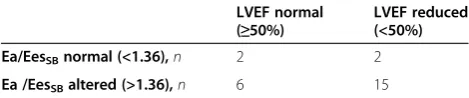
to 104; MAP = 70 mmHg (IQR = 67 to 71) with an LVEF of 54% (IQR = 47.5 to 58), ScvO2= 62% (IQR = 56.8 to 69) and Ea/EesSBratio = 1.07 (IQR = 0.95 to 1.14). Five of these patients had a calculated Ea/EesSB ratio >1.36, and the remaining twenty patients had Ea/EesSB ratios <1.36 (Figure 3). Table 1 shows comparisons of variables between septic and nonseptic patients. Compared with non–septic shock patients, septic shock patients had reduced Ea; lower EesSB, LVEF and MAP; and higher Ea/EesSB ratios and HRs. Table 2 shows the absence of correlation between Ea/EesSB status and normal or re-duced LVEF in septic shock patients (P= 0.5) in a 2 × 2 contingency table.
Discussion
Our data show that, using bedside monitoring tech-niques [15], most of our patients who presented to ICUs in septic shock had significant ventriculoarterial decoup-ling independent of the commonly seen reduced Ea, whereas our non–septic shock patients displayed pre-served ventriculoarterial coupling. This altered ventricu-loarterial coupling in septic patients occurred despite a preserved or even elevated CI. These data suggest that, in septic shock patients, decoupling was associated with
the observed impaired LV performance and reduced Ea. The lack of correlation between LVEF and EesSB sug-gests that LVEF cannot be considered a clinical index of contractility or ventricular performance. This conclusion is not surprising, because LVEF is a function not only of contractility but also of Ea. On the basis of these data, we speculate that the use of vasoconstrictors in septic shock with the aim of maintaining sufficient MAP according to the published Surviving Sepsis Campaign guidelines could be the cause of the reduced LVEF, as previously suggested [3].
[image:4.595.58.540.89.236.2]Ventriculoarterial decoupling is an important index of cardiovascular inefficiency. An adequate Ea/Ees ratio is fundamental for efficient cardiovascular performance and is a determinant of cardiac energetics [16]. Ea/Ees ratio reflects the interaction between cardiac function and the arterial system that is necessary to modulate the cardiovascular response to either physiological or pathological conditions. The changes in CO and vascular resistance in different physiological conditions (for example, age, exercise, rest) and pathological condi-tions (for example, hypertension, heart failure, diabetes) strictly depend on both LV function and the arterial system. Aging and cardiovascular diseases such as hypertension, Figure 3Graphs representing the distribution of ratios of arterial elastance to single-beat end-systolic elastance in newly diagnosed septic shock patients (A) and non–septic shock patients (B).
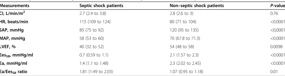
Table 1 Comparison of hemodynamic variables between septic shock and non–septic shock patientsa
Measurements Septic shock patients Non–septic shock patients P-value
CI, L/min/m2 2.7 (2.4 to 3.8) 2.8 (2.6 to 3) 0.76
HR, beats/min 115 (109 to 124) 80 (71 to 104) <0.0001
SAP, mmHg 85 (75 to 92) 120 (95 to 135) <0,0001
MAP, mmHg 58 (53 to 60) 70 (67.8 to 71.3) <0.0001
LVEF, % 40 (32 to 52) 54 (48 to 58) 0.0098
EesSB, mmHg/ml 0.7 (0.59 to 1.1) 2.1 (1.57 to 2.3) <0.0001
Ea, mmHg/ml 1.4 (1.1 to 1.48) 2.3 (2.02 to 2.45) <0.0001
Ea/EesSBratio 1.81 (1.49 to 2.03) 1.07 (0.95 to 1.18) 0.01
aCI,
[image:4.595.58.538.588.716.2]coronary artery disease, congestive heart failure and cardiac valve dysfunction can alter Ea/Ees coupling by reducing LV performance, increasing Ea or both [17].
Many researchers have shown that vasoactive drugs can affect ventriculoarterial coupling in several differ-ent clinical situations. The phosphodiesterase inhibitor E-1020 was found to increase heart mechanical effi-ciency by improving Ea/Ees coupling in heart disease patients [18]. Levosimendan, an inodilator, restored Ea/Ees coupling in patients with ischemic cardiomyopathy undergoing cardiac surgery [19], and enoximone impro-ved Ea/Ees better than dobutamine in subjects with dilated cardiomyopathy [20,21]. Recently, Martin et al. demonstrated a better prognosis in patients with poly-trauma using LV stroke work and ventriculoarterial coupling as targets of therapy [22].
Our study has some limitations. First, it is a retro-spective analysis of proretro-spectively collected data. Still, the data were collected in a consistent fashion from all participants. Second, the method we applied to measure Ees does not take into consideration the curvilinear shape of the elastance curve, which could be misleading if contractility was much reduced. However, estimated Ees values for our septic cohort were not depressed enough to reach this region of curvilinearity. Third, the definition of Ea was simplified as Ea = end-systolic pres-sure/SV, but this is routinely used as a valid surrogate. Fur-thermore, we measured arterial pressure from indwelling radial artery catheters that may either over- or underrepre-sent central aortic systolic pressure in septic patients. Dif-ferences of about 10 mmHg may exist between peripheral and central pressure sites. However, the mathematical ef-fect of systolic arterial pressure on the calculated Ea/EesSB would be small because it is used in the calculation of both parameters. Fourth, we did not reanalyze our septic patients following restoration of arterial pressure with vasopressor agents or after recovery to see if the as-sociated uncoupling was resolved. This latter limita-tion forms the basis of an ongoing clinical study.
We cannot deduce the clinical implications of this decoupling from our analysis; however, because most of our septic shock patients had uncoupled Ea/Ees at the time of diagnosis, we speculate that patients with septic
shock and decoupled Ea/Ees would benefit from vaso-active therapies aimed at normalizing the Ea/Ees ratio. For example, in a murine model of sepsis, Ducrocqet al. [23] demonstrated that increasing MAP by the use of selective α-adrenergic vasopressors such as phenyle-phrine will unmask LV failure. Use of a more balanced
α- and β-adrenergic agent, such as norepinephrine, may result in a better cardiovascular state with less potential for cardiac decompensation. Furthermore, the Surviving Sepsis Campaign guidelines suggest using inotropes as the last step in enhancing SvO2values >70% [14]. We did not find a correlation between SvO2values and ventriculoar-terial coupling in our septic shock patients, which is not surprising, because, owing to possible alterations in per-ipheral oxygen uptake, high venous oximetry cannot be considered a good index of perfusion or cardiovascular performance in these patients.
Conclusion
In septic shock patients, there is a higher percentage of ventriculoarterial decoupling compared to nonseptic pa-tients admitted to ICUs. This decoupling is associated with impaired LV performance. Because ventriculoarterial decoupling is an index of cardiovascular inefficiency and a determinant of cardiac energetics, we speculate that such “uncoupled”patients may benefit from therapies aimed at normalizing the Ea/Ees ratio. However, this hypothesis remains to be tested.
Key messages
Ea and LV Ees can be measured at the bedside in critically ill patients.
Septic shock affects both ventricular and arterial elastance.
Disproportionate changes in either ventricular or arterial elastance will lead to ventriculoarterial decoupling.
Therapies aimed at normalizing the Ea/Ees ratio may improve cardiovascular efficiency.
Abbreviations
CI:Cardiac index; CO: Cardiac output; Ea: Arterial elastance; EesSBES: Single-beat
end-systolic left ventricular elastance; Ees: End-systolic left ventricular elastance; EF: Ejection fraction; ESP: End-systolic pressure; ESPVR: End systolic pressure– volume relationship; HR: Heart rate; LV: Left ventricular; MAP: Mean arterial pressure; Ppao: Pulmonary artery occlusion pressure; ScVO2: Central venous
oxygen saturation; SV: Stroke volume; SvO2: Mixed venous oxygen saturation.
Competing interests
The authors declare that they have no competing interests.
Authors’contributions
[image:5.595.56.291.123.172.2]FG made substantial contributions to the conception and design of the work, interpretation of data and drafting the manuscript and revising it critically for important intellectual content. BF Acquired, analyzed and interpreted the data and drafted the manuscript. AM made substantial contributions to the design of the work and revised the manuscript critically
Table 2 Analysis of correlation between ventriculoarterial coupling and left ventricular ejection fraction in septic shock patientsa
LVEF normal (≥50%)
LVEF reduced (<50%)
Ea/EesSBnormal (<1.36),n 2 2
Ea /EesSBaltered (>1.36),n 6 15
aEa,
for important intellectual content. PB made substantial contributions to the conception or design of the work; acquired, analyzed and interpreted the data; and drafted the manuscript. RB made substantial contributions to the conception and design of the work and drafted the manuscript. MP Interpreted the data and revised the manuscript critically for important intellectual content. All authors read and approved the final manuscript.
Acknowledgements
We thank all the ICU physicians for their contributions in designing the study and interpreting the data, as well as all the nurses for their generous cooperation.
Author details
1Department of Anesthesia and Critical Care Medicine, University Hospital of
Pisa, Via Paradisa 2, Pisa 56124, Italy.2Department of Anesthesiology and
Intensive Care, University of Rome,“La Sapienza”, Viale del Policlinico 155, Rome 00161, Italy.3Department of Critical Care Medicine, University of
Pittsburgh, 606 Scaife Hall, 3550 Terrace St, Pittsburgh, PA 15213, USA.
Received: 23 November 2013 Accepted: 3 April 2014 Published: 24 April 2014
References
1. Werdan K, Müller-Werdan U:Elucidating molecular mechanisms of septic cardiomyopathy—the cardiomyocyte model.Mol Cell Biochem1996, 163–164:291–303.
2. Flierl MA, Rittirsch D, Huber-Lang MS, Sarma JV, Ward PA:Molecular events in the cardiomyopathy of sepsis.Mol Med2008,14:327–336.
3. Vieillard-Baron A:Septic cardiomyopathy.Ann Intensive Care2011,1:6. 4. Parker MM, McCarthy KE, Ognibene FP, Parrillo JE:Right ventricular
dysfunction and dilatation, similar to left ventricular changes, characterize the cardiac depression of septic shock in humans. Chest1990,97:126–131.
5. Sunagawa K, Maughan WL, Sagawa K:Optimal arterial resistance for the maximal stroke work studied in isolated canine left ventricle. Circ Res1985,56:586–595.
6. Sunagawa K, Maughan WL, Burkhoff D, Sagawa K:Left ventricular interaction with arterial load studied in isolated canine ventricle. Am J Physiol1983,245:H773–H780.
7. Burkhoff D, Sagawa K:Ventricular efficiency predicted by an analytical model.Am J Physiol1986,250:R1021–R1027.
8. Starling MR:Left ventricular-arterial coupling relations in the normal human heart.Am Heart J1993,125:1659–1666.
9. Chantler PD, Lakatta EG, Najjar SS:Arterial-ventricular coupling: mechanistic insights into cardiovascular performance at rest and during exercise.J Appl Physiol2008,105:1342–1351. A published erratum appears inJ Appl Physiol2009,106:1027.
10. Hatib F, Jansen JRC, Pinsky MR:Peripheral vascular decoupling in porcine endotoxic shock.J Appl Physiol2011,111:853–860.
11. Kim HK, Alhammouri MT, Mokhtar YM, Pinsky MR:Estimating left ventricular contractility using inspiratory-hold maneuvers.Intensive Care Med2007,33:181–189.
12. Takeuchi M, Igarashi Y, Tomimoto S, Odake M, Hayashi T, Tsukamoto T, Hata K, Takaoka H, Fukuzaki H:Single-beat estimation of the slope of the end-systolic pressure-volume relation in the human left ventricle. Circulation1991,83:202–212.
13. Chen CH, Fetics B, Nevo E, Rochitte CE, Chiou KR, Ding PA, Kawaguchi M, Kass DA:Noninvasive single-beat determination of left ventricular end-systolic elastance in humans.J Am Coll Cardiol2001,38:2028–2034. 14. Dellinger RP, Levy MM, Rhodes A, Annane D, Gerlach H, Opal SM, Sevransky
JE, Sprung CL, Douglas IS, Jaeschke R, Osborn TM, Nunnally ME, Townsend SR, Reinhart K, Kleinpell RM, Angus DC, Deutschman CS, Machado FR, Rubenfeld GD, Webb SA, Beale RJ, Vincent JL, Moreno R, Surviving Sepsis Campaign Guidelines Committee including the Pediatric Subgroup: Surviving Sepsis Campaign: international guidelines for management of severe sepsis and septic shock: 2012.Crit Care Med2013,41:580–637. 15. Guarracino F, Baldassarri R, Pinsky MR:Ventriculo-arterial decoupling in
acutely altered hemodynamic states.Crit Care2013,17:213. 16. Prabhu SD:Altered left ventricular-arterial coupling precedes pump
dysfunction in early heart failure.Heart Vessels2007,22:170–177.
17. Goto Y, Futaki S, Kawaguchi O, Hata K, Takasago T, Saeki A, Nishioka T, Suga H:Left ventricular contractility and energetic cost in disease models—an approach from the pressure–volume diagram. Jpn Circ J1992,56:716–721.
18. Takaoka H, Takeuchi M, Odake M, Hayashi Y, Mori M, Hata K, Yokoyama M: Comparison of the effects on arterial-ventricular coupling between phosphodiesterase inhibitor and dobutamine in the diseased human heart.J Am Coll Cardiol1993,22:598–606.
19. Guarracino F, Cariello C, Danella A, Doroni L, Lapolla F, Stefani M, Baldassarri R, Vullo C:Effect of levosimendan on ventriculo-arterial coupling in patients with ischemic cardiomyopathy.Acta Anaesthesiol Scand2007, 51:1217–1224.
20. Ishihara H, Yokota M, Sobue T, Saito H:Relation between ventriculoarterial coupling and myocardial energetics in patients with idiopathic dilated cardiomyopathy.J Am Coll Cardiol1994,23:406–416.
21. Beanlands RS, Bach DS, Raylman R, Armstrong WF, Wilson V, Montieth M, Moore CK, Bates E, Schwaiger M:Acute effects of dobutamine on myocardial oxygen consumption and cardiac efficiency measured using carbon-11 acetate kinetics in patients with dilated cardiomyopathy. J Am Coll Cardiol1993,22:1389–1398.
22. Martin RS, Norris PR, Kilgo PD, Miller PR, Hoth JJ, Meredith JW, Chang MC, Morris JA Jr:Validation of stroke work and ventricular arterial coupling as markers of cardiovascular performance during resuscitation. J Trauma2006,60:930–935.
23. Ducrocq N, Kimmoun A, Furmaniuk A, Hekalo Z, Maskali F, Poussier S, Marie PY, Levy B:Comparison of equipressor doses of norepinephrine, epinephrine, and phenylephrine on septic myocardial dysfunction. Anesthesiology2012,116:1083–1091.
doi:10.1186/cc13842
Cite this article as:Guarracinoet al.:Ventriculoarterial decoupling in human septic shock.Critical Care201418:R80.
Submit your next manuscript to BioMed Central and take full advantage of:
• Convenient online submission
• Thorough peer review
• No space constraints or color figure charges
• Immediate publication on acceptance
• Inclusion in PubMed, CAS, Scopus and Google Scholar
• Research which is freely available for redistribution