The F
1
-ATP synthase complex in bloodstream
stage trypanosomes has an unusual and essential
function
Achim Schnaufer
1,*, G Desmond
Clark-Walker
2, Alodie G Steinberg
1and Ken Stuart
1,3,*
1Seattle Biomedical Research Institute, Seattle, WA, USA,2Molecular Genetics and Evolution, Research School of Biological Sciences, The Australian National University, Canberra, ACT, Australia and 3
Department of Pathobiology, University of Washington, Seattle, WA, USA
Survival of bloodstream form Trypanosoma brucei, the agent of African sleeping sickness, normally requires mitochondrial gene expression, despite the absence of oxidative phosphorylation in this stage of the parasite’s life cycle. Here we report that silencing expression of the asubunit of the mitochondrial F1-ATP synthase complex is lethal for bloodstream stage T. brucei as well as for T. evansi, a closely related species that lacks mitochondrial protein coding genes (i.e. is dyskinetoplastic). Our results suggest that the lethal effect is due to collapse of the mitochondrial membrane potential, which is required for mitochondrial function and biogenesis. We also identified a mutation in the c subunit of F1 that is likely to be involved in circumventing the requirement for mitochon-drial gene expression in another dyskinetoplastic form. Our data reveal that the mitochondrial ATP synthase complex functions in the bloodstream stage opposite to that in the insect stage and in most other eukaryotes, namely using ATP hydrolysis to generate the mitochon-drial membrane potential.
The EMBO Journal(2005)24,4029–4040. doi:10.1038/ sj.emboj.7600862; Published online 17 November 2005 Subject Categories: cellular metabolism; microbiology & pathogens
Keywords: ATP synthase; dyskinetoplasty; mitochondria; RNA editing; trypanosome
Introduction
During its life cycle the unicellular eukaryoteTrypanosoma brucei, an important pathogen of humans and livestock (http://www.who.int/tdr/diseases/tryp/default.htm), alter-nates between a mammalian host and an insect vector, the tsetse fly. The environmental changes encountered by the parasite require significant morphological and physiological
adaptations, reflected, for example, by regulation of the activity of the organism’s single mitochondrion (Schneider, 2001; Schnauferet al, 2002; Matthews, 2005). Whereas the insect stage has a highly active mitochondrion and generates energy by both oxidative and substrate level phosphoryla-tion, the long slender (LS) bloodstream stage in the mamma-lian host generates energy through glycolysis (Coustouet al, 2003; Hannaertet al, 2003). As a consequence, the LS stage mitochondrion is devoid of cristae and cytochrome-contain-ing respiratory complexes and was thought to be largely inactive. Surprisingly, it was recently shown that replication of mitochondrial DNA (mtDNA) and editing of mitochondrial mRNAs, which is required for mitochondrial gene expression in these organisms, are essential processes, even in the LS stage (Timmset al, 2002; Stuartet al, 2005). These findings suggested the presence of unidentified mitochondrial func-tions in the bloodstream stage. They also represented a conundrum since dyskinetoplastic (Dk) bloodstream stage trypanosomes that lack mtDNA (kinetoplast DNA or kDNA in trypanosomatids) exist in the wild and have also been generated in the lab (Schnauferet al, 2002). The Dk organ-isms are incapable of differentiating into the insect stage, underscoring the requirement for a functional respiratory chain in that stage of the life cycle, and rely on direct mechanical transmission between mammalian hosts (Schnaufer et al, 2002; Timms et al, 2002). Hence, the LS stage parasites may undergo adaptations that compensate for the loss of mtDNA or its expression.
The F0F1-ATP synthase complex (respiratory complex V) is partially encoded in the mtDNA and is expressed in the LS stage (Opperdoes et al, 1976; Bienen and Shaw, 1991; Williams, 1994). In eukaryotic cells, it is located in the inner mitochondrial membrane and participates in ATP gen-eration through oxidative phosphorylation (Boyer, 1997). The F0part is membrane embedded while the F1part extends into the matrix. Each protozoan and yeast F0 complex is com-posed of one each of subunits 6 and 4 and a ring of multiple subunits 9. Subunit 6 is localized between the subunit 9 ring and subunit 4, which links F0and F1as a peripheral stalk. The function and location of a number of additional F0 subunits are less understood. F1 is comprised of a ring of six alternating a and b subunits that have ATP synthase catalytic sites at their interfaces. F1is linked to the subunit 9 ring via a central stalk that is composed of subunitsg,d, and e. ATP synthesis is achieved by utilizing the proton motive force that is generated by the respiratory proton pumps. Protons are proposed to flow through a channel formed by subunits 6 and 9, inducing rotation of the subunit 9 ring, which forces the central stalk to rotate within the F1 ring. This in turn drives ATP synthesis by inducing conformational changes in the a and b subunits. Some inhibitors of ATP synthase are specific for the assembled F0F1 complex (e.g. oligomycin, which binds to the OSCP Received: 4 May 2005; accepted: 10 October 2005; published online:
17 November 2005
*Corresponding authors. A Schnaufer, Seattle Biomedical Research Institute, 307 Westlake Ave N, Suite 500, Seattle, WA 98109-5219, USA. Tel.:þ1 206 256 7488; Fax: þ1 206 256 7229;
E-mail: [email protected] or K Stuart. Tel.:þ1 206 256 7316; Fax:þ1 206 256 7229; E-mail: [email protected]
EMBO
EMBO
JOURNAL
EMBO
subunit of F0) while others also affect the isolated F1moiety (e.g. azide).
The F0F1-ATP synthase can work in reverse and hydrolyze ATP to pump protons. Such a reverse function is well documented in prokaryotes, for example, in anaerobic bac-teria (Futai and Kanazawa, 1983), but demonstrated cases are rare in eukaryotes and involve hypoxic or anoxic conditions. For instance, when ischemic conditions in mammalian tis-sues result in a loss of respiration, F0F1-mediated ATP hydro-lysis leads to depletion of cellular ATP, with pathological consequences (St Pierre et al, 2000). For certain yeasts growing under anaerobic conditions, there is evidence that the complex functions as a proton pump in order to maintain a mitochondrial membrane potential (DCm) (Chen and Clark-Walker, 1999; Clark-Walker, 2003; Lefebvre-Legendre et al, 2003). The requirement for maintaining aDCm reflects the need to import the numerous essential mitochondrial proteins into the organelle (Neupert, 1997).
In the vast majority of eukaryotes, including trypano-somes, mtDNA encodes essential subunits of the respiratory proton pumps (complexes I, III, and IV) and of the F0part of complex V (Burgeret al, 2003). Hence, mtDNA loss would be expected to deprive the cell of both its usual mechanism for maintaining aDCm and using an F0F1-ATP synthase working in reverse. Petite-positive yeasts and some mammalian cells can survive the complete (r0) or functionally complete (r) loss of their mtDNA, apparently at least in part due to the electrogenic activity of the mitochondrial ATP/ADP carrier protein (AAC) (Giraud and Velours, 1997; Buchet and Godinot, 1998). In contrast, petite-negative yeasts cannot tolerate loss of mtDNA but mutations in F1 subunits can convert these yeasts into petite-positive forms (Clark-Walker et al, 2000).
These observations prompted us to investigate whether a similar scenario might occur in trypanosomes. Indeed, stu-dies using the F0F1-ATP synthase complex inhibitor oligo-mycin had suggested that this complex is involved in main-taining aDCm in the LS stage (Nolan and Voorheis, 1992; Vercesi et al, 1992; Divo et al, 1993; Bertrand and Hajduk, 2000). Here we present direct genetic evidence that the F0F1 -ATP synthase complex fulfills an essential role in the LS stage ofT. bruceiby generatingDCm using ATP hydrolysis. This is a reversal of its physiological role in the insect stage of the parasite and in the vast majority of eukaryotic organisms. We also show that the F1moiety of the complex is still required in Dk forms, whereas RNA editing is not. In addition, we identi-fied a mutation in a subunit of F1that appears to be involved in enabling trypanosomes to survive as Dk forms. Our findings provide insight into important questions regarding mitochon-drial function in the disease-causing stage of trypanosomes.
Results
Knockdown of ATP synthase subunitain LS stage T. brucei is lethal
We assessed the requirement for the F1 part of the ATP synthase complex in LS stageT. bruceiby inducible silencing of its essentialasubunit. A transgenic LS cell line, expressing the tet repressor protein (tetR) (Wirtz et al, 1999), was transfected with a construct containing inverted repeats of the first 530 bp of theT. brucei ATP synthaseagene placed downstream of a tetracycline (tet)-inducible promoter.
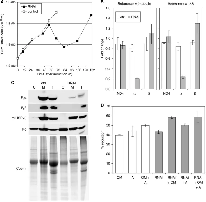
Addition of tet to the culture medium, which results in expression of double-stranded RNA and RNAi-mediated de-gradation of the target mRNA (Shiet al, 2000), led to massive lysis of parasites after 56 h (Figure 1A). No live parasites were detected between 60 and 85 h after induction of RNAi by microscopic inspection of the culture (not shown), but cul-ture growth resumed indicating escape from RNAi as com-monly seen with RNAi in trypanosomes (Chenet al, 2003).
To assess the effectiveness and specificity of the knock-down, we performed quantitative real-time RT–PCR on RNA that was prepared from parasites 43 h after RNAi induction as well as from uninduced control and parental cell line para-sites. The subunitamRNA was reduced byB80% while the level of mitochondrially encoded ND4 mRNA, serving as an additional control, was not significantly changed compared to the parental cell line whetherb-tubulin mRNA or 18S rRNA was used as RNA standard (Figure 1B). The slight increase in subunitbmRNA in the induced cells might be a response to loss of activity of the corresponding protein or complex (see below).
Western analysis of crude mitochondrial and cytosolic fractions prepared from 45 h RNAi-induced parasites revealed a substantial loss of F1subunitband F0subunit 4 compared to uninduced control; reagents for the a subunit were not available (Figure 1C, compare lanes 2 and 5). A crude mitochondrial fraction was prepared by permeabilizing cells with 0.015% (w/v) digitonin, which leaves the mitochon-drion intact (Tanet al, 2002), and was then lysed using 0.1% Triton X-100. Cells and other organelles that had escaped lysis together with insoluble proteins were pelleted in a subse-quent centrifugation step. Analysis with an antibody specific for mitochondrial HSP70 as well as Coomassie staining of a parallel gel showed that comparable amounts of sample were loaded with perhaps slightly more for the uninduced sam-ples. Analysis with an antiserum specific for the cytosolic ribosomal protein P0 showed cytosol was released by digito-nin treatment (lanes 1 and 4). It also indicated that lysis was incomplete (as expected) since P0 was also present in the organellar and pellet fractions. Samples from recovered cul-tures (e.g. 130 h) had normal levels ofb subunit, reflecting cell escape from RNAi (data not shown). The reduced levels of subunits 4 andbsuggest that, as observed for yeast (Lai-Zhang et al, 1999; Lefebvre-Legendre et al, 2001), in the absence of theasubunit the F1F0complex does not assemble properly and its unincorporated subunits degrade and/or mislocalize.
Mitochondrial ATPase activity was also substantially re-duced upon RNAi induction (Figure 1D). ATP hydrolytic activity was measured via release of free phosphate (Lawet al, 1995) in mitochondria prepared from 45 h RNAi-induced and unRNAi-induced cells using digitonin as described above. Oligomycin, a specific inhibitor of the F0F1complex, and azide, an inhibitor of F1 (Buchet and Godinot, 1998), reduced activity byB40 and B44%, respectively. This is similar to the degree of inhibition observed with other crude mitochondrial fractions from T. brucei (Bienen and Shaw, 1991) while 50–60% inhibition was observed with purified preparations ofT. bruceiF0F1-ATP synthase (Opperdoeset al, 1976; Williams, 1994), indicating the presence of other ATP hydrolytic activities in these crude mitochondrial fractions. Induction of RNAi for 45 h reduced ATPase activity by B43%, which is comparable to the reduction obtained with
oligomycin or azide. Addition of oligomycin and/or azide to these extracts resulted in minor further decreases of ATPase activity, presumably since none of these treatments alone is 100% effective. Together, these results demonstrate that the activity resulting from the mitochondrial ATP synthase com-plex in these extracts was reduced substantially, consistent with the results from the Western analysis. Overall, these results show that knockdown of ATP synthase subunitaby
RNAi resulted in substantial and specific reduction of the amount and activity of the ATP synthase complex, which subsequently resulted in cell death.
Inactivation of the F1-ATP synthase complex results in loss ofDWm in LS stage T. brucei
We assayedDCm in response to induction of RNAi since the ATP synthase complex in LS stageT. bruceiwas proposed to 1.E+02
1.E+03 1.E+04 1.E+05
0 12 24 36 48 60 72 84 96 108 120 132 Time after induction (h)
Cumulative cells ( × 10 3/ml) RNAi control F1α F0β mtHSP70 P0 1 2 3 4 5 6 ctrl RNAi C M I C M I Coom. Reference = β-tubulin 0.0 0.2 0.4 0.6 0.8 1.0 1.2 1.4 1.6 Fold change ct rl RNAi Reference = 18S ND4 α β ND4 α β 0 10 20 30 40 50 60 70 OM A OM + A RNAi RNAi + OM RNAi + A RNAi + OM + A % reduction A B C D
Figure 1 Knockdown of ATP synthase subunitaexpression is lethal to LS stageT. brucei. (A) Culture growth shown as cumulative cell numbers after normalization for dilution during cultivation. Expression of subunitawas silenced using tet-inducible RNAi (solid squares); uninduced cells were maintained in parallel (open squares). (B) Determination of mRNA levels for ATP synthase subunitsaandband for the mitochondrial transcript ND4 by real-time RT–PCR (DDCt method). Total RNA was isolated from induced cells after 43 h as well as from uninduced control cells. Primers for subunitawere located outside the region targeted by RNAi. Relative changes in mRNA levels with respect to the parental cell line are indicated, usingb-tubulin mRNA or 18S rRNA for normalization. Average numbers for four amplifications are shown, using RNA preparations from two independent RNAi experiments. (C) Western blot analysis of crude cytosolic (C), soluble mitochondrial (M), and insoluble (I) fractions prepared from RNAi-induced cells after 45 h and from uninduced control cells. The blots were probed with reagents detecting the indicated proteins. Coomassie staining (bottom panel) revealed the protein loading. (D) Analysis of ATPase activity in crude mitochondrial fractions, generated as in (C) and assayed by measuring release of free phosphate. ATP synthase inhibitors oligomycin (OM; 2.5mg/ml) and azide (A; 1 mM) were added where indicated. Average numbers for four assays are shown, using extract preparations from two independent RNAi experiments.
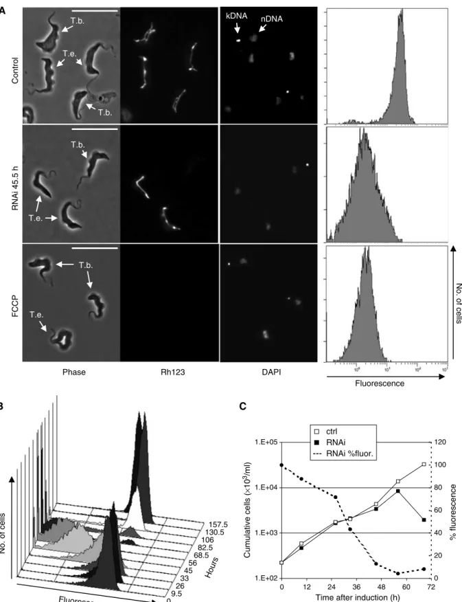
function in the generation of a DCm (Nolan and Voorheis, 1992; Divo et al, 1993). Live T. brucei were stained with 0.25mM rhodamine 123 (Rh123), a fluorescent dye that is a marker for energized mitochondria (Divoet al, 1993; Wilkes et al, 1997), and the resultant green fluorescence intensity was observed by epifluorescence microscopy and measured by flow cytometry (Figure 2A). Visual inspection by fluores-cence microscopy showed bright fluoresfluores-cence of the single tubular mitochondrion with very little background staining as reported (Divo et al, 1993). Inclusion of the uncoupler FCCP reduced the fluorescence substantially (bottom row). Induction of ATP synthase subunitaRNAi for 45.5 h resulted in fluorescence with an intensity that is comparable to that of FCCP-treated cells (center row; note that Dk T. evansi cells were included as controls for staining). Analysis of Rh123 fluorescence over the RNAi time course revealed continuously decreasing fluorescence for 56 h followed by emergence of a population of parasites with normal DCm due to escape from RNAi (Figure 2B). Comparison of the percentage of normal Rh123 fluorescence (and thus DCm) with the growth curve from Figure 1A showed that the decrease ofDCm set in just a few hours after induction of RNAi and reached 50% afterB30 h, or more than 24 h before the onset of an effect on growth (Figure 2C). This suggests that the decrease in DCm is a primary response to the inactivation of the F1-ATP synthase complex and not a consequence of other lethal events. In addition, the DCm decrease is not immediately lethal but may result in lethality such as by loss of mitochondrial protein import as discussed below. In conclusion, these results strongly support an essential role of the F1part of the ATP synthase in generation of aDCm.
Knockout of kinetoplastid RNA editing ligase 1 in Dk T. evansi
We investigated the requirement for the RNA editing complex in LS stage Dk trypanosomes since ATP synthase subunit 6 is encoded in the maxicircle component of mtDNA and its mRNA requires editing to be functional (Bhat et al, 1990). Analysis of two Dk strains showed that they contain functional editing complexes (Domingo et al, 2003). One of the strains, T. evansi Antat 3/3, has a single class of minicircles and lacks maxicircles (Borst et al, 1987; Songaet al, 1990) and thus lacks all mitochondrial protein coding genes, including that for ATP synthase subunit 6. We prepared null mutants of the kinetoplastid RNA editing ligase 1 (KREL1), which is essential in LS stage T. brucei (Schnaufer et al, 2001), using a gene replacement strategy (Wirtz et al, 1999). The first allele was replaced with the neomycin resistance marker and the second with a construct containing tetR and the hygromycin resistance marker. Clonal transfectants were obtained at normal frequency. Western analysis confirmed the absence of the KREL1 protein from these null mutants (Supplementary Figure S1A) and PCR analysis confirmed the absence of the KREL1 gene (data not shown). The growth rate of KREL1/cells was indistinguishable from that of wild-type and KREL1þ/
cells (Supplementary Figure S1B). Thus, in contrast toT. brucei, the KREL1 enzyme and, by implication, the editing complex and RNA editing do not have a vital function in T. evansiAntat 3/3.
Knockdown of ATP synthase subunitain Dk T. evansi Antat 3/3 is lethal
ATP synthase subunitaexpression was silenced by inducible RNAi expression in the T. evansi KREL/
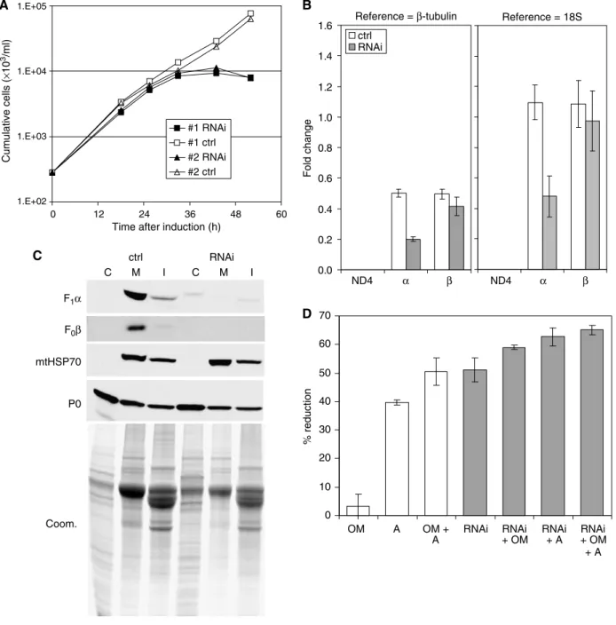
mutant, taking advantage of the expression of tetR in these cells. This knockdown was lethal to the parasite and no escape was observed, implying a robust knockdown (Figure 3A). Real-time RT–PCR analysis of RNAs from cells induced for 24 h showed that the knockdown was specific for subunit a (Figure 3B). No signal was observed for ND4 mRNA as expected since mtDNA—and hence this gene—is absent (note that our analysis compared transcript levels of RNAi and uninduced control cells with levels in T. brucei). The relative amounts ofa and b subunits in uninduced control cells were reduced byB50% whenb-tubulin was used as a reference, which was not the case when 18S rRNA was used for normalization. This indicates that the relative amounts of the two reference RNAs differ betweenT. evansiandT. brucei. Subcellular fractions from theT. evansiparasites in which RNAi was induced for 31 h and from uninduced control cells were prepared by sequential lysis with digitonin and Triton X-100, as described above for T. brucei, and analyzed by immunoblotting (Figure 3C). Interestingly, abundance of F0 subunit 4 appeared to be reduced inT. evansicompared to T. brucei, possibly as a result of decreased stability of F0in the absence of the mitochondrially encoded subunit 6. As in T. brucei, the amounts of F1 subunit b and F0 subunit 4 were substantially reduced upon RNAi induction. This again suggests that the ATP synthase complex cannot assemble in the absence of the a subunit and that unincorporated sub-units are degraded. The RNAi knockdown resulted in reduc-tion of ATP hydrolytic activity to a similar extent as in T. brucei using crude mitochondrial fractions obtained and assayed as described above (Figure 3D). Oligomycin had only a small effect, if any, which is consistent with the expected lack of a functional F0 part required to confer oligomycin sensitivity to the F0F1complex (Boyer, 1997). Insensitivity of mitochondrial ATPase activity isolated from various Dk trypanosomes to oligomycin was reported previously (Opperdoeset al, 1976).
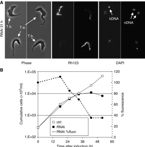
Knockdown of ATP synthase subunita expression in T. evansi resulted in substantial reduction of mitochondrial staining with Rh123 (Figure 4A). DCm decreased with a kinetic profile similar toT. bruceibut the onset of cell death occurred sooner in T. evansi (Figure 4B). Cell growth had already slowed when the Rh123 fluorescence reached 50% in T. evansi(atB35 h) while growth was unaffected at this stage (50% fluorescence at B30 h) in T. brucei. These apparent differences in part may reflect a somewhat lowerDCm in T. evansi, judged from results obtained by fluorescence microscopy using alternative dyes (data not shown). However, they may also reflect an altered role of F1 in generating theDCm (see below).
Effect of ATP synthase inhibitors on growth andDWm One prediction from the above results is that growth of LS T. bruceiandT. evansiAntat 3/3 should show very different susceptibilities to the F0F1 inhibitor oligomycin. Indeed, 125 ng/ml oligomycin was lethal to LS T. brucei while T. evansi was resistant to the inhibitor (Figure 5A and B). Growth of T. evansi did slow at concentrations above 500 ng/ml, perhaps due to nonspecific toxicity at high
No. of cells Fluorescence RNAi 45.5 h Control A 0 9.5 26 33 45 56 68.5 82.5 106 130.5 157.5 Fluorescence Hours No. of cells B C 1.E+02 1.E+03 1.E+04 1.E+05 0 12 24 36 48 60 72
Time after induction (h)
Cumulative cells ( × 10 3/ml) 0 20 40 60 80 100 120 % fluorescence ctrl RNAi --- RNAi %fluor. FCCP kDNA nDNA T.b. T.b. T.b. T.e. T.e. T.b. T.e. Phase Rh123 DAPI
Figure 2 Knockdown of ATP synthase subunitain LST. bruceiabolishesDCm. (A) Live cells were stained with the fluorescent mitochondrial marker Rh123 (0.25mM), immobilized, and analyzed by phase contrast and epifluorescence microscopy.DCm-related fluorescence was also measured by flow cytometry (right column). Top row: uninducedT. bruceicells, mixed with DkT. evansi, which can be distinguished by the absence of kDNA (DAPI staining; nDNA: nuclear DNA) and which serve as a control for Rh123 staining. Center panel: after induction of subunitaRNAi inT. bruceifor 45.5 h. Bottom panel: cells treated with the uncoupler FCCP. In all panels, the bar represents 20mm. (B) Change over time inDCm-related fluorescence after induction of subunitaRNAi. (C) Change inDCm-related fluorescence combined with growth analysis. Fluorescence is expressed as percent of average fluorescent signal compared to uninduced control cells.
concentrations (data not shown). In contrast, T. evansi showed about two-fold higher sensitivity than T. brucei to the F1 inhibitor azide (Figure 5C and D). The effects of oligomycin and azide on growth were consistent with the effects of these inhibitors on DCm: while 125 mg/ml oligo-mycin selectively abolishedDCm inT. brucei,T. evansicells were unaffected (Supplementary Figure S2, center rows). In
contrast, 0.5 mM azide abolished mitochondrial staining with Rh123 for bothT. bruceiandT. evansi.Insect formT. brucei, which, like most other organisms, generates DCm using respiratory complexes III and IV, remained unaffected (Supplementary Figure S2, bottom rows).
Incubation with 80mM bongkrekic acid, an inhibitor of the AAC, also selectively decreasedDCm in LST. bruceiand
#1 RNAi #1 ctrl #2 RNAi #2 ctrl mtHSP70 P0 Coom. 0.0 0.2 0.4 0.6 0.8 1.0 1.2 1.4 1.6 Fold change ctrl RNAi 0 10 20 30 40 50 60 70 % reduction OM A OM + A RNAi RNAi + OM RNAi + A RNAi + OM + A 1 2 3 4 5 6 F1α F0β ctrl RNAi C M I C M I 1.E+02 1.E+03 1.E+04 1.E+05 0 12 24 36 48 60
Time after induction (h)
Cumulative cells (
×
10
3/ml)
Reference = β-tubulin Reference = 18S
ND4 α β ND4 α β
A
C
B
D
Figure 3 Knockdown of ATP synthase subunitaexpression is lethal to DkT. evansiAntat 3/3. (A) Culture growth shown as cumulative cell numbers after normalization for dilution during cultivation. Expression of subunitawas silenced using tet-inducible RNAi (solid squares), and uninduced cells were maintained in parallel (open squares). Growth curves for two individual clones are shown. (B) Determination of mRNA levels for ATP synthase subunitsaandband for the mitochondrial transcript ND4 as described for Figure 1B. Total RNA was isolated from induced cells after 24 h and from uninduced control cells. Indicated are relative changes in mRNA levels with respect to LST. brucei. Average numbers for four real-time PCR experiments are shown, using RNA preparations from two independent RNAi experiments. (C) Western blot analysis of crude cytosolic (C), soluble mitochondrial (M), and insoluble (I) fractions prepared from RNAi-induced cells after 31 h and from uninduced control cells. The blots were probed with reagents detecting the indicated proteins. Coomassie staining (bottom panel) revealed the protein loading. (D) Analysis of ATP hydrolytic activity in crude mitochondrial fractions, generated as in (C) and assayed by measuring release of free phosphate. Average numbers for four assays are shown, using extract preparations from two independent RNAi experiments. See Figure 1D for abbreviations.
T. evansi (Supplementary Figure S3). Hence, exchange of ATP and ADP between cytosol and mitochondrial matrix is involved in maintaining DCm in the bloodstream forms, but not the insect form. This role of the AAC can be direct and/or indirect, as discussed below.
Identification of a potential compensatory mutation in a Dk trypanosome
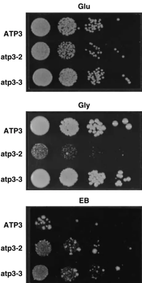
In the petite-negative yeast Kluyveromyces lactis, several mutations that can compensate for the loss of mtDNA are located in subunits of F1 (Clark-Walker et al, 2000). To investigate whether a similar mechanism might be responsi-ble for dyskinetoplasty in trypanosomes, we cloned and compared gene sequences for a, b, and g subunits from normalT. bruceistrains 427 and 164, the acriflavine-induced Dk strain 164 (Stuart, 1971), andT. evansistrain Antat 3/3. Among the three polymorphisms identified was a change from Leu to Pro at amino-acid residue 262 of thegsubunit of T. bruceiDk164 (Supplementary Figure S4). This mutation is in the C-terminal region of the g subunit (in the matrix proximal region of F1), close to suppressor mutations identified in K. lactis (atp3-1 and atp3-2 in Supplementary Figure S4) (Clark-Walkeret al, 2000). We therefore tested whether this mutation might be associated with increased tolerance to the loss of mtDNA. Conservation of this leucine between T. brucei and K. lactis enabled us to introduce
the homologous amino-acid change into the yeast, where the g subunit is encoded by the ATP3 gene. Yeast cells that harbor the Leu to Pro mutation (designated atp3-3) grew on standard glucose plates as well as wild type (Figure 6, top panel). Cells expressing atp3-2 grew a little slower. Cells expressing atp3-3 grew as well as wild type on a nonfermentable carbon source (glycerol), indicating that this mutation did not significantly affect the ability of the ATP synthase complex to function in oxidative phosphor-ylation (Figure 6, center panel). In contrast, as found before (Clark-Walkeret al, 2000), yeast cells expressing atp3-2 grew poorly on glycerol. When plated on glucose medium contain-ing ethidium bromide (EB), which causes deletions in mtDNA, the atp3-3 cells grew as well as the atp3-2 cells, while wild-type cells grew very poorly (Figure 6, bottom panel). Analysis of the individual EB-resistant atp3-3 colonies showed that they had lost mtDNA and did not grow on nonfermentable substrates (data not shown). The two resistant colonies obtained for the wild-type strain are EB-induced mutants containing suppressors of r0/r lethality (data not shown). Thus, the mutation identified in the ATP synthase subunit g from T. brucei DK164 can convert the petite-negative yeast K. lactis into a petite-positive form. Consequently, it seems likely that the Leu to Pro change has a similar role in compensating the loss of kDNA in trypanosomes. 0 20 40 60 80 100 120 % fluorescence ctrl RNAi ---RNAi %fluor. RNAi 31 h Phase Rh123 DAPI T.e. T.b. T.b. kDNA nDNA 1.E+02 1.E+03 1.E+04 1.E+05 0 12 24 36 48 60
Time after induction (h)
Cumulative cells ( × 10 3/ml) A B
Figure 4 Knockdown of ATP synthase subunitainT. evansiAntat 3/3 reducesDCm. (A) Rh123 staining of immobilized cells as described for Figure 2A.T. bruceicells served as staining controls. Note that theT. evansicell in the upper right corner has duplicated its nuclear genome. (B) Change inDCm-related fluorescence combined with growth analysis as described for Figure 2C. Fluorescence is expressed as percent of average fluorescent signal compared to uninduced control cells.
Suppressor mutations in K. lactis reduce the Km of F1 for ATP (Clark-Walker, 2003). Similarly, we found that the atp3-3 mutation reduced the Km from 2.0670.14 to 0.8970.14mmol/min mg protein (Supplementary Figure S5).
Discussion
The F1part of the mitochondrial ATP synthase complex was found to be essential for generatingDCm and for survival of LS bloodstream form T. brucei as well as its Dk relativeT. evansiAntat 3/3. We hypothesize that cell death is due to the collapse of critical transport processes between mitochon-drion and cytoplasm, which depend onDCm. These results indicate that the ATP synthase complex hydrolyzes ATP to generate theDCm in bloodstream forms, hence functioning opposite to its ATP synthesis mode that occurs in the insect stage and in most eukaryotes. A mutation was found in the nuclearly encoded ATP synthase g subunit of a Dk strain, which appears to help these cells evade the need for gene
products encoded in mtDNA, such as subunit 6 of the ATP synthase complex. The RNA editing complex, which, like mtDNA, is essential in normalT. brucei, was shown not to be essential inT. evansi, suggesting that its function inT. brucei is limited to its role in the expression of mitochondrial genes. The activity of the F0F1-ATP synthase complex is essential in LST. bruceisince both knockdown of expression of itsa subunit and addition of oligomycin or azide, inhibitors of this complex, were lethal within a few days (Figures 1A and 6A). RNAi knockdown of theasubunit resulted in reduced levels of subunits b and 4, confirming that the complex cannot assemble in the absence of thea subunit (Lai-Zhanget al, 1999; Lefebvre-Legendreet al, 2001).
Collapse ofDCm appeared to be the primary consequence of subunitaknockdown since its decrease began shortly after induction of RNAi and reached background levels prior to inhibition of growth (Figure 2C). This finding supports the conclusion of others that oligomycin sensitivity of theDCm reflects its generation by the ATP synthase complex in LS Oligomycin T. brucei 1.E+02 1.E+03 1.E+04 1.E+05 1.E+06 0 12 24 36 48 60 72 Time (h) 0 12 24 36 48 60 72 Time (h) 0 12 24 36 48 60 72 84 96 108 Time (h) 0 12 24 36 48 60 72 84 96 108 Time (h) Cumulative cells ( × 10 3/ml) 1.E+02 1.E+03 1.E+04 1.E+05 1.E+06 1.E+07 Cumulative cells ( × 10 3/ml) 1.E+02 1.E+03 1.E+04 1.E+05 1.E+06 Cumulative cells ( × 10 3/ml) 1.E+02 1.E+03 1.E+04 1.E+05 Cumulative cells ( × 10 3/ml) no drug 125 ng/ml 250 ng/ml 500 ng/ml Azide T. brucei no drug 0.01 mM 0.05 mM 0.1 mM 0.5 mM 1.0 mM Oligomycin T. evansi no drug 125 ng/ml 250 ng/ml 500 ng/ml Azide T. evansi no drug 0.01 mM 0.05 mM 0.1 mM 0.5 mM 1.0 mM A B C D
Figure 5 Comparison of the sensitivities of growth of LS formT. bruceiand DkT. evansito the F0F1inhibitor oligomycin (A, B) and the F1 inhibitor azide (C, D). Cells were grown in the presence of the indicated concentrations of inhibitor. Cumulative cell numbers reflect normalization for dilution during cultivation.
T. brucei(Nolan and Voorheis, 1992; Vercesiet al, 1992; Divo et al, 1993).DCm is absolutely required for mitochondrial import of proteins encoded in the nucleus as well as for other transport processes and is therefore essential for the vast majority of mitochondrial activities, including biogenesis of the mitochondrion itself (Neupert, 1997). Some mitochon-drial activities in LST. bruceiare repressed (Schneider, 2001), but recent findings have suggested important roles for the organelle in that stage of the life cycle (Schnauferet al, 2002). For instance, respiration in LS T. brucei is mediated by a mitochondrial trypanosome alternative oxidase and this en-zyme might be essential (Helfert et al, 2001). Collapse of DCm is also a key event in apoptosis and althoughT. brucei lacks components of the classical apoptotic pathway (Esseiva et al, 2004), it appears to have some form of programmed cell death (Welburnet al, 1997), which may have been triggered by inactivation of the ATP synthase complex.
MtDNA and its expression with the help of RNA editing have recently been shown to be essential for survival of LS bloodstream formT. brucei(Timmset al, 2002; Stuartet al, 2005). The fact that Dk trypanosomes can survive as blood-stream forms therefore presents an intriguing conundrum
(Schnauferet al, 2002), which is underscored by our finding that the normally essential RNA editing enzyme KREL1 can be knocked out in DkT. evansiwithout an obvious phenotype (Supplementary Figure S1). Findings presented in this paper help explain how LS Dk strains might generate theDCm and survive the loss of the normally required mitochondrial gene products. The Dk strains require F1-ATP synthase activity, like the wild type, as evident from the loss of DCm and growth inhibition resulting from knockdown of a subunit expression or addition of azide to the medium (Figures 3A, 4, and 5D). However, unlike the wild-type strain, ATP hydro-lysis activity and cell growth of DkT. evansiare essentially insensitive to oligomycin (Figure 5B; Opperdoeset al, 1976). This suggests that inT. evansi, the F1part, which by itself is insensitive to oligomycin, still functions in the generation of the DCm and that the F0F1 complex is altered. MtDNA of T. brucei encodes a protein with homology to ATP synthase subunit 6, the mRNA of which requires extensive RNA editing (Bhatet al, 1990). This subunit is an essential component of the proton channel of the F0 moiety (Boyer, 1997) and is therefore expected to be essential for the proton-pumping activity of F0F1-ATP synthase in LS T. brucei. This is also implied by collapse of theDCm inT. bruceifollowing treat-ment with acriflavine (Timmset al, 2002), which specifically deletes mtDNA. Based on our finding of a mutated ATP synthase gsubunit in a Dk strain of T. brucei, we suggest that mutations in subunits of the F0F1 complex may have altered the complex and compensated, at least in part, for the loss of mitochondrial gene products. Our finding resembles the identification of suppressor mutations in F1subunits in the petite-negative yeastK. lactis, which, likeT. brucei,does normally depend on the synthesis of mitochondrial gene products (Clark-Walkeret al, 2000). Indeed, introduction of the identified mutation into the gsubunit of K. lactis sup-pressed the petite-negative phenotype and enabled growth under conditions that induce loss of mtDNA (Figure 6). Like previously identified suppressor mutations in K. lactis, the mutation identified here results in a lower Km for ATP (Supplemental Figure S5; Clark-Walker, 2003). In addition, the mutation may increase the stability of F1in the absence of F0and this possibility remains to be investigated. The situa-tion in Dk cells therefore may be analogous tor0/r mam-malian cells and petite-positive yeasts, where ATP hydrolysis by F1 appears to result in a sufficiently low mitochondrial ATP concentration to allow the electrogenic exchange by the AAC of ADP3for ATP4. This exchange could generate a DCm that would sustain essential organelle functions, even in the absence of processes that require mitochondrial gene products (Giraud and Velours, 1997; Buchet and Godinot, 1998). Hence, the AAC would have a more indirect role for generatingDCm in the normal LS form (by supplying ATP for proton pumping by the F0F1complex) and a direct role in the Dk form. Indeed, DCm of both forms (but not the insect form) was reduced by the AAC inhibitor bongkrekic acid (Supplementary Figure S3). Additional studies are needed to determine whether F1-ATPases from Dk trypanosomes in general have a lower Km for ATP and if the mutation that we identified is sufficient to compensate for the loss of mitochondrial gene products inT. brucei. Considering that a singular evolutionary event may have given rise to stable Dk trypanosomes in nature (Brunet al, 1998) and the difficulty of generating such strains in the laboratory (Stuart, 1971; ATP3 Glu Gly EB atp3-2 atp3-3 ATP3 atp3-2 atp3-3 ATP3 atp3-2 atp3-3
Figure 6 The Leu262Pro mutation in the gene encoding the g
subunit of the F1moiety from a Dk strain ofT. bruceiconverts the petite-negative yeastK. lactisinto a petite-positive form. The Leu to Pro mutation was introduced into the conserved residue of the ATP synthasegsubunit ofK. lactis(Leu265 in ATP3) and the mutant allele (atp3-3) expressed in a strain lacking the endogenous ATP3 gene. Control strains expressed the wild-type ATP3 gene or a suppressor mutation identified previously (atp3-2). Growth was analyzed after serial dilution on plates containing the carbon sources glucose (Glu), glycerol (Gly), or glucose medium with 16mg/ml EB. Plates were incubated at 281C for 2, 6, and 9 days, respectively.
Timms et al, 2002), it seems more likely that multiple mutations may be required.
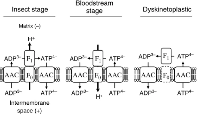
Our model for the generation ofDCm in trypanosomes is summarized in Figure 7. The F0F1-ATP synthase complex has the conventional role of ATP generation during oxidative phosphorylation in the insect stage but it does not appear to be essential, since oligomycin does not affect intracellular ATP levels and only moderately affects growth (Coustouet al, 2003). In the LS bloodstream stage, by contrast, the complex is normally essential, although much less abundant than in the insect stage (Brownet al, 2001), and has a role opposite to that of the conventional role, namely generation ofDCm at the expense of ATP consumption. The shift between life cycle stages and the need to control ATP hydrolysis in insect stage mitochondria suggests the existence of a critical regulatory system. The identification of a peptide that inhibits the enzymes’ ATP hydrolytic activity may be a component of such a regulatory system since its abundance appears to be upregulated in the insect stage (Chiet al, 1996). Intriguingly, our analysis of the T. brucei genome database (www.GeneDB.org) revealed three different genes for F0 subunit 9 (data not shown). Analysis of their expression or activity in different life cycle stages may indicate potential mechanisms that regulate the activity of the complex. The unusual but essential role of the mitochondrial ATP synthase complex in the disease-causing stage ofT. bruceimay open opportunities for drug development. An inhibitor selective for the ATP hydrolytic direction of the enzyme would be ex-pected to be lethal to the parasite but not the host, and such inhibitors may be adapted from those under development to prevent tissue damage under ischemic conditions (St Pierre et al, 2000).
Materials and methods
Trypanosome plasmid construction and transfection The inducible RNAi plasmid for silencing ATP synthase subunita
was generated using the pQuadra system (Inoueet al, 2005). Briefly, the first 530 bp of the subunitagene were amplified by PCR, using oligos with specifically designedBstXI sites. Ligation with Bst XI-digested pQuadra1 and pQuadra3 plasmids generated pQuadra-ATPa, containing inverted repeats of the PCR product separated by a spacer region. Knockout plasmids for replacing the two KREL1 alleles with T7RNAP plus neomycin resistance marker and tetR plus hygromycin resistance marker, respectively, have been described previously (Schnauferet al, 2001). Transfection ofNotI-linearized constructs into the LS bloodstream form ‘single marker’ cell line (Wirtzet al, 1999) orT. evansiAntat 3/3 (Borstet al, 1987) and selection of transgenic cell lines was carried out as described (Schnauferet al, 2001).
Growth analysis of trypanosomes
Throughout growth analyses, cells were maintained at exponential growth (between 105 and 106cells/ml). RNAi was induced by adding 1mg/ml tet to the medium.
Quantitative real-time RT–PCR analysis
Mid-log trypanosomes were harvested at room temperature (10 min, 1300g) and RNA was isolated using the Ultraspec RNA Reagent (Biotecx Laboratories Inc., Houston, TX). Quantitative real-time RT–PCR analysis was carried out as described (Carneset al, 2005), using 10ml of a mix of the specific primers at 1.5mM (see Supplementary data). Reactions were analyzed with the ABI Prism 7000 software (Applied Biosystems). Relative amounts of RNA template in the preparations were calculated using the DDCt method (Ingham et al, 2001). Parallel amplifications minus the reverse transcription step revealed only insignificant contamina-tions with genomic DNA.
Digitonin fractionation, ATPase assay, and Western blotting Crude mitochondrial preparations were obtained by fractionation with digitonin (Tan et al, 2002). ATPase activity was measured based on release of free phosphate (Law et al, 1995). Briefly, B4108mid-log trypanosomes were harvested by centrifugation (1300g, 10 min), washed, and permeabilized with 0.015% (w/v) digitonin. The crude cytosolic fraction was obtained as the supernatant from a 4000g/3 min spin at 41C. The pellet, represent-ing the crude mitochondrial fraction, was resuspended in ATPase assay buffer (10 mM Tris–HCl, pH 8.2; 0.2 M KCl; 2 mM MgCl2). Where indicated, oligomycin or sodium azide was added to 25mg/ ml (B10mg/mg protein) and 1 mM, respectively. The reaction was started by addition of ATP to a final concentration of 5 mM. After 0, 10, and 20 min, 95ml aliquots were added to 5ml 3 M trichloric acid and kept on ice for 30 min. The precipitate was pelleted (16 000g, 10 min), 90ml of the supernatant was added to 0.5 ml Sumner reagent (Lawet al, 1995), and absorbance was measured at 610 nm. For Western analysis, the crude mitochondrial fraction obtained above was lysed by addition of 0.1 volume of 10% Triton X-100. Soluble and insoluble fractions were obtained by centrifugation at 16 000gfor 10 min and separated by SDS–PAGE. Gels were either stained with Coomassie blue or transferred to PVDF membrane. Western blots were probed with reagents against the F1moiety of
Crithidia fasciculata(Speijeret al, 1997), which crossreacts only
with thebsubunit of theT. bruceicomplex, subunit 4 ofLeishmania
tarentolae (Nebohacova et al, 2003), mitochondrial HSP70 from
T. brucei(Panigrahi et al, 2003), and ribosomal protein P0 from
T. cruzi(Skeiky et al, 1994). For Western analysis of whole-cell
lysates, cells were harvested by centrifugation and lysed in SDS sample buffer. Extracts corresponding to 5107 cell equivalents were fractionated and transferred to PVDF membrane as described above and probed with monoclonal antibodies against editosome proteins KREPA1, KREPA2, KREL1, and KREPA3 (Panigrahiet al, 2001; Schnauferet al, 2001).
Analysis ofDWm by microscopy and flow cytometry
A 1 ml portion of mid-log trypanosomes was incubated in the presence of 250 nM Rh123 (Molecular Probes; plus 1mg/ml DAPI for microscopy) for 20 min at 371C, harvested (1300g, 10 min), and washed with 1 ml CytoMix (25 mM HEPES, pH 7.6; 120 mM KCl; Matrix (–)
Intermembrane space (+)
Insect stage Bloodstreamstage Dyskinetoplastic
H+ ADP3– ATP4– ATP4– AAC AAC F0 F1 ADP3– H+ ADP3– ATP4– ATP4– AAC AAC ADP3– ADP3– ATP4– ATP4– AAC AAC ADP3– F0 F0 F1 F1
Figure 7 Model for ATP synthase complex function in trypanosome mitochondria. The ATP synthase complex functions conventionally in the insect stage and generates ATP by utilizing the proton gradient produced by the respiratory chain. The ATP synthase complex works in reverse in the LS bloodstream stage, which lacks a cytochrome-mediated respiratory chain, and utilizes ATP to pump protons out of the matrix, generating a DCm. In Dk trypanosomes, the F1part is still essential for generating aDCm, presumably for ATP hydrolysis as in the wild type, but the F0part is absent or nonfunctional. The electrogenic ATP/ADP exchange that is catalyzed by AAC may be involved in the generation of aDCm as has been proposed forr0andrmammalian and yeast cells (see text). A high rate of ATP hydrolysis by F1may be required to keep the matrix ATP concentration sufficiently low, which may be achieved by mutations in F1 subunits like those identified here and in petite-negative yeasts. Note that each AAC catalyzes the exchange of one molecule of ADP for one molecule of ATP; two AAC proteins are shown solely to clarify ATP/ADP entry and exit.
0.15 mM CaCl2; 10 mM K2HPO4/KH2PO4, pH 7.6; 2 mM EDTA; 5 mM MgCl2; 6 mM glucose). For microscopy, cells were resus-pended in residual buffer, partially immobilized on HMI-9 medium containing 0.65% low-melting-type agarose, and analyzed using a Nikon Eclipse E600 fluorescence microscope. For flow cytometry, cells were resuspended in 0.5 ml CytoMix and analyzed for green fluorescence in a Beckman Coulter Epics XL-MCL flow cytometer. Sequence comparison of ATP synthase subunitsa,b, andc Genomic DNA was prepared from the following trypanosome strains:
T. brucei brucei427, procyclic and bloodstream stage (Cross, 1975);T.
brucei brucei164 and DK164 (Stuart, 1971);T. evansiAntat 3/3 (Borst
et al, 1987). Complete coding sequences for ATP synthase subunits
were amplified by PCR (see Supplementary data for primers), subcloned, and sequenced. In each case, sequences from at least three individual PCR reactions were analyzed to exclude PCR artifacts. Yeast strains and plasmids; expression of mutagenized ATP3 in yeast
See Supplementary data for details.
Supplementary data
Supplementary data is available atThe EMBO JournalOnline.
Acknowledgements
We thank R Benne (University of Amsterdam) for the F1antiserum, S Reed (Corixa Corp.) for the P0 antiserum, M Inoue (Kirume University) for the pQuadra plasmids, G Cross (Rockefeller University) for the single-marker cell line, A Schneider (University of Fribourg) for technical advice and David Pe´rez-Morga (Universite´ Libre de Bruxelles) for advice on immobilizing trypanosomes. We thank J Carnes for advice on real-time PCR analysis and other members of the Stuart lab for helpful discussions. LiJun Ouyang (RSBS, ANU) is thanked for skilled technical assistance. We also thank H Interthal (University of Washington) and G Domingo (SBRI) for critical reading of the manuscript. This work was supported in part by NIH grant AI014102-28 (KS) and the MJ Murdock Charitable Trust (SBRI).
References
Bertrand KI, Hajduk SL (2000) Import of a constitutively expressed protein into mitochondria from procyclic and bloodstream forms
ofTrypanosoma brucei.Mol Biochem Parasitol106:249–260
Bhat GJ, Koslowsky DJ, Feagin JE, Smiley BL, Stuart K (1990) An extensively edited mitochondrial transcript in kinetoplastids encodes a protein homologous to ATPase subunit 6. Cell 61: 885–894
Bienen EJ, Shaw MK (1991) Differential expression of the oligomy-cin-sensitive ATPase in bloodstream forms ofTrypanosoma brucei
brucei.Mol Biochem Parasitol48:59–66
Borst P, Fase-Fowler F, Gibson WC (1987) Kinetoplast DNA of
Trypanosoma evansi.Mol Biochem Parasitol23:31–38
Boyer PD (1997) The ATP synthase—a splendid molecular machine.
Annu Rev Biochem66:717–749
Brown BSV, Chi TB, Williams N (2001) TheTrypanosoma brucei mitochondrial ATP synthase is developmentally regulated at the level of transcript stability. Mol Biochem Parasitol 115: 177–187
Brun R, Hecker H, Lun ZR (1998) Trypanosoma evansi and
T. equiperdum: distribution, biology, treatment and phylogenetic
relationship (a review).Vet Parasitol79:95–107
Buchet K, Godinot C (1998) Functional F1-ATPase essential in maintaining growth and membrane potential of human mito-chondrial DNA-depleted rho degrees cells. J Biol Chem 273: 22983–22989
Burger G, Gray MW, Lang BF (2003) Mitochondrial genomes: any-thing goes.Trends Genet19:709–716
Carnes J, Trotter J, Ernst N, Steinberg AG, Stuart K (2005) An essential RNA editing insertion specific endonuclease in
Trypanosoma brucei.Proc Natl Acad Sci USA, (in press)
Chen XJ, Clark-Walker GD (1999) Alpha and beta subunits of F1-ATPase are required for survival of petite mutants in
Saccharomyces cerevisiae.Mol Gen Genet262:898–908
Chen Y, Hung CH, Burderer T, Lee GS (2003) Development of RNA interference revertants inTrypanosoma bruceicell lines generated with a double stranded RNA expression construct driven by two opposing promoters.Mol Biochem Parasitol126:275–279 Chi TB, Choi SY, Williams N (1996) The ATP synthase of
Trypanosoma bruceiis developmentally regulated by an inhibitor
peptide.Arch Biochem Biophys333:291–297
Clark-Walker GD (2003) Kinetic properties of F1-ATPase influence the ability of yeasts to grow in anoxia or absence of mtDNA.
Mitochondrion2:257–265
Clark-Walker GD, Hansbro PM, Gibson F, Chen XJ (2000) Mutant residues suppressing rho(0)-lethality in Kluyveromyces lactis occur at contact sites between subunits of F(1)-ATPase.Biochim
Biophys Acta1478:125–137
Coustou V, Besteiro S, Biran M, Diolez P, Bouchaud V, Voisin P, Michels PA, Canioni P, Baltz T, Bringaud F (2003) ATP generation in theTrypanosoma brucei procyclic form: cytosolic substrate level is essential, but not oxidative phosphorylation.J Biol Chem 278:49625–49635
Cross GAM (1975) Identification, purification and properties of clone-specific glycoprotein antigens constituting the surface coat ofTrypanosoma brucei.Parasitology71:393–417
Divo AA, Patton CL, Sartorelli AC (1993) Evaluation of rhodamine 123 as a probe for monitoring mitochondrial function in
Trypanosoma bruceispp.J Protozool40:329–335
Domingo GJ, Palazzo SS, Wang B, Panicucci B, Salavati R, Stuart KD (2003) DyskinetoplasticTrypanosoma bruceicontain functional editing complexes.Eukaryot Cell2:569–577
Esseiva AC, Chanez AL, Bochud-Allemann N, Martinou JC, Hemphill A, Schneider A (2004) Temporal dissection of Bax-induced events leading to fission of the single mitochondrion in
Trypanosoma brucei.EMBO Rep5:268–273
Futai M, Kanazawa H (1983) Structure and function of proton-translocating adenosine triphosphatase (F0F1): biochemical and molecular biological approaches. Microbiol Rev 47: 285–312
Giraud MF, Velours J (1997) The absence of the mitochondrial ATP synthase delta subunit promotes a slow growth phenotype of rho-yeast cells by a lack of assembly of the catalytic sector F1.Eur J
Biochem245:813–818
Hannaert V, Bringaud F, Opperdoes FR, Michels PA (2003) Evolution of energy metabolism and its compartmentation in Kinetoplastida.Kinetoplastid Biol Dis2:11
Helfert S, Estevez AM, Bakker B, Michels P, Clayton C (2001) Roles of triosephosphate isomerase and aerobic metabolism in
Trypanosoma brucei.Biochem J357:117–125
Ingham DJ, Beer S, Money S, Hansen G (2001) Quantitative real-time PCR assay for determining transgene copy number in transformed plants.BioTechniques31:132–140
Inoue M, Nakamura Y, Yasuda K, Yasaka N, Hara T, Schnaufer A, Stuart K, Fukuma T (2005) The 14-3-3 proteins ofTrypanosoma
bruceifunction in motility, cytokinesis and cell cycle.J Biol Chem
280:14085–14096
Lai-Zhang J, Xiao Y, Mueller DM (1999) Epistatic interactions of deletion mutants in the genes encoding the F1-ATPase in yeast
Saccharomyces cerevisiae.EMBO J18:58–64
Law RH, Manon S, Devenish RJ, Nagley P (1995) ATP synthase
from Saccharomyces cerevisiae. Methods Enzymol 260:
133–163
Lefebvre-Legendre L, Balguerie A, Duvezin-Caubet S, Giraud MF, Slonimski PP, di Rago JP (2003) F1-catalysed ATP hydrolysis is required for mitochondrial biogenesis inSaccharomyces cerevisiae growing under conditions where it cannot respire.Mol Microbiol 47:1329–1339
Lefebvre-Legendre L, Vaillier J, Benabdelhak H, Velours J, Slonimski PP, di Rago JP (2001) Identification of a nuclear gene (FMC1) required for the assembly/stability of yeast mitochon-drial F(1)-ATPase in heat stress conditions. J Biol Chem 276: 6789–6796
Matthews KR (2005) The developmental cell biology of
Nebohacova M, Maslov DA, Falick AM, Simpson L (2003) The effect of RNAi down regulation of RET1 30 TUTase on mitochondrial
de novoprotein synthesis and stability of respiratory complexes
inTrypanosoma brucei.J Biol Chem279:7819–7825
Neupert W (1997) Protein import into mitochondria. Annu Rev
Biochem66:863–917
Nolan DP, Voorheis HP (1992) The mitochondrion in bloodstream forms of Trypanosoma brucei is energized by the electrogenic pumping of protons catalysed by the F1F0-ATPase.Eur J Biochem 209:207–216
Opperdoes FR, Borst P, De Rijke D (1976) Oligomycin sensitivity of the mitochondrial ATPase as a marker for fly transmissibility and the presence of functional kinetoplast DNA in African trypano-somes.Comp Biochem Physiol B55B:25–30
Panigrahi AK, Allen TE, Haynes PA, Gygi SP, Stuart K (2003) Mass spectrometric analysis of the editosome and other multiprotein complexes inTrypanosoma brucei.J Am Soc Mass Spectrom14: 728–735
Panigrahi AK, Schnaufer A, Carmean N, Igo Jr RP, Gygi S, Ernst N, Palazzo SS, Weston D, Aebersold R, Salavati R, Stuart KD (2001) Four related proteins of theT. bruceiRNA editing complex.Mol
Cell Biol21:6833–6840
Schnaufer A, Domingo GJ, Stuart KD (2002) Natural and induced dyskinetoplastid trypanosomatids: how to live without mitochon-drial DNA.Int J Parasitol32:1071–1084
Schnaufer A, Panigrahi AK, Panicucci B, Igo Jr RP, Salavati R, Stuart K (2001) An RNA ligase essential for RNA editing and survival of the bloodstream form of Trypanosoma brucei. Science 291: 2159–2162
Schneider A (2001) Unique aspects of mitochondrial biogenesis in trypanosomatids.Int J Parasitol31:1403–1415
Shi H, Djikeng A, Mark T, Wirtz E, Tschudi C, Ullu E (2000) Genetic interference in Trypanosoma bruceiby heritable and inducible double-stranded RNA.RNA6:1069–1076
Skeiky Y, Benson DR, Elwasila M, Badaro R, Burns Jr JM, Reed SG (1994) Antigens shared byLeishmaniaspecies andTrypanosoma
cruzi: immunological comparison of the acidic ribosomal P0
proteins.Infect Immun62:1643–1651
Songa EB, Paindavoine P, Wittouck E, Viseshakul N, Muldermans S, Steinert M, Hamers R (1990) Evidence for kinetoplast and nuclear
DNA homogeneity inTrypanosoma evansiisolates.Mol Biochem
Parasitol43:167–180
Speijer D, Breek CK, Muijsers AO, Hartog AF, Berden JA, Albracht SP, Samyn B, Van Beeumen J, Benne R (1997) Characterization of the respiratory chain from cultured Crithidia fasciculata. Mol
Biochem Parasitol85:171–186
St Pierre J, Brand MD, Boutilier RG (2000) Mitochondria as ATP consumers: cellular treason in anoxia.Proc Natl Acad Sci USA97: 8670–8674
Stuart KD (1971) Evidence for the retention of kinetoplast DNA in an acriflavin-induced dyskinetoplastic strain of Trypanosoma
bruceiwhich replicates the altered central element of the
kineto-plast.J Cell Biochem49:189–195
Stuart KD, Schnaufer A, Ernst NL, Panigrahi AK (2005) Complex management: RNA editing in trypanosomes.Trends Biochem Sci 30:97–105
Tan TH, Bochud-Allemann N, Horn EK, Schneider A (2002) Eukaryotic-type elongator tRNAMet ofTrypanosoma brucei be-comes formylated after import into mitochondria.Proc Natl Acad
Sci USA99:1152–1157
Timms MW, van Deursen FJ, Hendriks EF, Matthews KR (2002) Mitochondrial development during life cycle differentiation of African trypanosomes: evidence for a kinetoplast-dependent dif-ferentiation control point.Mol Biol Cell13:3747–3759
Vercesi AE, Docampo R, Moreno SNJ (1992) Energization-depen-dent Ca2+accumulation inTrypanosoma bruceibloodstream and procyclic trypomastigotes mitochondria.Mol Biochem Parasitol 56:251–258
Welburn SC, Barcinski MA, Williams GT (1997) Programmed cell death in trypanosomatids.Parasitol Today13:22–26
Wilkes JM, Mulugeta W, Wells C, Peregrine AS (1997) Modulation of mitochondrial electrical potential: a candidate mechanism for drug resistance in African trypanosomes.Biochem J326:755–761 Williams N (1994) The mitochondrial ATP synthase of
Trypanosoma brucei: structure and regulation. J Bioenerg
Biomembr26:173–178
Wirtz E, Simone L, Claudia O, Cross GAM (1999) A tightly regulated inducible expression system for conditional gene knock-outs and dominant-negative genetics inTrypanosoma brucei.Mol Biochem
Erratum
The F1-ATP synthase complex in bloodstream stage
trypanosomes has an unusual and essential function
A Schnaufer, GD Clark-Walker, AG Steinberg and K Stuart
The EMBO Journal(2006)25,1175–1176. doi:10.1038/sj.emboj.7601013
Correction to:The EMBO Journal(2005)24, 4029–4040. doi:10.1038/sj.emboj.7600862; published online 3 November 2005 Owing to a typesetting error, Figures 1C and 3C of the above article were published incorrectly in print. The correct Figures 1 and 3 are reproduced below in their entirety.
The Publisher would like to apologize for any inconvenience this may have caused.
A D 1.E+02 1.E+03 1.E+04 1.E+05 0 12 24 36 48 60 72 84 96 108 120 132 Time after induction (h)
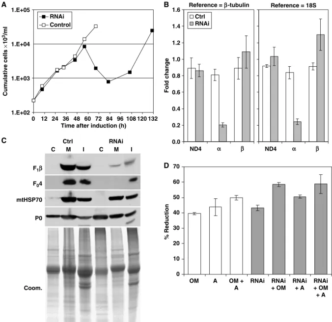
Cumulative cells × 10 3/ml RNAi Control C Reference = β-tubulin 0.0 0.2 0.4 0.6 0.8 1.0 1.2 1.4 1.6 Fold change Ctrl RNAi Reference = 18S ND4 ND4 0 10 20 30 40 50 60 70 OM A OM + A RNAi RNAi + OM RNAi + A RNAi + OM + A % Reduction F1β mtHSP70 P0 1 Ctrl F04 RNAi I M C I M C Coom. 6 5 4 3 2 α β α β B
Figure 1 For Figure Caption see next page.
EMBO
EMBO
JOURNAL
EMBO
D C 0 10 20 30 40 50 60 70 #1 RNAi #1 Ctrl #2 RNAi #2 Ctrl 1.E+02 1.E+03 1.E+04 1.E+05 Cumulative cells × 10 3/ml 0 12 24 36 48 60
Time after induction (h) Ctrl RNAi I M C I M C F1β mtHSP70 P0 F04 Coom. 1 2 3 4 5 6 OM A OM + A RNAi RNAi + OM RNAi + A RNAi + OM + A % Reduction ND4 α β ND4 α β 0.0 0.2 0.4 0.6 0.8 1.0 1.2 1.4 1.6 Fold change
Reference = β-tubulin Reference = 18S Ctrl
RNAi
A B
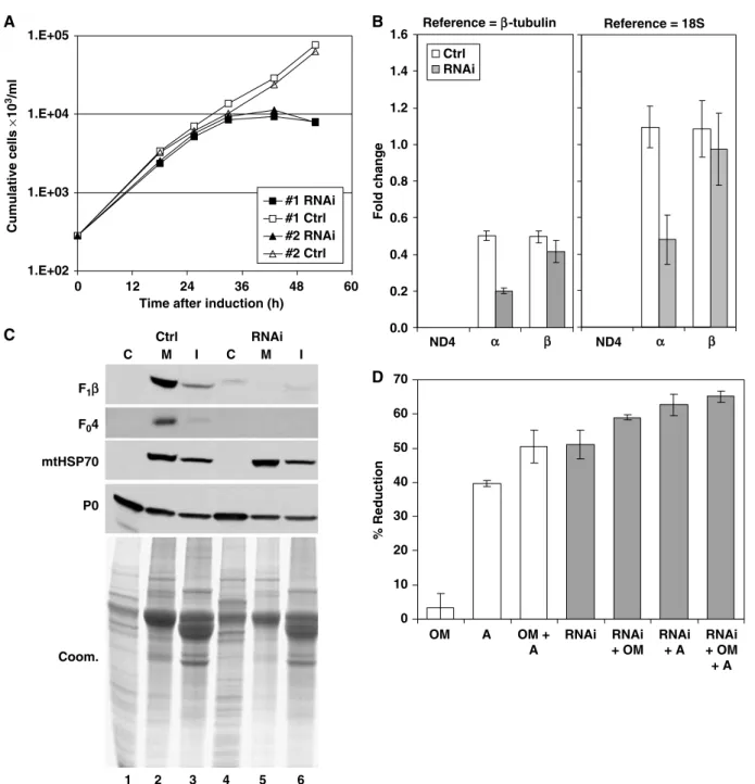
Figure 3 Knockdown of ATP synthase subunitaexpression is lethal to DkT. evansiAntat 3/3. (A) Culture growth shown as cumulative cell numbers after normalization for dilution during cultivation. Expression of subunitawas silenced using tet-inducible RNAi (solid squares), and uninduced cells were maintained in parallel (open squares). Growth curves for two individual clones are shown. (B) Determination of mRNA levels for ATP synthase subunitsaandband for the mitochondrial transcript ND4 as described for Figure 1B. Total RNA was isolated from induced cells after 24 h and from uninduced control cells. Indicated are relative changes in mRNA levels with respect to LST. brucei. Average numbers for four real-time PCR experiments are shown, using RNA preparations from two independent RNAi experiments. (C) Western blot analysis of crude cytosolic (C), soluble mitochondrial (M), and insoluble (I) fractions prepared from RNAi-induced cells after 31 h and from uninduced control cells. The blots were probed with reagents detecting the indicated proteins. Coomassie staining (bottom panel) revealed the protein loading. (D) Analysis of ATP hydrolytic activity in crude mitochondrial fractions, generated as in (C) and assayed by measuring release of free phosphate. Average numbers for four assays are shown, using extract preparations from two independent RNAi experiments. See Figure 1D for abbreviations.
Figure 1 Knockdown of ATP synthase subunitaexpression is lethal to LS stageT. brucei. (A) Culture growth shown as cumulative cell numbers after normalization for dilution during cultivation. Expression of subunitawas silenced using tet-inducible RNAi (solid squares); uninduced cells were maintained in parallel (open squares). (B) Determination of mRNA levels for ATP synthase subunitsaandband for the mitochondrial transcript ND4 by real-time RT–PCR (DDCt method). Total RNA was isolated from induced cells after 43 h as well as from uninduced control cells. Primers for subunitawere located outside the region targeted by RNAi. Relative changes in mRNA levels with respect to the parental cell line are indicated, usingb-tubulin mRNA or 18S rRNA for normalization. Average numbers for four amplifications are shown, using RNA preparations from two independent RNAi experiments. (C) Western blot analysis of crude cytosolic (C), soluble mitochondrial (M), and insoluble (I) fractions prepared from RNAi-induced cells after 45 h and from uninduced control cells. The blots were probed with reagents detecting the indicated proteins. Coomassie staining (bottom panel) revealed the protein loading. (D) Analysis of ATPase activity in crude mitochondrial fractions, generated as in (C) and assayed by measuring release of free phosphate. ATP synthase inhibitors oligomycin (OM; 2.5mg/ml) and azide (A; 1 mM) were added where indicated. Average numbers for four assays are shown, using extract preparations from two independent RNAi experiments.