Early Changes in Cortical Emotion Processing
Circuits after Mild Traumatic Brain Injury
from Motor Vehicle Collision
Xin Wang,1–3Hong Xie,2Andrew S. Cotton,1 Kristopher R. Brickman,4Terrence J. Lewis,3John T. Wall,2 Marijo B. Tamburrino,1William R. Bauer,2Kenny Law,1Samuel A. McLean,5and Israel Liberzon6
Abstract
Mild traumatic brain injury (mTBI) patients frequently experience emotion dysregulation symptoms, including post-traumatic stress. Although mTBI likely affects cortical activation and structure, resulting in cognitive symptoms after mTBI, early effects of mTBI on cortical emotion processing circuits have rarely been examined. To assess early mTBI effects on cortical functional and structural components of emotion processing, we assessed cortical activation to fearful faces within the first 2 weeks after motor vehicle collision (MVC) in survivors who did and did not experience mTBI. We also examined the thicknesses of cortical regions with altered activation. MVC survivors with mTBI (n=21) had sig-nificantly less activation in left superior parietal gyrus (SPG) (-5.9, -81.8, 33.8;p=10-3.623), left medial orbitofrontal gyrus (mOFG) (-4.7, 36.1,-19.3;p=10-3.231), and left and right lateral orbitofrontal gyri (lOFG) (left:-16.0, 41.4,-16.6;
p=10-2.573; right: 18.7, 22.7,-17.7; p=10-2.764) than MVC survivors without mTBI (n=23). SPG activation in mTBI survivors within 2 weeks after MVC was negatively correlated with subsequent post-traumatic stress symptom severity at 3 months (r= -0.68,p=0.03). Finally, the SPG region was thinner in the mTBI survivors than in the non-mTBI survivors (F=11.07, p=0.002). These results suggest that early differences in activation and structure in cortical emotion pro-cessing circuits in trauma survivors who sustain mTBI may contribute to the development of emotion-related symptoms.
Keywords:human studies; MRI, TBI
Introduction
M
ild traumatic brain injury (mTBI) is common world-wide, and is estimated to affect 600 of every 100,000 adults each year.1–3 Following mTBI, symptoms that include altered emotional responses, irritability, post-traumatic stress, and/or de-pression, may quickly develop.4–10Although these symptoms may spontaneously remit in some patients over subsequent weeks,7,8 emotion-related symptoms remain significantly more apparent in mTBI than in non-mTBI trauma survivors at months to years with trauma that results from combat or traffic accidents.4–6,9–12Themechanisms that link mTBI to the pathophysiology of these emotion-related symptoms remain unclear.
Functional neuroimaging studies suggest that prefrontal, cin-gulate, and insular cortical regions, as well as subcortical nuclei including the amygdala, are involved in emotion processing and regulation.13–15In addition, occipital and parietal cortical regions have been linked to processing of visual emotional stimuli, and the temporal cortex has been linked to processing of auditory
emo-tional stimuli.16–18It is thought that mTBI may rapidly compromise some or all these circuits to cause emotion-related post-mTBI symptoms.19However, to date, most post-mTBI functional mag-netic resonance imaging (fMRI) work has focused on memory and attention,20,21rather than on emotional responses or regulation. The findings suggest that mTBI rapidly affects cortical activation in the days to months after mTBI in ways that contribute to memory and attention deficits.20–23A few studies in chronic mTBI patients re-port deficits in negative emotion processing that may contribute to post-traumatic stress disorder (PTSD) or depressive symptoms, which suggests a link between mTBI and the development of these disorders.24–26 A recent study indicates that chronic mild and moderate TBI may impair cortical processing of affective facial features, possibly contributing to impaired interpersonal skills and development of mood disorders.27To date, studies have focused on chronic changes, and have not tested for early functional effects of mTBI. Consequently, there is little understanding of mTBI con-tributions to the development of early emotion processing or reg-ulation deficits.
Departments of1Psychiatry,2Neurosciences,3Radiology, and4Emergency Medicine, University of Toledo, Toledo, Ohio.
5
Department of Anesthesiology, University of North Carolina at Chapel Hill, Chapel Hill, North Carolina.
6
Department of Psychiatry, University of Michigan, Ann Arbor, Michigan.
DOI: 10.1089/neu.2015.4392
Advanced MRI approaches have begun to identify post-mTBI brain structural changes that were unrecognized with conven-tional radiological examination. Early mTBI effects on white matter (WM) integrity, microhemorrhaging,28–30 and hypo- and
hyperperfusion31–33have been reported. High resolution structural MRI (sMRI) has detected alterations in cortical thickness34–36and volume.37,38A few studies have simultaneously examined brain activation and structure after mTBI and suggested that structural changes might be associated with changes in activation of attention/ working memory tasks.39Although changes in the WM of emotion circuitry may explain some post-mTBI emotional symptoms,25,40–43 we recently reported that mTBI-related changes in right precuneus cortex thickness within days after motor vehicle collision (MVC) are associated with severity of acute post-traumatic stress symp-toms.35This finding raises the possibility that cortical thickness changes also contribute to post-mTBI alterations in emotion pro-cessing. However, cortical structural changes that contribute to emotional activity have not been examined.
In the current study, we assessed activation and structure of cortical regions involved in emotion processing within days after MVC in survivors who did or did not have mTBI. Survivors were asked to identify the gender of faces showing fearful expressions as contrasted with faces showing neutral expressions. These tasks activated cortical circuits that are involved in processing of emo-tional facial stimuli.17,18,44These circuits include early visual areas (V1–V4), fusiform face areas, lateral fusiform and superior tem-poral pathways involved in processing of gender and facial ex-pressions, and limbic frontal cortical regions involved in processing and regulating negative emotions.45,46One goal was to examine mTBI effects on cortical processing of emotional visual stimuli. In addition, we assessed relationships between cortical activation and thickness using our previously described cross-modal analytical approach.47Finally, we assessed post-traumatic stress symptoms at 3 months after MVC to test for potential correlations between persistent emotion-related symptoms and early cortical emotional activity after mTBI.
Methods
Participants and assessments
Forty-four adult (18–60 years old) MVC survivors were re-cruited from emergency departments (EDs) within 48 h of the ac-cident. The survivors were excluded if they were: pregnant, under the influence of alcohol or recreational drugs at the time of the accident, seriously injured (Abbreviated Injury Scale score>2), or had sustained moderate to severe TBI (Glasgow Coma Scale [GCS] scores<13, or any abnormality detected with conventional com-puterized tomography in the ED). All survivors gave written In-stitutional Review Board approved informed consent. All survivors were alert and oriented upon testing. Survivors completed self-reports in the ED that included the Rivermead Post-Concussion Symptoms Questionnaire,48as well as information about the MVC. Police traffic reports from the Ohio Department of Public Safety (https://ext.dps.state.oh.us) were used to confirm information on the MVC collected in EDs. Three months after MVC, 24 survivors completed the PTSD Checklist-Stressor Version (PCL) question-naire with the MVC specified as the index traumatic event.49,50
mTBI diagnosis
mTBI was diagnosed according to American Congress of Re-habilitation Medicine (ACRM) criteria using ED medical records and self-reported symptoms. Using this ED information, individ-uals who had experienced head impact or acceleration-deceleration
during MVC were considered to have sustained mTBI if they had loss of consciousness (LOC) for<30 min, post-traumatic amnesia (PTA) for<24 h, or severe neurological symptoms such as disori-entation, dizziness, or headache.51 MVC survivors who did not meet any ACRM criteria served as a non-mTBI control group.
MRI data acquisition
All participants completed MRI brain scanning within 2 weeks after MVC, except one participant who was scanned at 20 days. MVC survivors were scanned using a 3 T General Electric Signa HDX MRI scanner. Survivors were positioned in the scanner and their heads were comfortably restrained to reduce movement. Heart rate and respiration were monitored throughout the experiment. fMRI images were acquired with a T2*-weighted, Echo Planar Imaging pulse sequence (single-phase gradient echo pulse se-quence, repetition time [TR]=2000 ms, echo time [TE]=30 ms, flip angle [FA]=90 degrees, field of view [FOV]=240 mm, ma-trix=64·64, slice thickness=3.5 mm with no gaps, 34 axial in-terleaved slices to cover the whole brain, with 225 phases obtained in each run). A high-resolution T1-weighted sMRI image was also obtained with our previously used three dimensional (3D) Volume Inversion Recovery Fast Spoiled Gradient Recall Echo (IR-FSPGR) protocol (TR=7.9 ms, TE=3 ms, inversion time [TI]=650 ms, FOV=25.6·25.6 cm, matrix=256·256, slice thickness=1 mm with no gaps, voxel dimensions=1·1·1 mm, 164 contiguous axial slices to cover the whole brain).52An addi-tional T1- weighted sMRI scan for intermediate overlaying of fMRI data was obtained using a gradient recall echo (GRE) sequence (2 excitations, TR=250 ms, TE=3.6 ms, FA=90 degrees, FOV= 240 mm, matrix=64·64, slice thickness=3.5 mm, 34 axial slices to cover the whole brain).
fMRI paradigm
Blood oxygenation level dependent (BOLD) activation patterns associated with implicit emotion processing and regulation were probed using a Shifted-Attention Emotion Appraisal Task (SEAT). The SEAT paradigm is composed of trials that use a gray-scaled compound picture that presents an angry, fearful, or neutral emo-tional face on a background of an indoor or outdoor scene (Fig. 1). The subjects are cued to judge whether: 1) the face is male or female (Male/Female); 2) the background is of an indoor or out-door scene (Inout-door/Outout-door); or 3) they like or dislike the face (Like/Dislike). These three trial types activate different neural circuits and probe activation that is respectively associated with implicit emotion induction, emotion modulation by attention, or emotion modulation by cognitive appraisal.53,54Uncompounded pictures of either neutral faces or building scenes were used for control trials. Inter-trial intervals were randomized from 3 to 8 sec. Fourteen trials for each task and 10 control trials were organized into three runs of*7 min/run. The paradigms were created with E-Prime (PST, Inc., Pittsburgh, PA), and run on a Dell workstation. Stimuli were presented with a high resolution fMRI visual goggle presentation system (NordicNeuroLab). A fiberoptic button system (Psychology SoftwareTools, Inc.) was used to record responses. Time series of trials and survivor responses were logged in E-Prime.
Multi-plane surface-based integrative analysis of fMRI and sMRI data
A multi-plane surface-based approach was used to test mTBI effects on cortical activation and thickness in functionally defined cortical regions (http://homepages.utoledo.edu/xwang/index.html).47 Image processing was as follows.
measures covering each hemisphere). Individual MRI slices were visually inspected and inaccuracies in pial and WM borders were manually corrected if necessary.
Second, fMRI data were processed using FEAT from FSL 5.0.2.2 (FMRIB’s Software Library, www.fmrib.ox.ac.uk/fsl). fMRI images underwent removal of the initial four volumes, skull-striping, slice timing correction, motion correction, and high-pass temporal filtering (128 sec). fMRI data were not smoothed, to ensure spatial accuracy for surface rendering. Individual level activations were calculated with fixed effect in the FMRIB’s Improved Linear Model (FILM). Contrasts among different trial types were used to identify brain activation patterns associated with each trial type.
Subsequently, FLIRT/FSL was used to register each individual’s activation maps with high-resolution structural images, using the low-resolution structural image as an intermediate to create initial registration parameters. Initial registration parameters were fine-tuned by rerunning all steps using FLIRT plus FreeSurfer boundary-based corrections for cortical borders. fMRI contrast images were registered to individual high-resolution structural images using optimized registration parameters and then registered to the FreeSufer atlas using the surface-based registration param-eters of the individual high-resolution structural images.
Next, the FreeSurfer surface-based fMRI analysis procedure (http://surfer.nmr.mgh.harvard.edu/fswiki/FsTutorial/MultiModal/) was independently performed at five planes of thickness in cortex. The five planes were at 0, 25, 50, 75, and 100% of cortical thickness from the WM surface. Activation located inside a plane within a subject was normalized to the corresponding plane in the Free-Surfer atlas. The normalized individual activation was then spa-tially smoothed along the surface using a two-dimensional Gaussian kernel of full width at half maximum (FWHM) of 8 mm. Group comparisons of cortical activation were performed at every vertex using a general linear model (GLM) of two samplettests, controlling for age and gender. The maximalzscores across all five planes of each vertex were summed on the WM surface of the atlas. A cluster-wise correction for multiple comparisons within each hemisphere was performed by means of Z Monte Carlo simulations as implemented in FreeSurfer.56Differences were considered sig-nificant after a multiple comparison correction with a cluster-wise pvalue<0.05 and a surface area>100 mm2.57
Finally, cortical thickness and task-related activation were ex-tracted from functionally defined regions of interest (ROIs).
Sig-nificantly active clusters in each of the five planes in the FreeSurfer template were individualized to the individual spaces. The indi-vidualized supra-threshold vertices on any plane were summed to create a surface ROI for the individual. The thickness of vertices in the individual surface ROI was extracted from the subject’s thickness map, and a 3D ROI for measures of activation of the region was reconstructed. We projected the individualized supra-threshold vertices on each plane to a ribbon around the plane. The planes of the pial surface and of the WM surface were inwardly projected to 12.5% of cortical thickness. The other three planes were projected to 25% of cortical thickness centered at the plane. Each surface vertex was projected to a 1 mm3space in the ribbon, and any 3D 1 mm3voxel in the ribbon was included if it was hit by
the projection of any of the surface vertices. The hit voxels from all ribbons were merged to create a 3D volume ROI. The 3D volume ROI was registered on a segmentation map of gray matter to remove non-cortical voxels. The task-related activations in each run were re-presented by the average percent change in contrast of perimeter esti-mates (COPE) extracted from the 3D volume ROI using FSL/featquery. The extracted COPEs were then averaged across all three runs.
Statistical analysis
SPSS-21 was used for statistical analyses. Survivor age and the time between the MVC and MRI scan were compared for mTBI and non-mTBI groups using two samplettests. Univariate analyses of variance (ANOVA) of PCL scores and mean cortical thickness of ROIs were conducted using mTBI diagnosis as an independent variable, controlling for age and gender. Relationships between activation and PCL were examined with partial correlation analy-ses, controlling for age and gender. The response times and accu-racy of identifying gender of fearful and neutral faces were analyzed in a repeated measure factorial design of emotion by group. Results are reported as mean – standard deviation, with p<0.05 considered significant.
Results
Symptoms and demographics
Twenty-one survivors met diagnostic criteria for mTBI (mTBI group), whereas the remaining 23 survivors were free of mTBI
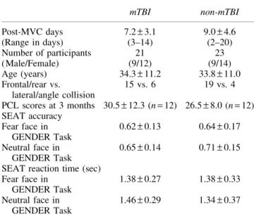
symptoms (non-mTBI group). We previously reported that the mTBI and non-mTBI groups did not significantly differ in age or the time between MVC and the MRI scan (ttest,p>0.1, Table 1).35 Gender and direction of collision were similarly distributed in both groups (Table 1). Twelve mTBI survivors and 12 non-mTBI survi-vors completed PCL questionnaires at 3 months after MVC, and PCL scores were not significantly different between groups (F[1, 20]= 1.40,p=0.251,g2=
0.07).
mTBI effects on cortical emotion processing
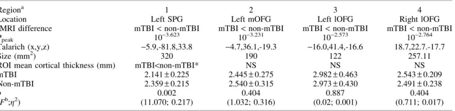
Activation associated with implicit emotional responses was revealed by contrasting the identification of the gender of fearful faces versus neutral faces. Compared with the non-mTBI group, the mTBI group had significantly less activation in response to fearful faces in clusters in the left superior parietal gyrus (SPG) and left medial orbitofrontal gyrus (mOFG), and bilaterally in the lateral orbitofrontal gyri (lOFG) (Fig. 2, Table 2).
Early cortical emotion activation relationships to persistent emotion-related symptoms
The above mentioned early decreased activation in the left SPG in the days after MVC was significantly negatively correlated with PCL scores 3 months after MVC in mTBI survivors (r= -0.68, p=0.03,df=8, Fig. 3). In contrast, this correlation was not sig-nificant in non-mTBI survivors (r= -0.37,p=0.29,df=8).
Table1. Demographic and Behavioral Information
mTBI non-mTBI
Post-MVC days 7.2–3.1 9.0–4.6
(Range in days) (3–14) (2–20)
Number of participants 21 23
(Male/Female) (9/12) (9/14)
Age (years) 34.3–11.2 33.8–11.0
Frontal/rear vs. lateral/angle collision
15 vs. 6 19 vs. 4
PCL scores at 3 months 30.5–12.3 (n=12) 26.5–8.0 (n=12) SEAT accuracy
Fear face in GENDER Task
0.62–0.13 0.64–0.17
Neutral face in GENDER Task
0.65–0.14 0.71–0.15
SEAT reaction time (sec) Fear face in
GENDER Task
1.38–0.27 1.38–0.33
Neutral face in GENDER Task
1.46–0.29 1.34–0.37
mTBI, mild traumatic brain injury; MVC, motor vehicle collision; PCL, PTSD Checklist-Stressor Version; SEAT, Shifted-Attention Emotion Appraisal Task.
mTBI effects on cortical thickness of functionally defined ROIs
The mean cortical thickness of the left SPG ROI with reduced activation was significantly thinner in the mTBI group than in the non-mTBI group (Table 2). Thicknesses in mOFG and lOFG ROIs were not different in the two groups.
Behavioral responses to identification of genders of emotional faces
In both groups, accuracy of identifying the gender of fearful faces was lower than on neutral face trials (F[1, 34]=4.737, p=0.037). The two groups did not differ in terms of accuracy (F[1, 34]=1.05, p=0.313,g2=0.03, Table 1) or response times (F[1, 34]=0.397,p=0.533,g2=
0.01).
Discussion
Emotional symptoms are often reported after mTBI; however, few studies have directly assessed mTBI effects on early post-trauma emotion processing in cortical circuits. The present study reports reduced early post-mTBI activation in the left SPG and
bilaterally in the orbitofrontal (OFG) in mTBI survivors during processing of fearful face visual stimuli. We also report for the first time structural thinning in the left SPG that co-localizes with duced SPG emotion activation in mTBI survivors. Finally, the re-sults provide evidence that supports an association between early reductions in activation in the left SPG and severity of post-traumatic stress symptoms 3 months after trauma in mTBI survi-vors. These findings suggest that mTBI leads to early cortical functional and structural states that contribute to later emotion-related symptoms.
Reduced early SPG activation in mTBI survivors compared with non-mTBI survivors
mTBI survivors in this study had reduced left SPG BOLD ac-tivation in a task involving viewing of fearful faces. Previous studies report that the SPG is active during working memory tasks, and that SPG activation is associated with recovery from cognitive and somatic symptoms in the weeks after a sport-related concus-sion.58,59SPG activation has been linked to spatial perception and spatially directed attention.60A bilateral intraparietal sulcus region that overlaps with the SPG cluster is active in eye gaze.61If di-minished SPG activation reflects altered processing of fearful ex-pressions or other dynamic features of the face, it is possible that mTBI impairs this processing and contributes to impairment of interpersonal skills and mood disorders following TBI.27 Early decreased SPG activation in response to fearful faces negatively correlated with post-traumatic stress symptoms at 3 months after MVC in mTBI survivors. This suggests that early SPG emotion-related activation states may be involved in or might herald the development of post-traumatic stress symptoms. Several factors, including a history of pre-injury mental health problems, have been linked to post-traumatic stress symptoms.8 The current findings suggest that mTBI effects on emotion processing may be an addi-tional contributor to post-traumatic stress symptoms.
Decreased SPG cortical thickness in mTBI survivors compared with non-mTBI survivors
In the mTBI group, cortical thinning occurred in the left SPG ROI where decreased activation was seen. Studies of a specific type of trauma report early alterations of cortical thickness,33,37and studies involving diverse traumas report cortical volume changes.36 Table2. Comparisons Between mTBI and Non-mTBI Groups in the Activation
of Identification of Genders of Fearful versus Neutral Faces, and ROI Cortical Thickness
Regiona 1 2 3 4
Location Left SPG Left mOFG Left lOFG Right lOFG
fMRI difference mTBI<non-mTBI mTBI<non-mTBI mTBI<non-mTBI mTBI<non-mTBI
Ppeak 10-3.623 10-3.231 10-2.573 10-2.764
Talarich (x,y,z) -5.9,-81.8,33.8 -4.7,36.1,-19.3 -16.0,41.4,-16.6 18.7,22.7.-17.7
Size (mm2) 320 190 122 257.11
ROI mean cortical thickness (mm) mTBI<non-mTBI* NS NS NS
mTBI 2.141–0.225 2.445–0.275 2.982–0.463 2.543–0.209
Non-mTBI 2.359–0.215 2.540–0.315 2.973–0.430 2.491–0.238
p 0.002 0.404 0.887 0.404
(Fb;g2) (11.070; 0.217) (1.032; 0.316) (0.02; 0.001) (0.711; 0.017)
aRegions are numbered as in Figure 2. b
Contrast, error degrees of freedom (df): (1, 40).
*Statistically significant atp<0.05 level; NS=not significant.
SPG, superior parietal gyrus; mOFG, medial orbitofrontal gyrus; lOFG, lateral orbitofrontal gyrus; fMRI, functional magnetic resonance imaging; mTBI, mild traumatic brain injury; ROI, region of interest.
SPG is vulnerable to coup/contrecoup injury in frontal/rear axis MVCs.62,63The present SPG thinning might reflect a structural underpinning of the reduced emotion-related activation in SPG. Co-localization of cortical thinning and lower functional activation has been reported in several diseases.64,65Mechanistically, acute hypoperfusion caused by microvasoconstriction following mTBI may reduce cortical thickness and affect fMRI activation.66,67From this thinking, multiple modality investigations of co-localization of cortical vascular, structural, and functional changes appear to offer promise for identifying early mechanisms by which mTBI affects cortical emotional functions and symptoms.
From our previous vertex-based, whole cortex analysis of the same subjects, we reported thickening in right precuneus and thinning in left posterior middle temporal gyrus.35Although left SPG thickness decreases did not reach significance in these pre-vious whole cortex analyses that used correction for multiple comparisons, decreased thickness was evident in the present anal-ysis of mean cortical thickness extracted from a single functionally defined SPG ROI. These different findings likely reflect differences in the previous exploratory versus the present targeted ROI ap-proaches. The observed thickness changes in right precuneus and left SPG and posterior middle temporal gyrus might also suggest that mTBI may have had different effects on the posterior left and right hemispheres. We suggest that right precuneus thickening may result from micro-edema,68,69and that thinning in the left SPG and posterior middle temporal gyrus may result from hypoperfusion, but these possibilities require further study.
Reduced early OFG activation in MVC survivors compared with non-mTBI survivors
mTBI survivors had reduced activation in the left mOFG and bilaterally in the lOFG during emotional processing of facial ex-pressions. However, OFG ROI thicknesses did not differ in mTBI versus non-mTBI survivors, nor was OFG ROI activation corre-lated with PCL symptom severity at 3 months. OFG has been implicated, for example, in integration of sensory, limbic, and prefrontal cortical inputs and in top-down inhibitory control of emotions.70–72Abnormalities in OFG function and structure have been reported in psychiatric disorders including PTSD.73Studies are needed to further elucidate the role of early OFG activation in post-mTBI recovery.
Limitations
This preliminary study has the following limitations. First, partly reflecting the difficulty in performing fMRI analyses during the early post-trauma stage, the sample was modest and further reduced before the 3 month follow-up was completed. Additional work is needed to confirm the present findings in larger samples. Similarly, replication in an independent sample is required to resolve the ap-parent inconsistency in SPG cortical thickness findings seen with our previous vertex-based whole brain analysis35and the current ROI analysis. Second, we studied mTBI and non-mTBI survivors who had experienced MVC and had similar physical injuries. This design did not permit assessment of changes that might be present in both mTBI and non-mTBI survivors as a consequence of MVC trauma. Inclusion of trauma-free healthy controls in future studies would be useful. Third, both groups had a low level of post-traumatic stress symptoms at 3 months; therefore, our study did not test if mTBI after MVC resulted in a higher incidence of PTSD. Finally, the current report did not address possible changes in subcortical (e.g., amygdala) or archicortical structures (e.g., hippocampus).
Conclusion
The current findings demonstrate mTBI effects on BOLD acti-vation and structure of cortical emotion circuits during the early post-mTBI period. The novel cross-modal analysis allowed ex-amination of potential structural mechanisms that may contribute to rapid cortical functional change after mTBI. The relationship be-tween early cortical changes and the severity of post-traumatic stress symptoms in mTBI survivors suggests potential targets for future attempts to reduce development of post-traumatic stress and other emotion-related symptoms.
Acknowledgments
The work is funded by NIH R21MH098198-01 and by a Pro-Medica Translational Research Stimulation Award to X.W. We thank Dr. Michael M. Dennis, Cindy Grey, Susan Yeager, Lindsey Katschke, Michelle Haunus, and the Department of Radiology at the University of Toledo for technical support; Dr. Joe Migliori Jr. for clinical support; Carol Brikmanis for editing the manuscript; and Karen Brenner of ProMedica Health System for survivor re-cruitment.
Author Disclosure Statement
No competing financial interests exist.
References
1. Cassidy, J.D., Carroll, L.J., Peloso, P.M., Borg, J., von Holst, H., Holm, L., Kraus, J., and Coronado, V.G. (2004). Incidence, risk fac-tors and prevention of mild traumatic brain injury: results of the WHO Collaborating Centre Task Force on Mild Traumatic Brain Injury. J. Rehabil. Med. 43 Suppl, 28–60.
2. Laker, S.R. (2011). Epidemiology of concussion and mild traumatic brain injury. PM R 3, S354–358.
3. Centers for Disease Control and Prevention (2016). Rates of TBI-related emergency department visits, hospitalizations, and deaths-United States, 2001–2010. In: Injury Prevention and Control: Traumatic Brain Injury & Concussion. CDC Center for Disease Control and Pre-vention. www.cdc.gov
4. Chossegros, L., Hours, M., Charnay, P., Bernard, M.N., Fort, E., Boisson, D., Sancho, P.-O., Yao, S.N., and Laumon, B. (2011). Pre-dictive factors of chronic post-traumatic stress disorder 6 months after a road traffic accident. Accid. Anal. Prev. 43, 471–477.
5. Mayou, R.A., Black, J., and Bryant, B. (2000). Unconsciousness, amnesia and psychiatric symptoms following road traffic accident injury. Br.J. Psychiatry 177, 540–545.
6. Zhang, S.R., Carroll, L.J., Cassidy, J.D., and Paniak, C. (2009). Fac-tors inflencing self-rated health in traffic-related mild trauma brain injury. J. Rehabil.Med. 41, 1062–1067.
7. Cancelliere, C., Hincapie´, C.A., Keightley, M., Godbolt, A.K., Coˆte´, P., Kristman, V.L., Sta˚lnacke, B.-M., Carroll, L.J., Hung, R., Borg, J., Nygren-de Boussard, C., Coronado, V.G., Donovan, J., and Cassidy, J.D. (2014). Systematic review of prognosis and return to play after sport concussion: results of the International Collaboration on Mild Traumatic Brain Injury Prognosis. Arch. Phys. Med. Rehabil. 95, S210–S229.
8. Carroll, L.J., Cassidy, J.D., Cancelliere, C., Coˆte´, P., Hincapie´, C.A., Kristman, V.L., Holm, L.W., Borg, J., Nygren-de Boussard, C., and Hartvigsen, J. (2014). Systematic review of the prognosis after mild traumatic brain injury in adults: cognitive, psychiatric, and mortality outcomes: results of the International Collaboration on Mild Trau-matic Brain Injury Prognosis. Arch. Phys. Med. Rehabil. 95, S152– S173.
9. Hoge, C.W., McGurk, D., Thomas, J.L., Cox, A.L., Engel, C.C., and Castro, C.A. (2008). Mild traumatic brain injury in U.S. Soldiers re-turning from Iraq. N. Engl. J. Med. 358, 453–463.
Collaboration on Mild Traumatic Brain Injury Prognosis. Arch. Phys. Med. Rehabil. 95, S230–237.
11. Lagarde, E., Salmi, L.R., Holm, L.W., Contrand, B., Masson, F., Ribereau–Gayon, R., Laborey, M. and Cassidy, J.D. (2014). Asso-ciation of symptoms following mild traumatic brain injury with post-traumatic stress disorder vs postconcussion syndrome. JAMA Psychiatry 71, 1032–1040.
12. Bryant, R.A., Nickerson, A., Creamer, M., O’Donnell, M., Forbes, D., Galatzer–Levy, I., McFarlane, A.C., and Silove, D. (2015). Trajectory of post-traumatic stress following traumatic injury: 6-year follow-up. Br. J. Psychiatry 206, 417–423.
13. Frank, D.W., Dewitt, M., Hudgens–Haney, M., Schaeffer, D.J., Ball, B.H., Schwarz, N.F., Hussein, A.A., Smart, L.M., and Sabatinelli, D. (2014). Emotion regulation: quantitative meta-analysis of functional activation and deactivation. Neurosci. Biobehav. Rev. 45, 202–211. 14. Kret, M.E., and Ploeger, A. (2015). Emotion processing deficits: a
liability spectrum providing insight into comorbidity of mental dis-orders. Neurosci. Biobehav. Rev. 52, 153–171.
15. Ochsner, K.N., Silvers, J.A., and Buhle, J.T. (2012). Functional im-aging studies of emotion regulation: a synthetic review and evolv-ing model of the cognitive control of emotion. Ann. N. Y. Acad. Sci. 1251, E1–24.
16. Neumann, D., Keiski, M.A., McDonald, B.C., and Wang, Y. (2014). Neuroimaging and facial affect processing: implications for traumatic brain injury. Brain Imaging Behav. 8, 460–473.
17. Haxby, J.V., Hoffman, E.A., and Gobbini, M.I. (2000). The distributed human neural system for face perception. Trends Cogn. Sci. 4, 223– 233.
18. Pallett, P.M., and Meng, M. (2013). Contrast negation differentiates visual pathways underlying dynamic and invariant facial processing. J. Vis. 13, 13–13.
19. Bryant, R. (2011). Post-traumatic stress disorder vs traumatic brain injury. Dialogues Clin. Neurosci. 13, 251–262.
20. McDonald, B.C., Saykin, A.J., and McAllister, T.W. (2012). Func-tional MRI of mild traumatic brain injury (mTBI): progress and per-spectives from the first decade of studies. Brain Imaging Behav. 6, 193–207.
21. Bryer, E.J., Medaglia, J.D., Rostami, S., and Hillary, F.G. (2013). Neural recruitment after mild traumatic brain injury is task dependent: a meta-analysis. J. Int. Neuropsychol. Soc. 19, 751–762.
22. Jantzen, K.J. (2010). Functional magnetic resonance imaging of mild traumatic brain injury. J. Head Trauma Rehabil. 25, 256–266. 23. Eierud, C., Craddock, R.C., Fletcher, S., Aulakh, M., King–Casas, B.,
Kuehl, D., and LaConte, S.M. (2014). Neuroimaging after mild trau-matic brain injury: Review and meta-analysis. Neuroimage Clin 4, 283–294.
24. Maki–Marttunen, V., Kuusinen, V., Brause, M., Perakyla, J., Polvi-vaara, M., dos Santos Ribeiro, R., Ohman, J., and Hartikainen, K.M. (2015). Enhanced attention capture by emotional stimuli in mild traumatic brain injury. J. Neurotrauma 32, 272–279.
25. Matthews, S.C., Strigo, I.A., Simmons, A.N., O’Connell, R.M., Re-inhardt, L.E., and Moseley, S.A. (2011). A multimodal imaging study in U.S. veterans of Operations Iraqi and Enduring Freedom with and without major depression after blast-related concussion. Neuroimage 54 Suppl 1, S69–75.
26. Shu, I.W., Onton, J.A., Prabhakar, N., O’Connell, R.M., Simmons, A.N., and Matthews, S.C. (2014). Combat veterans with PTSD after mild TBI exhibit greater ERPs from posterior-medial cortical areas while appraising facial features. J. Affect. Disord. 155, 234–240. 27. Radice–Neumann, D., Zupan, B., Babbage, D.R., and Willer, B.
(2007). Overview of impaired facial affect recognition in persons with traumatic brain injury. Brain Inj. 21, 807–816.
28. Benson, R.R., Gattu, R., Sewick, B., Kou, Z., Zakariah, N., Cavanaugh, J.M., and Haacke, E.M. (2012). Detection of hemorrhagic and axonal pathology in mild traumatic brain injury using advanced MRI: impli-cations for neurorehabilitation. NeuroRehabilitation 31, 261–279. 29. Wang, X., Wei, X.E., Li, M.H., Li, W.B., Zhou, Y.J., Zhang, B., and
Li, Y.H. (2014). Microbleeds on susceptibility-weighted MRI in de-pressive and non-dede-pressive patients after mild traumatic brain injury. Neurol. Sci. 35, 1533–1539.
30. Huang, Y.L., Kuo, Y.S., Tseng, Y.C., Chen, D.Y., Chiu, W.T., and Chen, C.J. (2015). Susceptibility-weighted MRI in mild traumatic brain injury. Neurology 84, 580–585.
31. Ge, Y., Patel, M.B., Chen, Q., Grossman, E.J., Zhang, K., Miles, L., Babb, J.S., Reaume, J., and Grossman, R.I. (2009). Assessment of
thalamic perfusion in patients with mild traumatic brain injury by true FISP arterial spin labelling MR imaging at 3T. Brain Inj. 23, 666–674. 32. Maugans, T.A., Farley, C., Altaye, M., Leach, J., and Cecil, K.M. (2012). Pediatric sports-related concussion produces cerebral blood flow alterations. Pediatrics 129, 28–37.
33. Doshi, H., Wiseman, N., Liu, J., Wang, W., Welch, R.D., O’Neil, B.J., Zuk, C., Wang, X., Mika, V., Szaflarski, J.P., Haacke, E.M., and Kou, Z. (2015). Cerebral hemodynamic changes of mild traumatic brain injury at the acute stage. PLoS One 10, e0118061.
34. Tate, D.F., York, G.E., Reid, M.W., Cooper, D.B., Jones, L., Robin, D.A., Kennedy, J.E., and Lewis, J. (2014). Preliminary findings of cortical thickness abnormalities in blast injured service members and their relationship to clinical findings. Brain Imaging Behav. 8, 102– 109.
35. Wang, X., Xie, H., Cotton, A.S., Tamburrino, M.B., Brickman, K.R., Lewis, T.J., McLean, S.A., and Liberzon, I. (2015). Early cortical thickness change after mild traumatic brain injury following motor vehicle collision. J. Neurotrauma 32, 455–463.
36. Ling, J.M., Klimaj, S., Toulouse, T., and Mayer, A.R. (2013). A prospective study of gray matter abnormalities in mild traumatic brain injury. Neurology 81, 2121–2127.
37. Depue, B.E., Olson–Madden, J.H., Smolker, H.R., Rajamani, M., Brenner, L.A., and Banich, M.T. (2014). Reduced amygdala volume is associated with deficits in inhibitory control: a voxel- and surface-based morphometric analysis of comorbid PTSD/mild TBI. Biomed. Res. Int. Article ID 691505.
38. Zhou, Y., Kierans, A., Kenul, D., Ge, Y., Rath, J., Reaume, J., Grossman, R.I., and Lui, Y.W. (2013). Mild traumatic brain in-jury: longitudinal regional brain volume changes. Radiology 267, 880–890.
39. Keightley, M.L., Chen, J.K., and Ptito, A. (2012). Examining the neural impact of pediatric concussion: a scoping review of multimodal and integrative approaches using functional and structural MRI tech-niques. Curr. Opin. Pediatr. 24, 709–716.
40. Niogi, S.N., Mukherjee, P., Ghajar, J., Johnson, C., Kolster, R.A., Sarkar, R., Lee, H., Meeker, M., Zimmerman, R.D., Manley, G.T., and McCandliss, B.D. (2008). Extent of microstructural white matter in-jury in postconcussive syndrome correlates with impaired cognitive reaction time: a 3T diffusion tensor imaging study of mild traumatic brain injury. AJNR Am. J. Neuroradiol. 29, 967–973.
41. Mayer, A.R., Ling, J., Mannell, M.V., Gasparovic, C., Phillips, J.P., Doezema, D., Reichard, R., and Yeo, R.A. (2010). A prospective diffusion tensor imaging study in mild traumatic brain injury. Neu-rology 74, 643–650.
42. Geary, E.K., Kraus, M.F., Pliskin, N.H., and Little, D.M. (2010). Verbal learning differences in chronic mild traumatic brain injury. J. Int. Neuropsychol. Soc. 16, 506–516.
43. Singh, M., Jeong, J., Hwang, D., Sungkarat, W., and Gruen, P. (2010). Novel diffusion tensor imaging methodology to detect and quantify injured regions and affected brain pathways in traumatic brain injury. Magn. Reson. Imaging 28, 22–40.
44. Bruce, V. and Young, A. (1986). Understanding face recognition. Br. J. Psychol. 77, 305–327.
45. Vuilleumier, P. (2005). How brains beware: neural mechanisms of emotional attention. Trends Cogn. Sci. 9, 585–594.
46. Lieberman, M.D., Inagaki, T.K., Tabibnia, G., and Crockett, M.J. (2011). Subjective responses to emotional stimuli during labeling, reappraisal, and distraction. Emotion 11, 468–480.
47. Wang, X., Garfinkel, S.N., King, A.P., Angstadt, M., Dennis, M.J., Xie, H., Welsh, R.C., Tamburrino, M.B., and Liberzon, I. (2010). A multiple-plane approach to measure the structural properties of func-tionally active regions in the human cortex. Neuroimage 49, 3075– 3085.
48. King, N.S., Crawford, S., Wenden, F.J., Moss, N.E., and Wade, D.T. (1995). The Rivermead Post Concussion Symptoms Questionnaire: a measure of symptoms commonly experienced after head injury and its reliability. J. Neurol. 242, 587–592.
49. McDonald, S.D., and Calhoun, P.S. (2010). The diagnostic accuracy of the PTSD Checklist: a critical review. Clin. Psychol. Rev. 30, 976– 987.
50. Wilkins, K.C., Lang, A.J., and Norman, S.B. (2011). Synthesis of the psychometric properties of the PTSD checklist (PCL) military, civil-ian, and specific versions. Depress. Anxiety 28, 596–606.
52. Wang, X., Gerken, M., Dennis, M., Mooney, R., Kane, J., Khuder, S., Xie, H., Bauer, W., Apkarian, A.V., and Wall, J. (2010). Profiles of precentral and postcentral cortical mean thicknesses in individual subjects over acute and subacute time-scales. Cereb. Cortex 20, 1513– 1522.
53. Sripada, R.K., Marx, C.E., King, A.P., Rampton, J.C., Ho, S.S., and Liberzon, I. (2013). Allopregnanolone elevations following pregnen-olone administration are associated with enhanced activation of emotion regulation neurocircuits. Biol Psychiatry 73, 1045–1053. 54. Liberzon, I., Ma, S.T., Okada, G., Shaun Ho, S., Swain, J.E., and
Evans, G.W. (2015). Childhood poverty and recruitment of adult emotion regulatory neurocircuitry. Soc. Cogn. Affect. Neurosci. 10, 1596–1606.
55. Fischl, B. (2012). FreeSurfer. Neuroimage 62, 774–781.
56. Hagler, D.J., Saygin, A.P., and Sereno, M.I. (2006). Smoothing and cluster thresholding for cortical surface-based group analysis of fMRI data. Neuroimage 33, 1093–1103.
57. Cruz–Orive, L.M., Gelsvartas, J., and Roberts, N. (2014). Sampling theory and automated simulations for vertical sections, applied to human brain. J. Microsc. 253, 119–150.
58. Pardini, J.E., Pardini, D.A., Becker, J.T., Dunfee, K.L., Eddy, W.F., Lovell, M.R., and Welling, J.S. (2010). Postconcussive symptoms are associated with compensatory cortical recruitment during a working memory task. Neurosurgery 67, 1020–1028.
59. Lovell, M.R., Pardini, J.E., Welling, J., Collins, M.W., Bakal, J., Lazar, N., Roush, R., Eddy, W.F., and Becker, J.T. (2007). Functional brain abnormalities are related to clinical recovery and time to return-to-play in athletes. Neurosurgery 61, 352–360.
60. Glover, S. (2004). Separate visual representations in the planning and control of action. Behav. Brain Sci. 27, 3–78.
61. Hoffman, E.A., and Haxby, J.V. (2000). Distinct representations of eye gaze and identity in the distributed human neural system for face perception. Nat. Neurosci. 3, 80–84.
62. Lillie, E.M., Urban, J.E., Lynch, S.K., Whitlow, C.T., and Stitzel, J.D. (2013). Evaluation of the extent and distribution of diffuse axonal injury from real world motor vehicle crashes – biomed 2013. Biomed. Sci. Instrum. 49, 297–304.
63. Urban, J.E., Whitlow, C.T., Edgerton, C.A., Powers, A.K., Maldjian, J.A., and Stitzel, J.D. (2012). Motor Vehicle crash-related subdural hematoma from real-world head impact data. J. Neurotrauma 29, 2774–2781.
64. Remy, F., Mirrashed, F., Campbell, B., and Richter, W. (2005). Verbal episodic memory impairment in Alzheimer’s disease: a combined structural and functional MRI study. Neuroimage 25, 253–266.
65. Rasser, P.E., Johnston, P., Lagopoulos, J., Ward, P.B., Schall, U., Thienel, R., Bender, S., Toga, A.W., and Thompson, P.M. (2005). Functional MRI BOLD response to Tower of London performance of first-episode schizophrenia patients using cortical pattern matching. Neuroimage 26, 941–951.
66. Tuor, U.I., Hudzik, T.J., Malisza, K., Sydserff, S., Kozlowski, P., and Del Bigio, M.R. (2001). Long-term deficits following cerebral hypoxia-ischemia in four-week-old rats: correspondence between be-havioral, histological, and magnetic resonance imaging assessments. Exp. Neurol. 167, 272–281.
67. Abu–Judeh, H.H., Parker, R., Aleksic, S., Singh, M.L., Naddaf, S., Atay, S., Kumar, M., Omar, W., El-Zeftawy, H., Luo, J.Q., and Abdel-Dayem, H.M. (2000). SPECT brain perfusion findings in mild or moderate traumatic brain injury. Nucl. Med. Rev. Cent. East. Eur. 3, 5–11. 68. Caner, B., Hou, J., Altay, O., Fuj, M., and Zhang, J.H. (2012).
Transition of research focus from vasospasm to early brain injury after subarachnoid hemorrhage. J. Neurochem. 123, 12–21.
69. Bederson, J.B., Levy, A.L., Ding, W.H., Kahn, R., DiPerna, C.A., Jenkins, A.L., 3rd, and Vallabhajosyula, P. (1998). Acute vasocon-striction after subarachnoid hemorrhage. Neurosurgery 42, 352–360. 70. Kringelbach, M. (2004). The functional neuroanatomy of the human
orbitofrontal cortex: evidence from neuroimaging and neuropsychol-ogy. Prog. Neurobiol. 72, 341–372.
71. Rolls, E.T., and Grabenhorst, F. (2008). The orbitofrontal cortex and beyond: From affect to decision-making. Prog. Neurobiol. 86, 216–244.
72. Vuilleumier, P., and Pourtois, G. (2007). Distributed and interactive brain mechanisms during emotion face perception: evidence from functional neuroimaging. Neuropsychologia 45, 174–194.
73. Jackowski, A.P., Araujo Filho, G.M., Almeida, A.G., Araujo, C.M., Reis, M., Nery, F., Batista, I.R., Silva, I., and Lacerda, A.L. (2012). The involvement of the orbitofrontal cortex in psychiatric disorders: an update of neuroimaging findings. Rev. Bras. Psiquiatr. 34, 207–212.
Address correspondence to: Xin Wang MD, PhD Department of Psychiatry University of Toledo 3120 Glendale Avenue Toledo, OH 43614