A genetic variant associated with post-MVC pain determines the extent
of miR-34a binding to ADRA2A
Maggie Walker
April 2015
University of North Carolina at Chapel Hill
Biology Department
Senior Honors Thesis
Principal Investigator: Dr. Sarah Linnstaedt
Abstract
Adrenergic alpha 2A receptors (ADRA2A) are known to play an important role in descending pain pathways and in the generation of stress-induced analgesia. However, the influence of ADRA2A genetic variants on acute pain severity after motor vehicle collision (MVC) has not been assessed. In this study we hypothesized that a genetic variant in the 3’UTR of ADRA2A, rs3750625, would be associated with acute pain outcomes following MVC. In addition, based on bioinformatics analyses, we
hypothesized that the allele at this location determines the efficiency of binding of miR-34a, a microRNA (miRNA) known to regulate pain and stress responses. The association between rs3750625 allele and acute post-MVC pain outcomes was assessed using a study of 948 European Americans who presented to the emergency department (ED) for
evaluation after MVC. The rs3750625 genotype was determined from patient blood ED samples using the Sequenom platform.miRNA binding was assessed using a luciferase reporter assay consisting of a miR-34a expression cassette and individual ADRA2A
3’UTR-luc reporters with either the minor or major allele. In regression models adjusted
for age, sex, and study site, the minor allele (A) of rs3750625 (n =91/942, 9.7%) was associated with greater ED pain severity (38.3 vs. 26.1), p=0.002). Luciferase reporter assays demonstrated that miR-34a binds the 3’UTR of ADRA2A and that the amount of knockdown is significantly more efficient when the minor allele is present. Additionally, knocking down miR-34a levels in neuroblastoma cells results in increased ADRA2A mRNA and protein expression. These results suggest that genetic variant rs3750625 in the 3’UTR of ADRA2A affects individual vulnerability to acute pain after traumatic events such as MVC, and that this influence may be mediated by miRNA binding.
Introduction
Small variants in the genome, such as single-nucleotide polymorphisms (SNPs), are sufficient to alter phenotypic outcomes.1 With increasingly more accurate technologies for genome sequencing, a number of polymorphisms have been found to be associated with pain.2, 3 However, it is often unclear what molecular mechanistic role these polymorphisms play in pain pathways.
The alpha-2A adrenergic receptor (ADRA2A) is important in negative feedback inhibition of catecholamines in the peripheral nervous system, and has been found to be associated with disorders such as ADHD and schizophrenia. Adrenergic receptors have also been found to be mediators of both pain and stress outcomes. Donello et al. found that ADRA2A plays a role in the development of stress-induced pain in preclinical models by measuring pain tolerance after stress in wild type and ADRA2A knockout mice. They present a model suggesting that without ADRA2A to function as a regulator of neurotransmission after stress, catecholamines accumulate and can sensitize peripheral nerves, leading to hyperalgesia.4 Additionally, Taneja et al. found that ADRA2A is involved in recovery after unexpected stress exposure.5 Genetic variants that regulate ADRA2A expression are therefore likely to affect pain and stress outcomes.
translation by binding to mRNA targets at a 6-8-nucleotide long seed region. The endogenous levels of miRNAs in a person’s body can change drastically due to environmental stimuli, including stress. A number of miRNAs have been shown to be associated with pain development.6 One miRNA, miR-34a, has previously been shown to
play a role in both stress and pain.7, 8
In our study, we hypothesized that genetic variants in ADRA2A affect pain development after motor vehicle collision, and that is mediated by a miR-34a interaction. My work focused most specifically on the effect of the rs3750625 polymorphism on the binding efficiency of miR-34a to ADRA2A, and how that binding impacts ADRA2A expression.
Methods
Initial Data Collection
Patient blood sample collection, genotyping, and statistical analyses were completed as part of a previously described MVC study.9 The cohort in the MVC study consisted of European Americans aged 18-65 years, presenting to one of eight emergency departments (EDs) within 24 hours of motor vehicle collision, but requiring no hospital admission. Patients were excluded who could not read and understand English, who were pregnant or prisoners, who were taking a β-adrenoreceptor antagonist, or who were taking opioids above a total daily dose of 30 mg of oral morphine or equivalent. Characteristics of study participates can be found in Table 1.
Blood samples were taken from study participants in the ED using PAXgene DNA tubes. Samples were purified with PAXgene blood DNA kit (QIAGEN) and genotyped using the Sequenom platform. Pain assessments and statistical analyses were completed by members of the UNC Anesthesiology Research Department.
Bioinformatics Analyses
The potential effect of rs3750625 on miRNA binding was found using bioinformatics analyses using the miRdSNP algorithm found at:
http://mirdsnp.ccr.buffalo.edu/search.php.10
Constructs
Two-step PCR was used to mutate the seed region and the compensatory site in the ADRA2A 3’UTR. Again, a2A-C was used as the parent construct, and primers with mismatched base pairs at either the seed region or compensatory site were used to create constructs a2A-C-seed (Primers 7-F, 7-R, Supplementary Table 1) and a2A-C-comp (Primers 6-F, 6-R, Supplementary Table 1), respectively.
Cell culture and generation of viral transductants
HEK 293 cells are neuroendocrine cells obtained by transformation of human embryonic kidney cells with sheared adenovirus 5 DNA. IMR-32 cells are human neuroblastoma cells. SH-SY5Y cells are cells derived from the subcloning of neuroblastoma cell line SK-N-SH. All cell lines were grown in Dulbecco’s Modified Eagle’s Medium (DMEM) with 10% fetal bovine serum (FBS) and 1% gentamicin.
To measure levels of ADRA2A mRNA and protein after miR-34a knockout, we used a third generation lentiviral packaging system as previously described.12 Briefly,
HEK 293 cells were transfected with vectors pMD2-VSVG, pRSV-REV, pMDLgp, and either pLCE s34 (miR-34a sponge), pLCE (control), or PLCE CXCR4 (CXCR4 sponge, control) using Fugene 6 Transfection Reagent (Promega). 48 hours after transfection, viral media was collected, filtered using Amicon Ultra centrifugal columns, and concentrated by centrifugation.
In preparation for transduction, IMR-32 and SH-SY5Y cells were plated and incubated until 70-90% confluency. On the day of transduction, growth media for both cell lines was replaced at half volume. Concentrated viral media was added to the supernatant covering the neuroblastoma cells. After 16 hours, media was replaced. After 72 hours transducted cells were checked for GFP expression and sorted using fluorescence-activated cell sorting (FACS).
qRT-PCR and Western blotting
qRT-PCR was used to analyze ADRA2A mRNA levels in IMR-32 cells after miR-34a knock-down. Random primers were used for reverse transcription as described (High Capacity Reverse Transcription Kit, Life Technologies) and SYBR reagents were used for qPCR with transcript-specific primers (Life Technologies).
Total protein was collected by lysing IMR-32 cells in RIPA buffer (Pierce, a subsidiary of Thermo Scientific, Rockford IL), protein concentration was measured with a BCA Protein Assay (Pierce), and analysis was completed by Western blotting.
Dual luciferase assays
After 48 hours of incubation, transfected cells were collected and lysed. RLUC and FLUC levels were read using a dual luciferase reporter assay (Promega). FLUC/RLUC ratios were calculated and normalized to ratios obtained from HEK 293 cells with a mutated miR-34a expression construct, pC-34a-mut.
Results
Association of ADRA2A polymorphisms with the severity of pain after MVC
As stated previously in the MVC study, it was found that patients with the minor allele (A) at rs3750625 in the 3’UTR of ADRA2A experience significantly greater pain
severity (body burden of pain) following MVC (p = 0.002, Table 2). The minor allele (A) at rs3750625, which occurs in 10% of the cohort, can then be referred to as the risk allele.
rs3750625 is located in the seed binding region of miR-34a
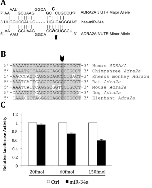
Given the significance of rs3750625 to pain outcomes, and its location in the 3’UTR of an adrenergic receptor, we wanted to see if this polymorphism had the potential to impact expression of ADRA2A. Using the miRdSNP algorithm,10 we found that rs3750625 is located in the seed binding region of miR-34a, where the risk allele creates a completely complementary site (Figure 1A).
Genetic conservation among species is a good predictor of functional importance, and we found that rs3750625 is highly conserved among species (Figure 1B).
miR-34a binds to the ADRA2A 3’UTR in a dose dependent fashion and the
efficiency of miR-34a binding is dependent on whether the risk allele is present at rs3750625
The presence of a miR-34a binding site in ADRA2A does not ensure that functional binding of miR-34a actually occurs. For this reason, I wanted to test functional binding of miR-34a to ADRA2A, first with the presence of the major allele (C) at rs3750625. Using a luciferase assay to visualize functional binding in HEK 293 cells, I found that miR-34a binds to the 3’UTR of ADRA2A in a dose dependent fashion (Figure 1C).
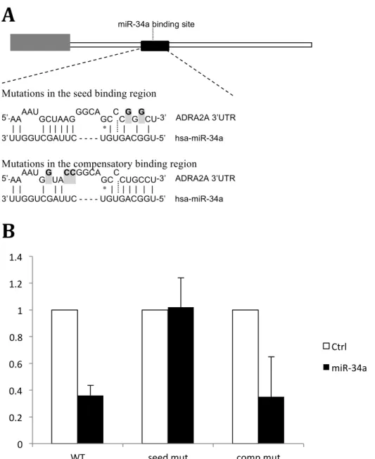
The seed region of the miR-34a binding site confers the majority of miR-34a specificity for ADRA2A
While the seed region of a miRNA is the most important region for binding, the
compensatory regions have also been shown to contribute to the gene silencing effects of miRNAs.13 miR-34a has a compensatory region just upstream of its seed binding site (Figure 2A). To see whether the compensatory site in miR-34a significantly contributes to its ability to bind to the 3’UTR of ADRA2A, I tested miR-34a binding using vectors with either seed region or compensatory region mutations. With a mutated seed region, we see luciferase knockdown comparable to the control, meaning miR-34a binding was effectively disrupted by the mutation. This is contrary to what we see with a mutated compensatory site, where the amount of binding is equivalent to when there are no mutations (Figure 2B). We can conclude, then, that the compensatory region in miR-34a does not significantly contribute to miR-34a binding to ADRA2A, and that the majority of binding specificity comes from the seed region where our polymorphism of interest is located.
Knockdown of miR-34a in an adrenergic neuroblastoma cell line affects ADRA2A mRNA and protein levels
Knowing that miR-34a can functionally bind to ADRA2A, I next wanted to see whether this miRNA functions as a gene silencer and down-regulates ADRA2A expression. In order to test the effect of miR-34a on ADRA2A expression, I knocked down miR-34a in a neuroblastoma cell line, IMR-32, and looked at both mRNA and protein levels. IMR-32 cells express miR-34a and ADRA2A (data not shown) and exhibit adrenergic
neurotransmitter properties, making them an ideal cell line to work in.14 miR-34a
knock-down was completed using a miR-34a-specific sponge (s34), and an unrelated sponge (sCXCR4) was used as a control. Using qPCR to measure mRNA levels, I saw a
significant increase in ADRA2A mRNA levels when miR-34a was knocked down (Figure 3A). In a similar fashion, with the use of a Western blot I saw a significant increase in ADRA2A protein levels when s34 was transfected into IMR-32 cells (Figure 3B). These results show that miR-34a has significant effects on the transcription and translation of ADRA2A in co-expressing cells.
Discussion
In this research, we found that ADRA2A expression can be regulated by miR-34a and that the interaction may explain the correlation between a genetic variation and
differences in pain severity after MVC. We identified a specific genetic polymorphism, rs3750625, which predicts pain severity after a stressful event. This may have clinical implications. New knowledge of polymorphisms and their roles in pain and stress pathways may change the way that individuals are treated in the emergency department after stress exposure. More broadly, a clearer understanding of how genetic variations change physiological outcomes can lead the way for the development of more
personalized medicine.
number of other diseases. If the role of miRNAs in disease pathologies can be elucidated, there is potential for new therapies that target specific miRNAs or miRNA populations.
Finally, this research gives us a fuller understanding of ADRA2A, it’s role in the mediation of post-stress pain, and the ways in which it is regulated. Since we know that adrenergic receptors play a vital role in many cellular pathways, our findings could help further elucidate the broader functions of these receptors in disease outcomes.
Troubleshooting
One aspect of this project that was particularly tough was the creation of a vector with the risk allele (A) at rs3750625. Vector a2A-C contained the major allele (C) at this location, so a point mutation, from (C) to (A), was necessary in order to create a new plasmid with the risk allele at rs3750625. In order to complete this mutation, I used the QuikChange II XL Site-Directed Mutagenesis kit (Agilent Technologies). Initial attempts to mutate a2A-C were unsuccessful due to failed Pa2A-CR, and I found that tweaking the primer design and the given PCR protocol were necessary to create the final product.
Primer design
The QuikChange method uses PCR and user-designed primers containing the allelic change, to mutate a parent plasmid (in my case, a2A-C) into the desired plasmid. Since the primers contain the mutant, they are not completely
complementary, however ideally this is not an issue because there are enough complementary nucleotides flanking the mismatch to induce binding. Initially, my initially primers were 32 nucleotides long with a melting temperature of 72.2°C, which is quite high for a melting temperature. The primers used during successful PCR were 25 nucleotides long with a 67°C melting temperature. From this, I learned that a 25-base pair primer contains enough complementary nucleotides to bind to the parent plasmid with one mismatch, and is preferable to a longer primer with a melting temperature above 70°C.
Protocol
Since initial PCR attempts were unsuccessful, even with new, shorter primers, I tweaked the given protocol in a few areas and found success.
The QuikChange protocol suggests using 125ng of each primer, a melting temperature of 60°C, and does not include DMSO in its PCR mix.
I found success using 140ng of each primer, an increased melting temperature of 68°C to match my primers, and 3% DMSO. DMSO is often used to enhance PCR outcomes, as it facilitates annealing by helping to break apart nucleic acid strands. Due to the high variability in uses of the QuikChange method, it is not surprising that a uniform protocol may not work for all users. Others experiencing issues using this kit may benefit from tweaking their primers design and melting
Model
This research supports the model of post-stress hyperalgesia put forth by Donello et al. Their work showed how knocking out ADRA2A in mice increases hyperalgesia after stress. The model presented by Donello et al. suggests that a decrease in ADRA2A receptors, and thus less feedback inhibition of catecholamines, leads to catecholamine buildup and subsequent sensitization of the sensory afferents. This sensitization makes ADRA2A knockout mice more susceptible pain after exposure to stress.4 Our work suggests that this model may be true in humans as well.
Future Direction
Other members of my research group have begun to look at miR-34a and ADRA2A expression in rat tissues, specifically adrenal gland, peripheral nerve, dorsal root ganglia, and superior cervical ganglion tissues. These tissues were chosen because of the function of ADRA2A in neurotransmission and regulation of catecholamine release. In order for miR-34a to interact with ADRA2A mRNA in-vivo, both must be present in the same tissues. Overlapping expression of miR-34a and ADRA2A was found in each of the aforementioned tissues. This further suggests that miR-34a plays a vital role in the nervous system as a regulator of ADRA2A expression.
Additionally, preliminary data has shown that stressed rats have dysregulated miR-34a and ADRA2A levels. Knowing this, it would be interesting to look more closely at the connection between miR-34a, ADRA2A, and catecholamine feedback. The model put forth by Donello et al. suggests that the dysregulation of ADRA2A disrupts normal catecholamine feedback in peripheral nerves, leading to sensitization and then
References
1. Shastry BS. SNPs: impact on gene function and phenotype. Methods Mol Biol 2009;578:3-22. 2. Bortsov AV, Smith JE, Diatchenko L, Soward AC, Ulirsch JC, Rossi C, Swor RA, Hauda WE,
Peak DA, Jones JS, Holbrook D, Rathlev NK, Foley KA, Lee DC, Collette R, Domeier RM, Hendry PL, McLean SA. Polymorphisms in the glucocorticoid receptor
co-chaperone FKBP5 predict persistent musculoskeletal pain after traumatic stress exposure. Pain 2013.
3. Nicholl BI, Holliday KL, Macfarlane GJ, Thomson W, Davies KA, O'Neill TW,
Bartfai G, Boonen S, Casanueva FF, Finn JD, Forti G, Giwercman A, Huhtaniemi IT, Kula K, Punab M, Silman AJ, Vanderschueren D, Wu FC, McBeth J, European Male Ageing Study G. Association of HTR2A polymorphisms with chronic widespread pain and the extent of musculoskeletal pain: results from two population-based cohorts. Arthritis and rheumatism 2011;63(3):810-818.
4. Donello, J. et al. A peripheral adrenoceptor-mediated sympathetic mechanism can transform stress-induced analgesia into hyperalgesia.Anesthesiology114, 1403–16 (2011).
5. Taneja M, Salim S, Saha K, Happe HK, Qutna N, Petty F, Bylund DB, Eikenburg DC. Differential effects of inescapable stress on locus coeruleus GRK3, alpha2-adrenoceptor and CRF1 receptor levels in learned helpless and non-helpless rats: a potential link to stress resilience. Behavioural brain research 2011;221(1):25-33.
6. Niederberger E. KK, Lotsch J.,Geisslinger G. MicroRNAs as new players in the pain game. Pain 2011;152:1455-1458.
7. Haramati S et al. MicroRNA as repressors of stress-induced anxiety: the case of
amygdalar miR-34. [Internet]. The Journal of neuroscience : the official journal of the Society for Neuroscience. 2011;31(40):14191–203.
8. von Schack D et al. Dynamic changes in the microRNA expression profile reveal
multiple regulatory mechanisms in the spinal nerve ligation model of neuropathic pain. [Internet]. PloS one. 2010;6(3):e17670.
9. Platts-Mills TF BL, Bortsov AV, Soward A, Swor RA, Jones JS, Lee DC, Peak DA, Domeier RM, Rathlev NK, Hendry PL, McLean SA. Using emergency department-based inception cohorts to determine genetic characteristics associated with long term patient outcomes after motor vehicle collision: methodology of the CRASH study. BMC Emerg Med 2011;11(14).
10. Bruno AE, Li L, Kalabus JL, Pan Y, Yu A, Hu Z. miRdSNP: a database of disease-
associated SNPs and microRNA target sites on 3'UTRs of human genes. BMC genomics 2012;13:44.
11. Gottwein E, Cullen BR. A human herpesvirus microRNA inhibits p21 expression and attenuates p21-mediated cell cycle arrest. Journal of virology 2010;84(10):5229-5237. 12. Dull T., Zufferey R., Kelly M., Mandel R. J., Nguyen M., Trono D., Naldini L.
(1998) A third-generation lentivirus vector with a conditional packaging system. J. Virol. 72:8463–8471.
13. Hibio, N., Hino, K., Shimizu, E., Nagata, Y. & Ui-Tei, K. Stability of miRNA
5′terminal and seed regions is correlated with experimentally observed miRNA-mediated silencing efficacy. Sci. Rep.2, 996 (2012).
14. West GJ, Uki J, Herschman HR, Seeger RC (1977) Adrenergic, cholinergic, and
Table 1. Baseline characteristics of study participants
Characteristic
Enrolled, n 948
Age, years, mean (SD) 36 (13)
Females, n(%) 575 (61)
Education, n(%)
8-11 yrs 42 (4)
HS 184 (19)
Post-HS 57 (6)
Some college 311 (33)
College 237 (25)
Post-college 113 (12)
Overall pain, 0-10 NRS, mean(SD)
Past month 0.4 (0.5)
ED 5.5 (2.4)
rs3750625, n(%)
CC 854 (90)
CA or AA 91 (10)
Table 2. Association of the ADRA2A genotype with pain severity in the ED following motor vehicle collision (n = 878)
Pain Severity (Body burden of pain)
SNP Alleles C/C C/A + A/A p-value
rs3750625 C/A 26.1 38.8 0.002
Figure 1: Specifications of miR-34a binding to ADRA2A and experimental assessment of functional binding.
[A] rs3750625 (black arrow, bolded nucleotides) is found within a predicted seed binding site of miR-34a (underlined nucleotides). Binding between miR-34a and ADRA2A with either the (C) allele or the (A) allele is shown with vertical black lines. A mismatch exists at rs3750625 when the (C) allele is present.
[B] The genetic variant rs3750625 (indicated by the bolded arrow) is highly conserved among seven mammals.
[C] Relative luciferase activity (y-axis) is shown after co-transfection of miR-34a and a2A-C in HEK 293T cells. miR-34a binding (black bars) is compared to binding of a control miRNA (open bars). The assay was completed in triplicated and error bars show standard error of the mean. [D] Relative luciferase activity (y-axis) is shown after transfection with miR-34a and either a2A-C (grey line, grey squares) or a2A-A (black line, black circles) in HEK 293T cells. The assay was completed in triplicated and error bars show standard error of the mean.
A
5’-AAAAUGCUAAGGGCAGCCCUGCCU-3’ ADRA2A 3’UTR Major Allele
| | | | || | | | | | | | |
3’UUGGUCGAUUC - - - - UGUGACGGU-5’ hsa-miR-34a
| | | | || | | || | | | | |
5’-AAAAUGCUAAGGGCAGCACUGCCU-3’ ADRA2A 3’UTR Minor Allele
B
C
0 0.2 0.4 0.6 0.8 1 1.2
20fmol 60fmol 150fmol
R
el
at
iv
e
Lu ci fe ra se A ct iv it
y
Ctrl miR-‐34a
5’-AAAATGCTAAGGGCAGCCCTGCCT-3’ Human ADRA2A
5’-AAAATGCTAAGGGCAGCCCTGCCT-3’ Chimpanzee Adra2a
5’-AACCCACTCAGGGCAGCCCTGACT-3’ Rhesus monkey Adra2a
5’-AAATCATTCAGGGCAGCCCTGCCT-3’ Rat Adra2a
5’-AAATCATTCAGGGCGGTCCTGCCT-3’ Mouse Adra2a
5’-CAAATGCTCAGGGCAGCCCTGCCC-3’ Dog Adra2a
5’-CAAACGCTAGGGGCAGCCCTGCCT-3’ Elephant Adra2a
D
0.4 0.5 0.6 0.7 0.8 0.9 1 1.1
20fmol 60fmol 150fmol
R
el
at
iv
e
Lu
ci
fe
ra
se
A
ct
iv
it
y
miR-‐34a
ADRA2A-‐3'UTR with major allele ADRA2A-‐3'UTR with minor allele
**
**
Figure 2: Specificity of miR-34a binding shown using mutation of compensatory and seed regions.
[A] Relative location of seed and compensatory binding regions of miR-34a in 1.4kb ADRA2A 3’UTR. Grey boxes and bolded nucleotides represent experimental mutations made to test binding specificity.
[B] Relative luciferase activity (y-axis) after transfection with miR-34a and ADRA2A 3’UTR with no mutations (WT), with mutations in the seed region (seed mut) or with mutations in the
compensatory region (comp mut). miR-34a binding (black bars) is compared to binding of a control miRNA (open bars). Experiment was completed in triplicate and error bars show standard deviation.
A
Mutations in the seed binding region
5’-AAAAUGCUAAGGGCAGCCCGGGCU-3’ ADRA2A 3’UTR
| | | | | | | | | | | |
3’UUGGUCGAUUC - - - - UGUGACGGU-5’ hsa-miR-34a
Mutations in the compensatory binding region
5’-AAAAUGGUACCGGCAGCCCUGCCU-3’ ADRA2A 3’UTR
| | | | | | | | | | |
3’UUGGUCGAUUC - - - - UGUGACGGU-5’ hsa-miR-34a
B
0 0.2 0.4 0.6 0.8 1 1.2 1.4
WT seed mut comp mut
Ctrl
miR-‐34a
A
Figure 3: Effect of miR-34a knockdown on ADRA2A mRNA and protein levels in neuroblastoma cells. miR-34a knockdown was completed using a miR-34a sponge, pLCE-s34. [A] Relative ADRA2A mRNA levels (y-axis) in IMR-32 cells expressing pLCE-s34 were measured using RT-qPCR. mRNA expression in cells expressing pLCE-s34 was compared to cells expressing an empty vector (pLCE) and a control miRNA sponge (sCXCR4). Error bars show standard deviation.
[B] ADRA2A protein levels were measured in IMR-32 cells expressing pLCE-s34 using Western Blotting. Quantification of band intensity was calculated using band intensity measurements from the NIK image analysis program.
0
0.5
1
1.5
2
2.5
3
pLCE
s34
sCXCR4
Re
la
%v
e
AD
RA2
A
m
RN
A
le
ve
ls
B
*
1.0 1.6 1.1
A
Supplementary Table 1
Primer
Name Primer Sequence (5’ à 3’) Use
Added
R.E. site Species
1-F ATGATGCTCGAGACTCAGAAACCCGGGCGC To make a2A-C XhoI human
1-R ATGATGAATTCCCATAAAATCAGATGTTCCCA
GAG To make a2A-C EcoRI human
2-F GCTAAGGGCAGCACTGCCTGCCCTC For mutating rs3750625 to make
a2A-A - human
2-R GAGGGCAGGCAGTGCTGCCCTTAGC For mutating rs3750625 to make a2A-A - human
3-F AGAAGTGGTACGTCATCTCGT qPCR Detection of ADRA2A mRNA - human
3-R CGCTTGGCGATCTGGTAGA qPCR Detection of ADRA2A mRNA - human
5-F TACTACACCGGTATGTTCCGCCAGGAGCAGCC
G
ADRA2A over-expression
construct AgeI human
5-R TACTACCTCGAGTCACACGATCCGCTTCCTGTC
C
ADRA2A over-expression
construct XhoI human
6-F AAAATGGTACCGGCAGCCCTGCCTGCCCTCC pL-GL3-ADRA2A-comp KpnI human
6-R GCTGCCGGTACCATTTTTCTTTAAAAAGAGA pL-GL3-ADRA2A-comp KpnI human
7-F GGCAGCCCGGGCTGCCCTCCCCATCCCCCGCT pL-GL3-ADRA2A-seed SmaI human
7-R GGGCAGCCCGGGCTGCCCTTAGCATTTTTCTT pL-GL3-ADRA2A-seed SmaI human