CHANGES IN MICROBIOTA, MORPHOHISTOCHEMICAL,
BIOCHEMICAL SHIFTS IN MICE ON THE SODIUM DEXTRAN
SULPHATE – INDUCED NONSPECIFIC ULCERATIVE COLITIS
AND THE EFFECTS OF FREE PROBIOTICS AND
IMMOBILIZED FORMS WITH ZEOLITE
A. A. Agababova
[a], N. Kh. Alchujyan
[a], A. M. Hakobyan
[a]*, A. G. Gevorkyan
[b]and V. H.
Barsegyan
[a]Keywords: Nonspecific ulcerative colitis, sodium dextran sulphate, gastrointestinal tract, probiotic, zeolite, lipid peroxidation, microbiota, intestinal –cerebral axis, inflammation.
Acute, inflammatory processes contribute to the fact that conventionally pathogenic and pathogenic microorganisms colonize the mucous membranes of the small intestine and form biofilms, can become a source of bacterial toxin, which, when the epithelial layer breaks, penetrates to the lymphatic and blood systems, contributing to the formation of sepsis. The barrier function of the epithelium is critical in the development of inflammatory bowel diseases, while normal functioning requires a constant balance between reactivity and tolerance to microorganisms of the intestinal lumen. Increased permeability of the intestinal mucosa is the main risk factor for the spread of bacteria. Epithelium, being an essential element of tissue barriers, provides selective transport for the movement of ions and macromolecules, and also creates an obstacle for their penetration into the underlying tissues. Control of the permeability of the epithelial layer is carried out by the apical intercellular complex - tight contacts, which comprise proteins of the claudine family. Intestinal flora affects the sensory, motor and immune functions of the intestine, and also interacts with higher nervous centers. Immunosuppressive processes are one of the main causes of destabilization of the barrier function, intestine, and brain.
*Corresponding Authors Fax:
E-mail: [email protected]
[a] H. Buniatian Institute of Biochemistry NAS RA, 5/1 P. Sevak St., 0014, Yerevan, Republic of Armenia.
[b] M. Herazi Yerevan State University, Republic of Armenia.
INTRODUCTION
The intestinal - cerebral axis (ICA) is a bi-directional communicative system through which the brain models the functions of the gastrointestinal tract (GIT) and vice versa. ICA is based on neuronal, endocrine and immunological mechanisms connected with each other at the organism, organ, cellular and molecular level.1 The relationship between cerebral microbiota of the intestine is one of the examples of the cooperation of the endocrine, nervous systems and nonspecific natural immunity, the importance of which for the organism in norm and in pathology is difficult to overestimate.
The gastrointestinal tract is closely related to the hypothalamic-pituitary tract through the release of peptides that control the brain's response, and also through neuroendocrine and sensory signals from the intestine. The microbiota affects the development of cognitive functions and the hypothalamic-pituitary response to the stress. Intestinal bacteria in the process of metabolism form serotonin, melatonin, amino acids, catecholamine, histamine, acetylcholine, partially being the most important neurotransmitters .1,2,3
The intestinal-cerebral axis is also traced in infections and inflammations that lead to mood changes and cognitive dysfunction. In particular, the brain is able to identify the
introduction of pathogenic microbes in the digestive tract and respond with a sense of anxiety and restlessness. The only mechanism capable signalize the brain of danger is hematological. The sensory neurons of the vagus nerve are contacted by submucosal immune cells and project the nerve endings into the mucous membrane of the gastrointestinal tract, the site of direct interaction between the organism and the pathogen.
The detection of Fos protein, synthesized by neurons including and markers of functional activity, in the sensory neurons of the nerve after inoculation of bacteria, testified that this connection is also accomplished by the vagus nerve.4,5 The death of intestinal neurons and glia damage with alteration of the expression of the acidic fibrillar protein and increased expression of the molecules of the main histocompatibility complex of the second class were noted in Crowns’ disease, ulcerative colitis, necrotizing enterocolitis and diabetes.6
The purpose of this work was to study the effect of the complex positions of dextran sodium sulfate on the intestinal microbiota of mice, to obtain a model of nonspecific ulcerative colitis (NUC), to interrelate the axis of the intestine-blood-brain, the ways of affecting some zeolites of this domestic production, the immune active and anti-inflammatory properties of zeolites, found in the regions of Armenia in the experimental models of the NUC.
EXPERIMENTAL
Germany). Sodium citrate anticoagulant, thiobarbituricacid, HEPES, 1.4-dithiothreitol, hematoxylin and eosin were purchased from Sigma-Aldrich Chemical Co.
Animals and treatmants: All procedures involved animals were in accordance with the International Laboratory Animal Care and the European Communities Council Directive (86/809/EEC) and approved by the respective local committee on biomedical ethics (H. Buniatian Institute of Biochemistry, Yerevan, NAS RA). The 2-to 3-month-old male mice from our breeding colony were used. All animals were maintained on a 12 h light/dark cycle at normal room temperature and housed in groups of 6/cage with free access to food and tap water.
Experimental design: To induct nonspecific ulcerative colitis induction, the animals were randomly divided into groups (n=12). Group I was contol group of native mice. Other groups were experimental groups with DSS-induced nonspecific ulcerative colitis.
Group II - mice received ad libitum 2.5% DSS dissolved in regular tap water for 7 days and examined immediately at the end of sodium dextran sulphate treatment.
GroupIII - a group of sodium dextran sulphate-induced nonspecific ulcerative colitis mice left untreated for the duration of 2 weeks in parallel to treated ones and referred as self-recovery to assess self-healing mechanism
Group IV-VI were of mice that were fed with 2.5 % DSS dissolved in regular tap water for 7 days and then given daily per os (through a flexible polypropylene gavage) separately probiotics, and/or probiotics immobilized with zeolites (PBZ). Control mice were given water only. On day 8, mice were sacrificed and monitored for colitis.
Probiotic strains and growth conditions: A commercially available probiotic, a concentrated source of naturally occurring microorganisms were used. L. rhamnosus strain
В-6778 and Bifidobacterium bifidum strain AC-1666 (RF), as well as L. plantarum strain В-2353 (RF), L. acidophilus Er317/402 "Narine" (RA), L. salivarius B-7701 possessing fungicidal activity and E. coli Nissle1917 strain were rehydrated in sterile 0.85% NaCl and routinely propagated at 37° C in MRS medium (Hi Media, India) pH of 6.5 ± 0.2. Limiting dilution assay (based on the method of McCrady) was used for the separation, characterization, and quantification of bacteria.7 Viability was confirmed by culturing 1 mL of the hydrated mixture on plates supplemented with MRS + agar at 37°C for 24 h. For immobilization of probiotics the culture medium was enriched with 5% of the chemically modified micronized natural mineral composition (vide infra). The probiotic mixture contained equal quantities the mentioned microorganisms (6 x 10⁹ CFU/mL in total).
Natural mineral composition. Natural minerals mined in Armenia were used viz., zeolites from Noyemberyan, bentonite from Ijevan and diatomite from Jadzor. Their multi-elemental composition at the trace and ultra trace levels have been determined and previously modified chemical composition (zeolite (80%), diatomite (10%), and bentonite (10%)) used as efficient stimulators and carriers of the probiotics.8 High pressure micro powder mill was used
to produce about 50 μm powder from the mentioned
minerals.
Evaluation of experimental colitis. The animals were weighed and monitored for the appearance of diarrhea and blood in the stool throughout the experimental period. The overall disease severity was assessed by a clinical scoring systemand the disease activity index score was calculated for each animal.9 Assessment includes 3 parameters, difference in weight before and after receiving DSS (0 =
weight gain ≥ 1g: 1 = weight gain <1g; 2 = weight loss <1g; 3 = weight loss ≥ 1g); stool consistency (0 = normal stool; 1
=soft but formed stool, 2 = liquid stool); the presence of blood in the perianal region (0 = no sign of blood; 1 = signs of blood in perianal region; 2 = presence of blood in perianal region). The minimum score is 0, maximum is 7. After decapitation colon is excised opened longitudinally, rinsed with saline, and the length was measured and considered as
an additional index for colitis, as well as mаcroscopic
damage of the colonic mucosa scored based on the degree of inflammation and the presence of edema and/or ulcerations (0 = normal; 1 = slight inflammation; 2 = moderate inflammation and/or edema and 3 = heavy inflammation and/or ulcerations).10 Toward the end of the treatment, before decapitation, all of the animals underwent a behavioral testing in open field (OP) and elevated plus-maze (EPM).
Behavioral examination: For open field test the mice were placed singly into an open field (diameter 1 m, divided by 2 concentric circles into 16 equal sections on the floor of the arena) and observed in 3 min to measure locomotor activity (the number of sectors crossed with all paws (crossing), the number of rears (posture sustained with hind-paws on the floor), grooming (including washing or mouthing of forelimbs, hind-paws, body and genitals) (exploratory behavior) and boluses (anxiety) counted manually/visually.11
For elevated plus-maze test, immediately after the open field test the mice were placed singly into a common central platform (10 cm × 10 cm) of elevated plus-maze comprised two open and two closed arms (45 cm x 10 cm x 10cm) and elevated to a height of 80 cm above the floor During a 3-min observation period, the following parameters were measured: number of open arms entries and number of closed arm entries. Exploration (grooming and rearing) and risk assessment (number of hanging over the open arms) were also examined. At the end of each trial, the open field and elevated plus maze were wiped clean with ethanol.
culture media, agar plates (Endo, sucrose, and blood agar), and incubated for 24 h; blood samples incubated for 7 days, monitored daily, to facilitate a growth of microbes.12 The following nutritional environments are used: sugar bouillon, blood agar, yolk-salt agar, Endo, Ploskirev and Saburo agars.
The identification of separated bacteria is carried out with morphological, biochemical properties, by the method of spotted series of Hiss. After the discovery of the biochemical character of microbes, which determines the family of culture and its type, is made of serological identification of microorganisms with serums until serotypes.
Histological examination: Samples for histology were excised from the brain and distal 6–8 cm of the colon, fixed in 10 % buffered formalin, and embedded in paraffin blocks.
Slices with 6 μm sections were stained with hematoxylin and eosin (H&E), and scored for histological damage.13 Pathological diagnosis of each specimen was assessed and analyzed by specialized histopathologist in a blinded manner.
Indices of oxidative stress referring to lipid peroxidation processes were established by measuring malondialdehyde (MDA) using thiobarbituric acid.14 Samples were deproteinized with 10 % TCA, centrifuged at 15000 rpm for 3 min, and supernatants were mixed with 0.6 N HCl and 0.72 % TBA, heated for 15 min in boiling water bath with the resultant formation of pink-colored secondary product of MDA which absorbance was measured at 535 nm wavelength against reagent blank containing all the reagents minus the sample. Protein was determined using crystalline bovine serum albumin as standard.15
Statistical analysis: All data were analyzed using a one-way analysis of variance (ANOVA) followed by post hoc Holm-Sidak test (SigmaStat3.5 for Windows). Data are expressed as the mean±S.E.M. Statistical significance is accepted at the level p< 0.05.
Results and Discussion
Effect of sodium dextran sulphate in mice, histopathological shifts
In the control group of mice, there were no abnormalities in the brain tissue (Figure 1). However, n the brain tissue of the second group mice proliferation of neurocytes, significant edema and dilated vessels were observed (Figure 2).
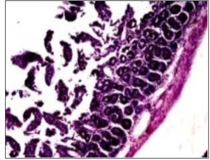
There was no pathological process in colon tissue of the control group mice. Clearly visible mucosa of the large intestine with small vill, not wide submucosal layer with small vessels were observed (Figure 3).
Histological examination of colon preparations of the experimental group mice showed changes in the intestinal mucosa. There was a focal glandular hyperplasia of the mucosa, up to the development of small polyps (Figure 4). In the mice of this group in the submucosal layer there was edema with an expansion of the submucosa and an increase in the number of small vesicles in this zone (Figure 5).
Figure 1. Brain tissue of the control group mice stained with hematoxylin-eosin
Figure 2. Brain tissue of mice of the second group stained with haematoxylin – eosin.
Figure 3. Colon tissue of the control group mice stained with hematoxylin-eosin.
Figure 4. Large intestine of the experimental group mice stained with haematoxylin - eosin.
Figure 5. Submucosal layer of the experimental group mice, with hematoxylin-eosin.
A new concept in gastroenterology is that a wide range of diseases associated with impaired peristalsis, including IBD (inflammatory bowel disease), can be diagnosed as intestinal neuropathy. It is still unclear whether neuropathy is a consequence of the disease or its cause. Given the similarity of intestinal glia and astroglion of the brain, the concept deserves special attention. It is the concept of unified mechanisms of intestinal and blood-brain barrier regulation.16 Glial cells of the intestine are morphologically, immunohistochemically and functionally equivalent to astrocytes of the central nervous system. Intestinal flora affects the sensory, motor and immune functions of the intestine and also interacts with higher nervous centres.
The purpose of this study is to identify some opportunistic microbes, in particular E. coli, the pro-inflammatory St. aureus, and the associated fungi of the genus Candida in dextran sulphate induced non specific ulcerative colitis. Increasing the concentration of microbial toxins contributes to the development of a variety of pathological processes in the human body. It is shown that thermolabile enterotoxins of various conditionally pathogenic enterobacteria, including E. coli, cause an increase in the permeability of the mucous membranes of the gastrointestinal tract. Changes in the appearance and behaviour of the animals viz., a depleted species, a discharge of blood from the perianal region, thin and cyanic peritoneum of the mice was
observed as a result of the experiments.17 On sowing in nutrient broths and subsequent re-crossings on solid nutrient media single colonies of E. coli were isolated from brain washings. Similarly, St. aureus and hemolytic E. coli were separated from the cultures of the contents of the large intestine. Coliforms were not found in the blood, so the phenomenon of translocation of microbes is ruled out. Ample growth of St. aureus, that resembled sepsis, was revealed following the studies in the blood. Fungi of the genus Candida were also observed in significant numbers throughout the all samples
The barrier function of the epithelium is the key in the development of inflammatory bowel diseases. While the normal functioning requires a constant balance between reactivity and tolerance to the microorganisms of the intestinal lumen, increased permeability of the intestinal mucosa is the main risk factor for the spread of bacteria. Epithelium being an important element of tissue barriers provides selective transport for the movement of ions and macromolecules, and also creates an obstacle for their penetration into the underlying tissues. The permeability of the epithelial layer is controlled by the apicalinter cellular complex - dense contacts, consisting of proteins of the claudine family.18 In acute inflammatory processes such as NUC caused by DSS, conditionally pathogens and pathogenic microorganisms, colonizing the small intestine mucous surfaces, forming bioplasts and can become a source of bacterial toxin. When the epithelial layer breaks they penetrate into the lymphatic and circulatory system and cause sepsis.
A two-week treatment of the mice with the mixture of probiotics, in free and immobilized forms, following DSS induced non specific ulcerative colitis (DSS-NUC), indicated that both probiotics and PBZ completely normalized microbiota in 14 days. The probiotics used in our studies contribute to the restoration of the intestinal microbiota, in particular by a sharp decrease in the number of fungi of the genus C. albicans, which resulted from a sharp drop in the body's immune status. In mice with NUC, probiotics destroy pro-inflammatory staphylococcus and restores the proportion of E. coli, which appropriately correlates with the intestinal microbiom preventing the further development of sepsis.
Colon length and disease activity index were also significantly improved suggesting on-going recovery from the disease. On day 14 post-SDS, the colon length of ulcerative colitis mice was (6.2 ± 0.3 cm) shorter than that of control mice (8.9 ± 0.8 cm). Treatment with probiotics attenuated colon shortening about equally (7.13 ± 0.32 cm and 7.41 ± 0.43 cm respectively), while it was normalized by PBZ. The DAI calculated by diarrhea, visible fecal blood
weight loss, as well as colon length and mаcroscopic
damage additional indices for colitis (see Material and methods) reached up to 8-11 in the DSS-treated group, it was significantly lowered to about one-third in the self healing processes on day 14 post-DSS, when all three treatment groups showed complete recovery. The above mentioned parameters correlated with histological grading of colitis based on the amount and depth of inflammation, as well as the amount of crypt damage or regeneration, and the percentage involvement by the disease process.
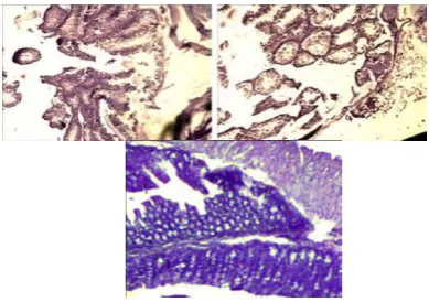
Histopathological changes observed in the colon and brain regions of corticolimbic system were examined using H&E staining. As in many other findings microscopic features of sodium dextran sulphate-induced nonspecific ulcerative colitis was characterized by epithelial damage, disruption of crypt architecture, and inflammation.22 Inflammatory cell infiltration in the areas of focal lesions, and edema in the colonic submucosal layer were detected (Figure 6 A, B and C).
Figure 6. Histopathological changes in the mouse colon following sodium dextran sulphate-induced nonspecific ulcerative colitis (upper row) and 14 days post-colitis (lower row). Erosions in the colonic mucosa, edema and inflammation in the subepithelial layer (A, B) x100. The intestinal wall with the mucous glandular hyperplasia and formation of the hyperplastic polyp (C) x100.
It should be noted that acute inflammatory or infectious process are accompanied by vasodilatation with increased capillary permeability and extravasation of protein-aqueous fluid in a loosely packed submucosal layer resulted in the edema relative to a thickened echogenic submucosal layer, there to mucosal hyperplasia seen in nonspecific ulcerative colitis mice is an initial step in the formation of polyps, which were also found, indicating an acute disease process.23
Two weeks after stopping sodium dextran sulphate, the recovery phase of sodium dextran sulphate-induced nonspecific ulcerative colitis was associated with epithelial apoptosis and disruption of the mucosa, the submucosa contained some macrophages, and lymphoid micro follicles were seen in lamina propria, as well as the colonic epithelial cell proliferation that involved in the regeneration
processes.24 However, in some places edema and erosions and focal glandular hyperplasia in the colonic mucosa were observed, epithelium was incompletely regenerated (Figure 7 D, E and F).
Figure 7. Histopathological changes in the mouse colon following sodium dextran sulphate-induced nonspecific ulcerative colitis (upper row) and 14 days post-colitis (lower row).Focal glandular hyperplasia of the mucosa (D) x100, H&E stain; ero-sions in the colonic mucosa, edema and inflammation in the subepithelial layer (E) x100 H&E stain; intact mouse colonic mucosa with not wide subepithelial layer (F), x100 H&E stain.
It is of interest to note that the sodium dextran sulphate-induced non specific ulcerative colitis caused edema and proliferation of glial cells in the mouse brain tissues lasted for the post-SDS period with manifestation of gangliomatous atypical nervous cells (Figure 8).
Figure 8. Histopathological changes in the mouse brain following sodium dextran sulphate -induced colitis (upperrow) and 14 days post-colitis (lower row) . Edema and proliferation in the brain tissues (A, B ), intensive focal proliferation of glial cells, and edema (D, E), and manifestation of gangliomatous cells (F). (H&E stain,100 X).
Figure 9. Effect of probiotic-treatment on the histopathological changes in colon and brain following sodium dextran sulphate-induced ulcerative colitis. (A) x 40, H&E stain. (B), x 100, H&E stain (C), x 40, H&E stain.
On the preparations of the PBZ-treated mice slight inflammation and proliferation of epithelial cells were observed, and some place deformed crypt structures occurred (Figure 10 A and B).
Figure 10. (A) PBZ-treatment presented the colonic wall x 40, H&E stain; the lymphoid macrofollicles in the colonic mucosa (B), x 100 H&E stain and in the subepithelial layer (C) x 100 H&E stain.
However, the layers of the mucosa are preserved, and in the colonic lamina propria the clusters of lymphoid structures are presented both in the form of follicles and in the form of diffusely located lymphoid elements, among which plasma cells and macrophages were found (Figure 10 C). Gut-associated lymphoid tissue is known play a pivotal role in there pair mechanisms. In inflammatory conditions, increased number, diameter and density of isolated lymphoid follicles suggest their involvement in immune surveillance, their presence is also indispensable in normal mucosal regeneration of the colon, as they serve as a regenerative pool of stem cells in case of mucosal damage, and/or contribute to the optimal cytokine milieu for the differentiation of immigrating stem cells reviewed elsewhere.26
Beneficial effect of PBZ treatment was found in the brain, where in some areas the ganglion cells surrounding the nerve structures that play a protective role in nervous tissues were detected. At the same time, in the brain of some PBZ-treated mice we observed signs of a negligible dystrophy, and someplace still widened full-blooded vessels pointed to the possibility of development of the per vascular edema that may be involved in the formation of the per vascular exudative cuff, which plays dual role, on one side it can be implicated in disruption of the blood-brain barrier, on the other can be considered as a protective mechanism of strengthening vascular wall from the outside.27 Notably, the similarity between perivascular spaces in human and rodent brains and their significance as anatomical routes for edema fluid drainage from damaged brain tissues has been demonstrated.28
Effect of the selected probiotics on the behavior and mood of mice
Depression-like behavior of mice was observed immediately after sodium dextran sulphate–induced nonspecific ulcerative colitis. Animals displayed a decreased motor activity in the open field mimicking human psychomotor retardation.29 Another major symptom of depression, detected in the open field and the elevated plus-maze, was a decreased number of rearing relevant to exploratory behavior showing a “refractory loss of interest”.30 Reduced grooming was also observed and reflects decreased self-care and motivation, another trait of depression-like behavior.31 Nonspecific ulcerative colitis mice exhibited also a decreased number of hanging (a
risk-assessment) in the elevated plus-maze. It has been shown that sodium dextran sulphate nonspecific ulcerative colitis modifies the behavioral responses in mice via impact on cerebral expression of stress related neuropeptide systems and level of pro-inflammatory cytokines in the limbic system.31 We have previously reported that behavior was no longer impacted at 14 days post-SDS in mild-to-moderate ulcerative colitis, but in severe colitis post-SDS non-treated animals still exhibited attenuated levels of depression and anxiety that disappeared completely in all the three groups of treated mice (Table 1, 2). Number of entries into open arms, percentage of open/total arm entries might also be earned to anxiety resembling human anxiety/depression, while number of enclosed arm entries, total number of arm entries and rearing probably related to motor activity.32
Table 1. Effect of treatment on the mice behavior in the open field following DSS-NUC.
Groups Motor activity
Rearing Gruming Defecati-on boli
Control 47.29±5.68 7.18±1.15 4.33±1.56 1.19±0.65 DSS-NUC 15.3±1.4 1.1±0.3 1.03±0.2 0.02±0.02 Post
self-recovery
17 ±2.85 1.6±0.8 1.45±0.71 0.69±0.58
Probiotic -treated
50.33±6.34 2.42±1.08 2.75±1.55 0.42±0.52
PBZ treated 64.83±16.7 7.9±2.5 6.08±1.13 0.69±0.93
According to above mentioned, anxiety/depression was for PB> PBZ (44.8, 47.5, and 59.3 % respectively), and motor activity assessed as a sum of variables the OF and the elevated plus-maze was for PBZ > PB (96.06, 80.92, 66.92, respectively).The number of defecation boli of treated mice from post-SDS group was similar to those of from non-treated group in the open field test. Contrary, both non-treated and non-treated mice from post-SDS groups showed normalized number of boli in the elevated plus-maze test. Thus, hardly to explain psycho-emotional activity of tested animals with respect to this variable, because of its opposite values determined in two tests used.
However, the other variables in both tests suggested the modulatory effects of all the probiotics, and the most beneficial effect of PBZ on motor and psycho-emotional activity of nonspecific ulcerative colitis-mice. This is in line with findings on regulation of emotional behavior by Lactobacillus strain in a mouse via the vagus nerve.33 Correlation between the changes in microbiota and depression has also been shown.34 As we mentioned above probiotics via correction of microbiota may enhance gut barrier, modulate immune response, as well as prevent gut-brain alterations relevant to mood and emotions. Probiotic bacteria lactobacillus and bifidobacterium attenuate inflammation in DSS-NUC in mice and may be used for induction of remission in nonspecific ulcerative colitis.35
Table 2. Effect of treatment on the mice behavior in the elevated plus-maze following DSS-NUC.
Groups Rearing Gruming Hanging over the arms
Defecation boli
Control (healthy)
6.6±1.04 3.62±0.5 4.93±0.88 1.14± 0.34
DSS-NUC 0.18±0.4** 0 0 0
Post self-recovery
2.5±0.76* 0.18±0.4** 0 0
Probiotic -treated
4.0±0.85* 2.67±1.5# 2.75±
1.29#
0.67± 0.49#
PBZ treated 8.91±3.32# 2.83±0.85# 5.66±1.4# 0.59±0.64#
Notes:M ± SD (n=12). # p >0.05, * p<0.05, ** p <0.01, *** p <0.001 (compared to control)
Reactive oxygen species produced by mucosa-resident cells or by newly recruited innate immune cells are essential for antimicrobial responses and regulation of signalling pathways including processes involved in wound healing. Over production of reactive oxygen species due to up-regulation of oxidases or altered mitochondria l function is linked to nonspecific ulcerative colitis, DSS-NUC is also accompanied by stimulation of lipid peroxidation processes i.e., system-wide oxidative stress response. The colonic MDA level was significantly increased in the DSS-NUC group (0.29 ±0.023 nmol MDA/mg protein) compared to control (0.017 ± 0.008 nmol MDA/mg protein) (p< 0.001). Colonic MDA content decreased in post-SDS group, remaining 5.6 times higher than control values. Treatment with probiotics reduced it approximately equally by half compared to self-healing processes, while PBZ interfered with suppression of lipid peroxidation in self-recovery group which requires further study.
Notably, probiotics, PBZ practically normalized MDA levels increased by 6 and 2.6 times in the blood leucocyte and plasma following NUC. At the same time, MDA level has increased by 7.5, 2.5, 2.1, and 2.1 times in the prefrontal cortex, striatum, hippocampus and hypothalamus respectively. Two weeks after stopping DSS, it was normalized in the striatum and hippocampus, but was by 3.6 and 1.3 times high in the prefrontal cortex and hypothalamus compared to control. Of interest probiotics normalized the MDA content in prefrontal cortex, and dropped it drastically below the control: PB from 2.5 to 3.5 times in the striatum, hippocampus and hypothalamus respectively). This may cause a decrease in the physiological level of oxidant challenge that is essential for governing life processes through redox signaling.36 In this respect the antioxidant effect of PBZ is more acceptable.
However, probiotics significantly inhibit lipid peroxidation preventing oxidative damage in the regions of corticolimbic system that may also contribute to their attenuation of histopathological changes in the brain and prevention mood disturbances following DSS-NUC.Immune-inflammatory processes are one of the main causes of destabilization of the barrier function of intestine and brain. Despite the known protective role of intestinal glee in natural immunity, it can be activated by any
pro-inflammatory stimulus, acting as an antigen present in cells and provoking the synthesis of a variety of cytokines. Moreover, the intestinal glia expresses iNOS and L-arginine necessary for the synthesis of nitric oxide (NO), which possesses both protective functions against foreign antigens (viruses and bacteria) and pro-inflammatory properties. The protective role of the intestinal nervous system is determined by the ability of glee and neurons to express Toll receptors (TLR) that recognize microbial and viral pathogens. Binding to the receptor initiates the activation of NF-kB and the transcription of the inflammatory response genes responsible for the synthesis of mediators, including cytokines, which ensure the elimination of the infectious agent, and also regulates epithelial proliferation and its permeability. TLR3 and TLR7 recognizing viral RNA and TLR4 recognizing LPS are detected in intestinal fibrillation, dorsal spinal ganglia of mice and in the ganglion of the vagus nerve. Thus, the intestinal nervous system can be directly activated by bacterial and viral components, carrying out the first line of intestinal wall protection and signaling to the brain in the presence of a threat. Deregulation of inflammation leads to hyper production of cytokines, oxygen metabolites and disruption of the barrier function both of the intestine and the brain. This occurs with systemic inflammation, an indicator of which is an increase C-reactive protein in the blood. Systemic inflammation, as a rule, leads to an increase in LPS in the brain, in those areas where the blood-brain barrier is absent, in the ventricular zones and nearby structures rich in vascular plexuses.37 Circulating LPS first reaches organs lacking a blood-brain barrier and induces transcription of its own CD14 receptor in the parenchymal structures surrounding the ventricles of the brain and then reaches distant brain structures (in the most severe lesion). Chronic endotoxinemia forms a stable inflammatory condition in the circulatory zones of the brain with subsequent destabilization of the blood-brain barrier and the spread of inflammation to other parts of the brain, which results in the development of neuron-degeneration.38
CONCLUSION
In conclusion, the selected strains of probiotics with psychological, antifungal activities in free form and immobilized on the micronized zeolite based composition of chemically modified natural minerals may decrease NUC disease activity index score, improve some of the symptoms associated with NUC, effectively restore gut microbiota balance, alleviate symptoms associated with depression/anxiety and histopathological changes in colon and brain, and protect against oxidative stress via inhibition of lipid peroxidation in the colon, blood and regions of corticolimbic system following DSS-NUC.
References
1Bondarenko V.M., Ryabichenko, E. V., Intestinal – brain axis. Neuronal and immune-inflammatory and mechanisms of brain and intestinal pathology, Zh. Microbiol. Epidemiol. Immunobiol., 2013(2), 112-120.
3Buresh, Ya., Bureshova, O., Houston, P., Techniques and basic
experiments on the study of the brain and behaviour, Elsevier, Amsterdam, 1991, 399.
4Neufeld, K. M., Kang, N., Bienenstock, J., Foster, J. A., Reduced anxiety-like behavior and central neurochemical change in germ-free mice, Neuro-gastroenterol. Motil., 2011, 23, 255-264. doi: 10.1111/j.1365-2982.2010.01620.x.
5Willis, C. L., Glia-induced reversible disruption of blood-brain barrier integrity and neuropathological response of the neurovascular unit, Toxicol. Pathol., 2011,39(1), 172–185. DOI 10.1177/0192623310385830
6Porfenov, A. I., Bondarenko, V. M., What we gained from acentary of investigation of symbiotic intestinal microflora, Аrch. Pathol., 2012, 2, 21-25.
7Aristovskaya, T. V., Vladimirskaya, M. E., Gollerbach, M. M., Katanskaya, G. A., Kashkin, P. N., (editor Seliber G. L.), Bolshoi praktikum po mikrobiologi, Moskva, Vysshaya Shkola, 1962, 491.
8Mery, C., Guerrero, L., Alonso-Gutierrez, J., Figueroa , M., Lema, J. M., Montalvo, S., Mena, C., Borja, R., Evaluation of natural zeolite as microorganism support medium in nitrifying batch reactors: influence of zeolite particle size,. J Environ Sci. Health A: Toxic. Hazard. Subst. Environ. Eng., 2012, 47(3), 420-427. DOI:10.1080/10934529.2012.646129
9Melgar. S., Karlsson, A., Michaëlsson, E., Acute colitis induced by dextran sulfate sodium progresses to chronicity in C57BL/6 but not in BALB/c mice: correlation between symptoms and inflammation, Am. J. Physiol. Gastrointest. Liver Physiol, 2005, 288(6), G1328–G1338. DOI:10.1152/ajpgi.00467.2004
10Mitrovic, M., Shahbazian, A., Bock, E., Pabst, M. A., Hol-zer, P., Chemo-nociceptive signalling from the colonis enhanced by mild colitis and blocked by inhibition of transient receptor potential ankyrin 1 channels, Brit. J. Pharmacol., 2010,160, 1430-1442.
11Brenes Saenz, J. C., Villagra, O. R., Fornaguera Trias, J. ,Factor analysis of Forced Swimming test, Sucrose Preference test and Open Field test on enriched, social and isolated reared rats, Behav. Brain Res., 2006, 169, 57–65. https://www.sciencedirect.com/science/article/pii/S016643
12Pokrovsky, V. I., Guidelines for microbiological diagnosis of enterobacteria-associated diseases (Metodicheskie ukazaniya po mikrobiologicheskoy diagnostike zabolevaniy, vyzyvaemukh enterobakteriyami, MU 04-723/3, Moscow, MH, USSR , 1984 (in Russian).
13Dieleman, L. A., Palmen, M. J., Akol, H., Bloemena, E., Pena, A. S., Meuwissen, S. G., Van Rees, E. P., Chronic experimental colitis induced by dextran sulphate sodium (DSS) is characterized by Th1 and Th2 cytokines, Clin. Exp. Immunol., 1998,114, 385–91. https://www.semanticscholar.org
14Vladimirov, Yu. A., Archakov, A. I., Lipids peroxidation in
biological membranes, Nauka, Moscow, 1972, 252.
15Lowry, O. H., Rosebrough, N. J., Farr, A. L., Randall, R. J., Protein measurement with the Folinphenol reagent, J. Biol Chem., 1951,193, 265-275.
http://www.jbc.org/content/193/1/265.long
16Savide, T. C., Sotroniew M. V., Labor. Investig., 2007, 87, 731-736.
17Bacsoti, G., Villanacci, V., Gastroenterology, 2007, 18(30), 4035-4041.
18Sun, Y., Zheng, Y. H., J. Virology, 2008, 82(11), 5562-5571.
19Byrnes, J. J., Gross, S., Effects of the ACE2 inhibitor GL1001 on acute dextran sodium sulfate-induced colitis in mice, Inflamm. Res., 2009,58, 819–827.
https://www.researchgate.net/publication/26283348
20Mantegazza, C., Molinari, P., D’Auria, E., Sonnino, M., Morelli, L., Zuccotti, G. V., Probiotics and antibiotic associated diarrhea in children: A review and new evidence on Lactobacillus rhamnosus GG during and after antibiotic treatment, Pharmacol. Res., 2018, 128, 63-72. doi: 10.1016/j.phrs.2017.08.001.
21Shi, L. H., Balakrishnan, K., Thiagarajah, K., Mohd. Ismail, N. I., Yin O. S., Beneficial Properties of Probiotics, Trop. Life Sci. Res., 2016,27, 73–90.
22Aviello, G., Knaus, U. G., ROS in gastrointestinal inflammation: Rescue or Sabotage? Brit. J. Pharmacol., 2017, 174(12), 1704-1718.
23Sipos, F., Muzes, G., Galamb, O., Spisák, S., Krenács, T., Tóth, K., Tulassay, Z., Molnár, B., Thepossible role of isolated lymphoid follicles in colonic mucosal repair, Pathol. Oncol. Res., 2010, 16(1), 11-18. https://doi.org/10.1007/s12253-009-9181-x
24Edelblum, K. L., Washington, M. K., Koyama, T., Robine S., Baccarini, M., Polk, D. B., Raf. protects against colitis by promoting mouse colon epithelial cell survival through NF-κB, Gastroenterology, 2008, 135(2), 539–551. https://www.gastrojournal.org/article/S0016-5085(08)007
25Porth, C. L., Essentials of Pathophysiology: Concepts of Altered
Health States, Lippincott Williams & Wilkins,Philadelphia, U. S. A., 2011.
26Frisoli, J. K., Desser, T. S., Jeffrey, R. B., Thickened sub-mucosal layer: a sonographic sign of acute gastrointestinal abnormality representing submucosal edema or hemorrhage.2000 ARRS Executive Council Award II. American Roentgen Ray Society, Am. J. Roentgenol, 2000, 175(6), 1595-1599.
27Semenov, I. V., Theoretical questions of etiology, pathophysiology, pathomorphopia and culturology of spiritual and psychosomatic diseases Edema, violation of the BBB and pathology of the vascular wall, 1984.
28Kida, S., Kato, T., Microendophenotypes of Psychiatric Disorders: Phenotypes of Psychiatric Disorders at the Level of Molecular Dynamics, Synapses, Neurons, and Neural Circuits, Curr. Mol. Med., 2015, 15(2), 111-118. http://www.eurekaselect.com/129012
29Wilner, P., Validity, reliability and utility of chronic mild stress model of depression: 10-year review and evaluation, Psychopharmacology, 1997, 134, 319–329
30Katz, R. J., Roth, K. A., Carrol, B. J., Acute and chronic stress effects on open field activity in the rat: implications for a model of depression, Neurosci. Biobehav., 1981, 5(2), 247-251.
https://www.sciencedirect.com/science/article/pii/0149763
31Cruz, A. P. M., Frei, F., Graeff, F. C., Ethopharmacological analysis of rat behavior on the plus-maze, Pharmacol. Biochem. Behav., 1994, 49, 171–176. https://eurekamag.com/pdf/008/008628931.
32Reichmann, F., Hassan, A. M., Farzi, A., Jain, P., Schuligoi , R., Holzer, P., Dextran sulfate sodium-induced colitis alters stress-associated behaviour and neuropeptide gene expression in the amygdala-hippocampus network of mice.
Sci. Rep., 2015, 5, 9970.
https://www.researchgate.net/publication/278301316
33Bravo, J. A., Julio-Pieper, M., Forsythe, P., Kunze, W., Dinan, T. G., Bienenstock, J., Cryan, J. F., Inges-tion of Lactobacillus strain regulates emotional behavior and central GABA receptor expres-sion in a mouse via the vagus nerve. Curr. Opin. Pharmacol., 2012, 12 (6), 667-672.
doi:10.1016/j.coph.2012.09.010.
35Mallon. P., McKay, D., Kirk, S., Probiotics for induction of remission in ulcerative colitis, Cochrane Database Syst. Rev., 2007, 17, CD005573.
36Sies, H., Hydrogen peroxide as a central redox signaling molecule in physiological oxidative stress: Oxidative eustress. Redox Biology, 2017, 11. 613-619.
https://doi.org/10.1016/j.redox.2016.12.035
37Lee ,K. J., Tack, J., Altered intestinal microbiota in irritable bowel syndrome, Neurogastroenterol. Motil., 2010, 22(5), 493-498.
https://onlinelibrary.wiley.com/doi/abs/10.1111/j.1365-2982.2010.01482.x
38De Legge, M. H., Smoke A., Neurodegeneration and inflammation, Nutr. Clin. Pract.,2008, 23(1), 35-41.