Quantitative Analysis of Dynamic Protein Interactions during
Transcription Reveals a Role for Casein Kinase II in
Polymerase-associated Factor (PAF) Complex
Phosphorylation and Regulation of Histone H2B
Monoubiquitylation
*
□SReceived for publication, March 17, 2016, and in revised form, April 30, 2016 Published, JBC Papers in Press, May 3, 2016, DOI 10.1074/jbc.M116.727735
Lynn Glowczewski Bedard‡§, Raghuvar Dronamraju¶, Jenny L. Kerschner¶储1, Gerald O. Hunter§, Elizabeth DeVlieger Axley§2, Asha K. Boyd‡§, Brian D. Strahl¶储**, and Amber L. Mosley§‡‡3
From the‡Department of Biology, DePauw University, Greencastle, Indiana 46135, the§Department of Biochemistry and Molecular Biology and‡‡Center for Computational Biology and Bioinformatics, Indiana University School of Medicine, Indianapolis, Indiana 46202, and the¶Department of Biochemistry and Biophysics,储Lineberger Comprehensive Cancer Center, and**Curriculum in Genetics and Molecular Biology, University of North Carolina School of Medicine,
Chapel Hill, North Carolina 27599
Using affinity purification MS approaches, we have identified a novel role for casein kinase II (CKII) in the modification of the polymerase associated factor complex (PAF-C). Our data indi-cate that the facilitates chromatin transcription complex (FACT) interacts with CKII and may facilitate PAF complex phosphorylation. Posttranslational modification analysis of af-finity-isolated PAF-C shows extensive CKII phosphorylation of all five subunits of PAF-C, although CKII subunits were not detected as interacting partners. Consistent with this, recombi-nant CKII or FACT-associated CKII isolated from cells can phos-phorylate PAF-Cin vitro, whereas no intrinsic kinase activity was detected in PAF-C samples. Significantly, PAF-C purifica-tions combined with stable isotope labeling in cells (SILAC) quantitation for PAF-C phosphorylation from wild-type and CKII temperature-sensitive strains (cka1⌬cka2– 8) showed that PAF-C phosphorylation at consensus CKII sites is significantly reduced incka1⌬cka2– 8strains. Consistent with a role of CKII in FACT and PAF-C function, we show that decreased CKII functionin vivoresults in decreased levels of histone H2B lysine 123 monoubiquitylation, a modification dependent on FACT and PAF-C. Taken together, our results define a coordinated
role of CKII and FACT in the regulation of RNA polymerase II transcription through chromatin via phosphorylation of PAF-C.
Transcription elongation by RNA polymerase II (RNAPII)4is a coordinated process that is regulated to ensure the proper expression of protein-coding genes. Numerous protein com-plexes play a role in aiding RNAPII loading onto a target gene promoter through the formation of preinitiation complexes. Following initiation, RNAPII proceeds into productive tran-script elongation, during which the enzyme must cope with a chromatin landscape that can have an inhibitory effect on RNAPII passage. The polymerase-associated factor complex (PAF-C) plays a central role in the regulation of RNAPII elon-gation and co-transcriptional histone methylation at histone H3 lysine residues 4 and 36 as well as monoubiquitylation of histone H2B at lysine 123 (H2B-K123ub1, (1–5)). In the model organismSaccharomyces cerevisiae,PAF-C is composed of five subunits: Cdc73, Ctr9, Leo1, Paf1, and Rtf1 (6). The human Paf1 complex contains an additional subunit, Ski8, which has been shown to be important in 3⬘-5⬘mRNA degradation (7). Human PAF-C has been shown to interact directly with RNAPII (8).
Various studies in yeast have linked PAF-C function to the facilitates chromatin transcription (FACT) complex, a histone chaperone that facilitates removal of a H2A/H2B dimer during transcription and replacement of that dimer following RNAPII passage (9 –11). The FACT complex is composed of Spt16 and Pob3. FACT makes contacts with the H2A/H2B dimer and has also been shown to interact with histones H3/H4, histone tails, and intact nucleosomes in some contexts (10, 12–15). Spt16 associates with all five subunits of yeast PAF-C as well as casein *This work was supported by National Institutes of Health Grants R01
GM099714 (to A. L. M.) and R01 GM110058 (to B. D. S.) and a biomedical research grant from the Indiana University School of Medicine. Summer support for part of the work on this project was provided by the DePauw University Student Faculty Summer Research Fund and Professional Devel-opment Fund (to L. G. B. and A. K. B.). The authors declare that they have no conflicts of interest with the contents of this article. The content is solely the responsibility of the authors and does not necessarily represent the official views of the National Institutes of Health.
□S This article containssupplemental Fig. S1 and Tables S1–S5.
1Supported by a postdoctoral fellowship awarded by University of North Carolina Lineberger Comprehensive Cancer Center Basic Mechanisms of Viral and Chemical Carcinogenesis Training Grant 5 T32 CA009156. 2Supported by the summer undergraduate program for prospective
physi-cian scientists at the Indiana University School of Medicine.
3To whom correspondence should be addressed: Dept. of Biochemistry and
Molecular Biology, Indiana University School of Medicine, 635 Barnhill Dr., Indianapolis, IN 46202. Tel.: 317-278-2350; Fax: 317-274-4686; E-mail: [email protected].
4The abbreviations used are: RNAPII, RNA polymerase II; PAF-C, polymer-ase-associated factor complex; H2B-K123ub1, histone H2B lysine 123 monoubiquitylation; FACT, facilitates chromatin transcription; CKII, casein kinase II; NSAF, normalized spectra abundance factor; TAP, tan-dem affinity purification; SAINT, significance analysis of interactome; SILAC, stable isotope labeling in cells; PSM, peptide spectral match; LS, low-salt.
kinase II (CKII), as determined by qualitative mass spectrome-try analysis (16). Affinity purification experiments have shown that PAF-C interacts genetically and physically with conserved transcription elongation factors, including Spt6, 5,6-dichloro-1--D-ribofuranosylbenzimidazole sensitivity-inducing factor, and FACT (16, 17). It has been proposed that Paf1 mediates the interaction between FACT and RNAPII in yeast (18). In higher eukaryotes, human PAF-C has been shown to interact with the general transcription elongation factor Transcription factor SII, the superelongation complex, and the FACT complex, con-sistent with its known interactions in yeast (8).
CKII is an abundant and constitutively active serine kinase that phosphorylates many targets in yeast and mammalian cells (19, 20). CKII contributes to the pathology of many human cancers (21–23). Multiple complexes containing CKII have been identified, including the transcription elongation factor FACT (16, 24). In mammalian cells, FACT co-purifies with CKII in a complex that phosphorylates p53 on serine 392 in response to DNA damage (24). In addition, deletion mutants of CKII exhibit defective transcription of specific cell-cycle genes, which results in a delay in entrance into S phase (25).
Quantitative analysis of dynamic protein interactions re-mains a significant challenge for proteomics because transient interaction partners are obtained at substoichiometric levels relative to bait proteins (reviewed in Ref. 26). Here we focused on the transient interactome of FACT and PAF-C and found that they are interaction partners with CKII. The use of hierar-chical clustering and normalized spectra abundance factor (NSAF) values from multiple baits readily identify reciprocal interactions between FACT/CKII and PAF-C/RNAPII. In-depth mass spectrometry analysis using MudPIT of biological replicate purifications of the FACT complex (Spt16-TAP) iso-lated under low-salt conditions followed by significance analy-sis of interactome (SAINT) resulted in the identification of sta-tistically significant interactions between FACT, CKII, PAF-C, and histones. Additionally, we show that all five subunits of PAF-C are targeted for phosphorylation by CKII in vivo. Although PAF-C is subjected to extensive phosphorylation by CKII, reciprocal interactions between CKII and PAF-C were not observed. However, reciprocal interactions were detected between CKII and FACT, suggesting that the FACT complex may facilitate CKII modification of PAF-C. In support of this idea, we show that CKII copurified with Spt16-TAP readily phos-phorylates PAF-Cin vitro, whereas no detectable kinase activity copurified with PAF-C through Ctr9-FLAG. In addition, we purified PAF-C from WT and CKII-defective cells using a SILAC approach and definitively show that CKII activity is required for phosphorylation of multiple residues across the subunits of PAF-C. Finally, and consistent with a role for CKII in PAF-C and FACT function, we show that temperature-sen-sitive mutants of CKII display reduced levels of histone H2B-K123ub1.
Experimental Procedures
Yeast Strains and Growth Conditions—All yeast strains are listed in supplemental Table S1. Gene deletions were per-formed using gene replacement (27). All expression plasmids contained the endogenous gene promoter. Mutagenesis of
plas-mids was performed with the QuikChange Lightning Multi site-directed mutagenesis kit (Agilent Technologies) with the primers and plasmids described insupplemental Table S2. Plas-mids were transformed into deletion strains using standard methods (28). Endogenous Paf1 and Ctr9 were C-terminally tagged with the 3⫻-FLAG epitope using the p3FLAG plasmid (29). All strains were confirmed by Western blotting and PCR. TAP-tagged cells used for standard purifications were grown to anA600⫽2.0 –2.5 in yeast extract, peptone, dextrose medium and pelleted by centrifugation. For stable isotope labeling in cells (SILAC) experiments, precultures of Ctr9-FLAG WT and
cka1⌬cka2– 8were grown in yeast nitrogen base 2% glucose without arginine or lysine overnight. The cka2– 8allele is a temperature-sensitive mutant with defective function at 25 °C and 37 °C (30 –32). The precultures were used to inoculate 3-liter cultures of Ctr9-FLAG WT cells in yeast nitrogen base 2% glu-cose containing [13C
6, 15N
2]lysine and [ 13C
6, 15N
4]arginine, referred to as heavy medium. The Ctr9-FLAG mutantcka1⌬ cka2– 8was grown in YNB 2% glucose containing standard 12C,14N-containing lysine and arginine, referred to as light medium. For the SILAC experiment, cells were grown to an
A600⫽1.5–2.0 at 30 °C and then subjected to a 2-h heat shock at 37 °C.
purification elutions were TCA-precipitated overnight at 4 °C, followed by overnight digestion with endoproteinase Lys-C and then another overnight digestion with Trypsin Gold (Promega) at 37 °C. All digestions were quenched with formic acid to a final concentration of⬃5%.
MudPIT Mass Spectrometry and Database Searching— Pep-tide mixtures were analyzed by MudPIT mass spectrometry as described previously (37). Each sample was pressure-loaded onto a 100-m fused-silica nanospray column pulled to an ⬃5-m tip using a P-2000 laser puller. The microcapillary col-umns contained two C18 reverse phases (Aqua, Phenomenex) separated by strong cation exchange resin (Luna, Phenome-nex). Each MudPIT column was placed in line with the LTQ Velos ion trap or LTQ Velos Orbitrap mass spectrometer, and a spray voltage of 2.0 kV was applied to the nanocolumn. The automated MudPIT cycles consisted of four to ten 120-min steps with increasing concentrations of ammonium acetate. Four-step MudPIT consisted of 8-l injections of 50, 100, 200, and 300 mMammonium acetate, respectively, followed by a 20-min wash with buffer A (5% acetonitrile and 0.1% formic acid) and then a 90-min organic gradient of 5– 80% buffer B (80% acetonitrile and 0.1% formic acid) to facilitate peptide elu-tion from the reverse-phase resin. Four MudPIT steps were used for the SILAC-labeled Ctr9-FLAG purifications. Ten-step MudPIT consisted of 8-l injections of 25, 50, 75, 100, 150, 200, 250, 300, and two 350 mMammonium acetate steps, followed by a buffer A wash and buffer B gradient as described above. Ten-step MudPIT was performed for all other AP-MS samples. Each full scan (at a resolution of 60,000 in the Orbitrap) was followed by 10 –15 MS/MS scans using data-dependent acquisition in the ion trap, where the most intense precursor ions were indi-vidually fragmented by collision-induced dissociation (collision energy⫽35).
Database searching of the RAW files was first done with SEQUEST HT (version 1.4.1.14) in Proteome Discoverer (1.4.0.288) using trypsin as the enzyme restriction and the fol-lowing parameters: two missed cleavages for trypsin, a precur-sor mass tolerance of 1.4 Da for ion trap data and 10 ppm for Orbitrap data, a fragment mass tolerance of 1.0 daltons, a⌬CN value of [mteq]0.15, and a fixed modification of⫹57 Da on cysteine residues and variable modifications of ⫹16 Da on methionine and⫹80 daltons on serine, tyrosine, and threonine. The SILAC experiments were searched as above but included dynamic modifications for [13C
6, 15N
2]lysine and [ 13C
6, 15N
4 ]ar-ginine. A customS. cerevisiaeFASTA database was used for a database search that contained 6631 protein sequences, includ-ing the entire yeast proteome from Uniprot (downloaded on February 27, 2014), and⬃150 common contaminant proteins, including proteolytic enzymes, human keratins, and common laboratory contaminants. Additionally, we included the peptide sequence for the TAP tag used for isolation of all protein com-plexes studied. The PSM counts for the TAP tag were manually added to the PSM count of the bait (i.e.TAP-tagged) subunit for subsequent quantitative analysis. The msf files from Proteome Discoverer were imported into Scaffold 4, and the peptides from Scaffold were used for subsequent quantitative and post-translational modification analysis for SAINT. The data obtained for the SILAC experiments were analyzed by the
quantitation module within Proteome Discoverer 1.4 (Thermo) to calculate the total peak area for each precursor ion and the relative ratio of heavy to light precursor ions. For posttransla-tional modification analysis of Cdc73-TAP, spectra were ana-lyzed with X!Tandem with the addition of protein N-terminal acetylation (⫹42 Da) as a dynamic search option. Each protein is required to have at least two peptides to be considered iden-tified. Additionally, site-specific modifications are reported from manual interpretation of spectra to confirm the fragment ion coverage of the specific phosphorylation site. Peptides used for phosphorylation mapping were also required to haveⱕ150 ppm from the LTQ Velos ion trap orⱕ10 ppm from the LTQ Velos Orbitrap. Peptide spectrum matches (PSMs) used for protein interaction analysis were identified at a peptide false discovery rate of less than or equal to 1% as calculated by Scaf-fold. Hierarchical clustering analysis was performed as described previously (35, 36). The peptide and protein identifi-cations for the Spt16-TAP (n⫽4), and Cdc73-TAP (n⫽3) purifications are available upon request. Additionally all RAW data files, Scaffold data files, and peak list files have been depos-ited into the MassIVE data repository under the title “FACT, PAF-C, and CKII.”
Label-free Quantitative Proteomics Approaches for Interac-tome Analysis—The total number of PSMs passing the criteria listed above were used for relative quantitation using the fol-lowing approaches. Two empirical –fold change scores were calculated (38). The first, referred to as FC-A, calculates the -fold enrichment of affinity purifications over control purifica-tions using the average mean of the PSMs per protein across replicates. The second score, referred to as FC-B, calculates the -fold change over control using the geometric mean of repli-cates. SAINT probability scores were also calculated for Spt16-TAP replicates as described previously using the Contaminant Repository for Affinity Purification (CRAPome) web site as detailed in multiple publications (37– 42). Finally, we also nor-malized the total spectral abundance for proteins of interest using NSAF calculations as described previously (35, 36, 43, 44).
In Vitro Kinase Assays—For kinase reactions, ⬃300 ng of low-salt (100 mM) purified Spt16-TAP-associated proteins and/or Ctr9-FLAG-associated proteins were incubated alone, in combination, or with recombinant CKII (Millipore) for 2 h at 30 °C in kinase buffer (40 mMHEPES (pH 7.5), 10 mMMgCl2, 5 mMdithiothreitol, and 10Ci of [␥-32P]ATP (6000 Ci/mmol, PerkinElmer Life Sciences)). Kinase reactions were stopped by the addition of 2⫻SDS-PAGE loading buffer and boiling at 100 °C for 10 min. Reactions were then separated by SDS-PAGE on a 10 –20% precast gel (Bio-Rad). The gels were dried under a vacuum prior to exposure to a PhosphorImager screen forⱖ1 h prior to scanning on a Fuji scanner.
clone 12CA5, 1:5000), anti-glyceraldehyde 6-phosphate dehy-drogenase (G6PDH) (Sigma-Aldrich, A9521, 1:100,000), anti-histone H2B-K123ub1 (Cell Signaling Technology, 5546S, 1:1000), anti-histone H2B (Active Motif, 39237, 1:50,000), and anti-histone H3 lysine 4 trimethylation (EpiCypher, 13-0004, 1:5000). HRP-conjugated anti-rabbit or anti-mouse secondary (1:10,000) antibody was used, and the signal was detected using ECL Prime or ECL (Amersham Biosciences).
Results
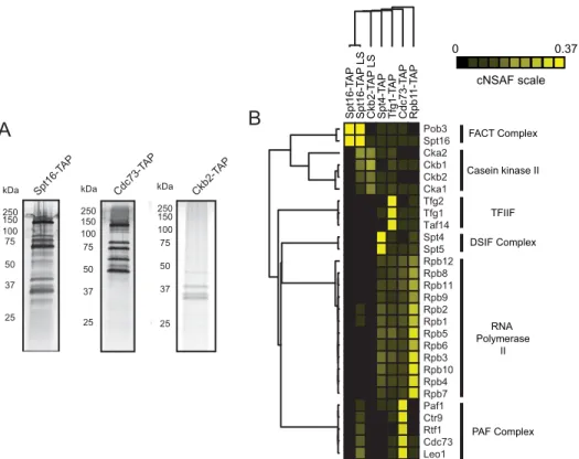
During the transcription cycle, RNAPII interacts with numerous accessory proteins to facilitate transcription initia-tion, elongation through chromatin, co-transcriptional RNA processing and transcription termination. Unlike RNA poly-merase I and III, which have “built-in” elongation factor activ-ities, all RNAPII-associated elongation factors interact in a dynamic fashion (35, 45). One approach to study transient interaction partners using AP-MS is to perform reciprocal puri-fication of low-level prey proteins to confirm the interaction with the original bait protein of interest (Fig. 1). Although this approach is common, the coordinated analysis of these purifi-cations using quantitative approaches is not often performed in favor of more standard qualitative (presence or absence) anal-ysis. In this study, we chose to perform four biological replicate purifications of Spt16-TAP under low-salt (LS, 100 mMNaCl) conditions to facilitate the capture of transient interacting pro-teins because FACT was the lowest-level RNAPII interaction partner known from previous studies identified in Rpb3-TAP
purifications (35, 36). A representative Spt16-TAP LS purifica-tion was used for hierarchical clustering analysis (Fig. 1,A,left panel, andB,second column). A high-salt (350 mMNaCl) purifica-tion was also performed for Spt16-TAP for comparison (Fig. 1B,
first column). In addition, we performed MudPIT analysis of Ckb2-TAP (a subunit of casein kinase II) and Cdc73-TAP (a sub-unit of PAF-C) and included datasets published previously for Spt4-TAP, Tfg1-TAP (a subunit of TFIIF), and Rpb11-TAP for comparison (36) (Fig. 1). For each dataset, NSAF values were cal-culated and analyzed by hierarchical clustering.
As shown in Fig. 1B, hierarchical clustering readily separates the known protein complexes into groups based on the AP-MS data (see dendrogram on the left and compare with protein complex labels on theright). Using these data, the Spt4/Spt5 heterodimer is readily identified as a complex based on the high abundance of the complex in Spt4-TAP samples. Overall, the cluster analysis identified two main sets of elongation factors. The first set includes TFIIF, Spt4/Spt5, and PAF-C as interac-tion partners that readily co-purify with RNAPII (Fig. 1B, bot-tom3⁄4of the cluster). The second set includes FACT and CKII, which appear to interact with PAF-C under low-salt conditions when using Spt16-TAP as bait (Fig. 1B, top1⁄4of the cluster). These data support two previously reported models: FACT can act as a complex with CKII (24), and PAF-C can serve as a scaffold to recruit FACT to RNAPII (18).
SAINT calculates interaction probabilities for proteins that are isolated through affinity purification approaches (referred
Pob3 Spt16 Cka2 Ckb1 Ckb2 Cka1 Tfg2 Tfg1 Taf14 Spt4 Spt5 Rpb12 Rpb8 Rpb11 Rpb9 Rpb2 Rpb1 Rpb5 Rpb6 Rpb3 Rpb10 Rpb4 Rpb7 Paf1 Ctr9 Rtf1 Cdc73 Leo1
Spt16-T
A
P
Spt16-T
A
P
LS
Ckb2-T
AP
LS
Spt4-T
AP
Tfg1-T
A
P
Cdc73-T
AP
Rpb1
1-T
A
P
FACT Complex
Casein kinase II
TFIIF
DSIF Complex
RNA Polymerase
II
PAF Complex
150 100 75
50
37
25 kDa
250 150
100 75
50
37
25 kDa
250
Ckb2-T AP
Cdc73-TAP
150 100 75
50
37
25 kDa
250
Spt16-TAP
A
B
0 0.37
cNSAF scale
FIGURE 1.Analysis of the FACT, PAF-C, and CKII interactome.A, TAP-tagged Spt16, Cdc73, and Ckb2 were immunoprecipitated using IgG-Sepharose and calmodulin-sepharose resins and eluted via 2 mMEGTA. Approximately 12.5% of a representative elution was electrophoresed on a Bio-Rad Mini Protean TGXTMgel and stained with silver nitrate. Band sizes were determined by comparison with Precision Plus protein standards (Bio-Rad) as indicated.B, hierar-chical clustering analysis of RNAPII elongation factor interactions. The heat map displays NSAF values for the indicated baits (top) and preys (right), withblack
squaresrepresenting proteins that were not detected corresponding to the color (NSAF) scale. A dendrogram is shown at thetopandleft, based on hierarchical
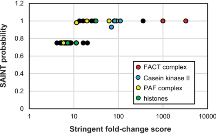
to as prey), such as TAP and FLAG purifications. In our analy-sis, the specific bait purifications (Spt16-TAP LS) are compared with mock purifications from untagged parental strains. Using SAINT, we analyzed the four Spt16-TAP LS biological repli-cates to identify significant interaction partners of FACT
(supplemental Table S3). In addition to the FACT complex
itself, histones H2A, H2B, and H4 (Htb1, Htb2, and Hht1, respectively), all four subunits of CKII, and the five subunits of PAF-C had SAINT scores ofⱖ0.75 (Fig. 2). Additional signifi-cant interaction partners of Spt16 are listed insupplemental
Table S3. There are also low-level PSM values identified in
rep-licate Spt16-TAP samples for the two largest subunits of RNA-PII (Rpb1 and Rpb2), but these interactions were not signifi-cantly enriched over controls. The interaction values from this purification span 4 orders of magnitude, perhaps revealing the true dynamic nature of protein interactions with the FACT complex.
The data presented so far clearly suggest a reproducible yet dynamic relationship between FACT, CKII, and PAF-C. The data obtained from Spt16-TAP low-salt purifications provide convincing evidence for protein interactions between FACT, CKII, and PAF-C. However, reciprocal purifications of PAF-C did not result in high levels of co-purifying FACT or CKII (Fig. 1). We next chose to further characterize the potential dynamic protein interaction between PAF-C and CKII in more detail. CKII is a constitutively active serine/threonine kinase that has been implicated in the phosphorylation of a number of tran-scription-related proteins including RNAPII itself (46). Multi-ple consensus motifs have been defined that are phosphory-lated by CKII, including [S/T]XX[E/D] (47), SDXE, SXX[E/D], and [D/E]S[D/E]X[D/E] and many other similar motifs (48, 49). Kinase-substrate interactions have also been characterized as highly transient, and previous studies using AP-MS to charac-terize these interactions have used approaches, including kinase overexpression, to increase the overall chance of catch-ing a snapshot of these quick reactions (41). To determine whether PAF-C and/or FACT subunits are substrates of CKII, we performed dynamic posttranslational modification searches
for serine, threonine, or tyrosine phosphorylation on Spt16-TAP and Cdc73-Spt16-TAP purifications. Using this approach, we found that all five subunits of PAF-C are putative CKII targets, suggesting that PAF-C and CKII are not only direct interacting partners but that the interaction rates are likely too rapid to capture high levels of CKII subunits in PAF-C purifications
(supplemental Table S4, Fig. 3). Two of the phosphorylation
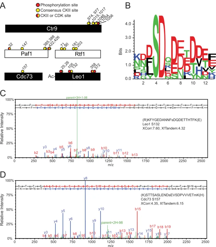
sites, Ser422 in Paf1 and Ser358 in Leo1, consist of a SPD/E sequence, which suggests that these sites could be targets of cyclin-dependent kinases or CKII based on their consensus motifs (49). Three of the PAF-C subunits (Ctr9, Paf1, and Rtf1) are putatively modified by CKII at multiple sites (Figs. 3,A,C, andD, with representative phosphosite mapping spectra pro-vided insupplemental Fig. S1). Serine 132 in Leo1, a putative CKII consensus site, is modified in over 80% of the PSMs iden-tified that contain that amino acid.
To define the consensus motifs for the PAF-C amino acids modified by CKII, we performed sequence enrichment analysis using Seq2Logo (50) (Fig. 4B). The major consensus sequence (n⫽10 sites) for PAF-C phosphorylation sites is SDX[D/E][D/ E]XD, which strongly resembles previously defined CKII phos-phorylation motifs (48, 49) (Fig. 3 andsupplemental Table S4). These data, in light of our protein interaction analysis, suggest that FACT may facilitate CKII recruitment to PAF-C for its subsequent phosphorylation (18). Although we performed full phosphorylation analyses of the FACT subunits Spt16 and Pob3, we did not identify any phosphorylated peptides in either protein. However, the C-terminal domain sequence of Spt16 was not detected in our analyses, suggesting that alternate pro-tease digestions may be needed to fully analyze potential mod-ification sites in FACT.
We next performed a series of experiments to confirm that the subunits of PAF-C arebona fidesubstrates of CKII using bothin vitroandin vivoapproaches. Using [␥32P]ATP, we first performedin vitrokinase assays using LS Spt16-TAP-purified FACT and Ctr9-FLAG-purified PAF-C as substrates in the presence or absence or recombinant CKII. As shown in Fig. 4A,
lane 2, proteins corresponding to the molecular weight of both subunits of the FACT complex can be phosphorylatedin vitro
in reactions containing recombinant CKII (Fig. 4A,Spt16and
Pob3). Recombinant CKII is also autophosphorylated in this experiment, as visualized in Fig. 4A,lane 5, which only contains recombinant CKII and ATP. Interestingly, phosphorylated Spt16 and Pob3 bands were also visualized in reactions lacking recombinant CKII, suggesting that CKII (or potentially some other co-purifying kinase) can also phosphorylate FACTin vitro(Fig. 4A). Importantly, we found that PAF-C was not phos-phorylated in the absence of recombinant CKII, suggesting that CKII is not a stable interacting partner of PAF-C (Fig. 4A,lane 3). These data are also in agreement with our AP-MS studies (Figs. 1 and 2). In contrast, four bands corresponding to the molecular weights of PAF-C subunits are readily phosphory-lated in the presence of recombinant CKII (Fig. 4A, lane 4). Notably, we did find that four of the five PAF-C subunits were phosphorylated in our in vitroreaction following incubation with Spt16-TAP and ATP (Fig. 4B,lane 3). These data clearly show that the FACT-CKII complex will readily phosphorylate PAF-Cin vitro.
0 0.2 0.4 0.6 0.8 1 1.2
1 10 100 1000 10000
FACT complex
PAF complex Casein kinase II
histones
SAINT probabilit
y
Stringent fold-change score
FIGURE 2.Identification of significant Spt16-TAP interaction partners.
Shown are stringent -fold change scoresversusSAINT interaction proba-bilities for FACT, CKII, and PAF-C complex members from analysis of Spt16-TAP samples (n⫽4). The specific subunit names and their interaction scores are listed insupplemental Table S3. A legend including a color code for the subunits of FACT, CKII, PAF-C, and nucleosomes is shown at the
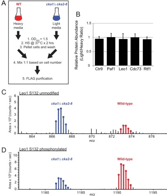
To test whether CKII is responsible for phosphorylation of PAF-C using anin vivoapproach, we performed quantitative proteomics analysis of Ctr9-FLAG-isolated PAF-C complexes
from WT and CKII temperature-sensitive strains (cka1⌬ cka2– 8) using SILAC-based quantitation (Fig. 5). The experi-mental scheme used for these studies is shown in Fig. 5A. Both
Ctr9
Paf1
Cdc73
Rtf1
Leo1
Phosphorylation site Consensus CKII site203 181 90
372 358 132
105 25,26
Ac-1054 1046 1015,1017 974, 977
157
147 385,388422,426
A
CKII or CDK site
1056
32
parent+3H+1-98
b2 y3
b3b4y4 b5
y5 y6 b6
b7 y7b8
y8b9 b10 y10
b11y11b12 b13
m/z
Relative Intensit
y
0% 25% 50% 75% 100%
0 250 500 750 1000 1250 1500 1750 2000 2250 2500
(R)KFYGEDANNFsDQDETTHTFK(E) Leo1 S132
XCorr:7.60, X!Tandem:4.32
parent+2H-98
b4 y3
b5 y4
b6 y5
b7 y6
b8
y7b9
y8
b10 y9
y10
b11 y11
b12y12 b13
b14 y13
b15
y14 y15 b17
y16 b18
y17 b19
S T T S A S L E N D S+80 E V S D P V V V E T M+16 K K M+16 T E V V V P D S V E S+80 D N E L S A S T T S
m/z
Relative Intensit
y
0% 25% 50% 75% 100%
0 250 500 750 1000 1250 1500 1750 2000 2250 2500
(K)STTSASLENDsEVSDPVVVETmK(H) Cdc73 S157
XCorr:4.35, X!Tandem:6.15
B
C
D
71
K F Y G E D A N N F S+80 D Q D E T T H T F K
K F T H T T E D Q D S+80 F N N A D E G Y F K
N
S
0.01.0 2.0 3.0 4.0
Bits
R D
LS
N
K
2
G
HS
T
N
D
Y
L
F
D
4
D
6ND E LK
S
Q
S
E
D
8
D
E
L
K
D
E
T
10
Y
T
D
N
H
V
D
12
N
D
V
T
E
H KY
V
E
F
FIGURE 3.The PAF-C complex is substantially phosphorylatedin vivo.A, schematics of PAF-C subunits with the location of MS-identified phosphorylation sites (red circles) and MS-identified CKII consensus sites (yellow circles) on PAF-C members. Putative CKII or cyclin-dependent kinase (CDK) sites are shown as
half-red/half-yellow circles.B, Seq2Logo analysis of consensus CKII phosphorylation sites identified in MudPIT analysis.C, representative spectra of Leo1
WT and CKII temperature-sensitive strains were grown to an
A600⫽1.5 and then heat shocked at 37 °C for 2 h prior to mixing of the cell pellets, lysis, and PAF-C purification. The full set of significantly changed peptides identified in the Ctr9-FLAG SILAC dataset is included insupplemental Table S5. As shown
insupplemental Table S5, we readily identified peptides for 10
phosphorylation sites across four subunits of PAF-C for which the levels of the phosphorylated peptides decreased, whereas the unmodified peptides increased incka1⌬cka2– 8(light) ver-susthe WT (heavy). Although we observed multiple decreases in the level of phosphopeptides incka1⌬cka2– 8, the relative protein abundance of all five PAF-C subunits was unchanged (Fig. 5B). The MS1 precursor intensity for the isotopic peaks are shown for representative Leo1 peptides KFYGEDANNFSDQ-DETTHTFKEENVELVR and KFYGEDANNFSPO4 DQDETT-HTFKEENV-ELVR containing unmodified or phosphorylated serine 132, respectively (Fig. 5,CandD). The MS1 intensity for peptide ions containing unmodified Leo1 serine 132 increased 2.5-fold following heat shock ofcka1⌬cka2– 8strains (Fig. 5C). The opposite trend was observed for Leo1 serine 132, with a 3-fold decrease in the abundance of MS1 area (Fig. 5D). Similar trends were observed for the other putative CKII sites in Ctr9, Paf1, and Rtf1, suggesting that these are CKII targets (
supple-mental Table S5). Overall, these data convincingly show that
PAF-C is abona fidesubstrate of CKIIin vivo. Interestingly, we also observed increased threonine 127 phosphorylation in Paf1. These data suggest that other kinases may partially compensate for the loss of CKII activity following heat shock of cka1⌬ cka2– 8strains to regulate Paf1 phosphorylation (supplemental
Table S5).
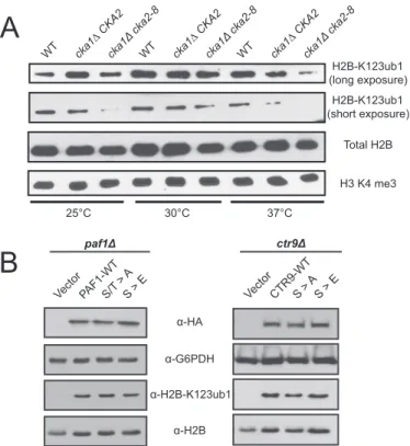
We next assessed the functional consequences of CKII dis-ruption on the biological pathways that are known to require PAF-C and FACT. Both complexes are known to regulate post-translational histone modifications, including H2B-K123ub1. Histone H2B-K123ub1 occurs during RNAPII transcription elongation and is a regulatory event for histone H3 lysine meth-ylation at Lys4and Lys79(11, 51–55). Because CKII is responsi-ble for extensive modification of PAF-C, we analyzed the effect of CKII disruption on H2B-K123ub1 levels in WT, cka1⌬ CKA2, andcka1⌬cka2– 8strains (Fig. 6A). At restrictive tem-peratures of 25 °C and 37 °C, where the activity of CKII is com-promised incka1⌬cka2– 8(32), the levels of H2B-K123ub1 are decreased incka1⌬cka2– 8strains relative to the levels of total histone H2B. Additionally, the levels of histone H2B-K123ub1 were decreased incka1⌬ CKA2 following incubation at the restrictive temperatures (Fig. 6A). No changes in histone H3K4 methylation levels were observed, which is in line with studies that show that reduced H2B-K123ub1 levels can occur without significant effects on H3K4 methylation (56). Taken together, these data show that CKII is an important regulator of histone H2B-K123ub1, likely through its regulation of PAF-C and FACT. Additional analysis of phosphosite mutants of the PAF-C subunits Paf1, Ctr9, and Cdc73 did not individually reveal defects in histone modification levels at H2B-K123ub1, histoneH3lysine4trimethylation,orhistoneH3lysine36trimeth-ylation (Fig. 6B). In addition, although strains with defective PAF-C are sensitive to the transcription elongation inhibitor 6-azauracil or hydroxyurea (16, 17, 57– 60), the phosphosite mutants did not display growth defects when exposed to these drugs (data not shown). These data suggest that changes in the phosphorylation of multiple subunits of PAF-C and/or FACT may be required to recapitulate the phenotype observed in
cka1⌬cka2– 8.Regardless, these findings suggest that CKII acts upstream of PAF-C and FACT as well as to regulate H2B-K123ub1 levels. However, the precise mechanisms through which this regulation occurs remain to be determined.
Discussion
In this study, we have performed an in-depth proteomics and genetic analysis of two transiently associated RNAPII elonga-tion factors that are recruited to target genes during active gene transcription. Although PAF-C subunits were not identified as significant interaction partners of RNAPII when using Rpb3-TAP as bait in previous studies, purification of PAF-C through Cdc73-TAP revealed that RNAPII is a major interaction part-ner of PAF-C (Fig. 1). However, purifications of FACT through Spt16-TAP under low-salt conditions revealed a significant interaction between FACT and PAF-C (supplemental Table S3 and Fig. 2). These data suggest that PAF-C may be involved in FACT recruitment to RNAPII, as has been hypothesized previ-ously, because PAF-C readily interacts with RNAPII in affinity purifications (18). FACT readily co-purifies with CKII in low-recombinant CKII
Spt16-TAP Ctr9-FLAG
+ + - -
+
- - +
- + - + +
-Spt16-PO4
Pob3-PO4
CKII α-PO4
32P-autoradiogram
Lane 1 2 3 4 5
Ctr9-PO4
Rtf1-PO4
Leo1-PO4
Paf1-PO4
CKII α-PO4
A
Spt16-TAP Ctr9-FLAG
+ - +
+
- +
Lane 1 2 3
Spt16-PO4
Pob3-PO4
Ctr9-PO4
Rtf1-PO4
Leo1-PO4
Paf1-PO4
recombinant recombinant
32P-autoradiogram
B
salt purifications. Although Cdc73-TAP samples did not con-tain significant levels of FACT or CKII subunit peptides, we found that four of five subunits of PAF-C are phosphorylated by CKIIin vitroandin vivo(Figs. 3–5). In addition, we found that Spt16-interacting CKII can readily phosphorylate purified PAF-Cin vitro(Fig. 4B). Together, these findings implicate the FACT complex in phosphorylation of PAF-C by CKII. The human FACT complex has been proposed previously to play a similar role in the regulation of p53 phosphorylation by CKII (24).
Previous studies have addressed the challenge of transient interaction partners by overexpression of bait proteins (specif-ically kinases) to increase the relative abundance of significant protein-protein interactions (41). However, this approach could result in a large number of false positive interactions as a result of overexpression effects on the biological system in
question. Additionally, single affinity purifications have been suggested as an approach to capture dynamic interaction part-ners. Unfortunately, single affinity purifications also have sig-nificantly higher interactions with cellular contaminants that, in our experience, are not removed by algorithms such as SAINT (37). Dynamic or transient interaction partners present even more of a challenge to existing statistical programs than small proteins (discussed above). This study suggests that inclusion of a large number of replicates in combination with follow-up reciprocal purifications and functional studies is the most robust approach for characterization of dynamic interac-tions such as those with kinases like CKII. However, it must also be acknowledged that the low detection frequency of transient interaction partners is a significant challenge for data-depen-dent acquisition-based approaches and their related statistical interpretation. For instance, confirmation of PAF-C
modifica-1180 1185 1190 1195
m/z 0
2 4 6 8 10
864 866 868 870 872 874 876
m/z 0
1 2 3 4 5 6
Area x 10
6 (counts • sec)
Area x 10
6 (counts • sec)
cka1∆ cka2-8 WT
1. OD600 = 1.5 2. HS @ 37°C x 2 hrs 3. Pellet cells and wash Heavy
media
Light media
4. Mix 1:1 based on cell number
5. FLAG purification
Leo1 S132 unmodified
cka1∆ cka2-8 Wild-type
cka1∆ cka2-8 Wild-type
Leo1 S132 phosphorylated
0 0.5
1 1.5
Ctr9 Paf1 Leo1 Cdc73 Rtf1
Relative Protein Abundance
(Light/Heavy Ratio)
A
A
B
C
D
tion by CKIIin vitroandin vivoshould result in the functional annotation of PAF-C and CKII as having both a transient pro-tein-protein interaction and an enzyme-substrate relationship. Our data show that CKII is a novel upstream regulator of H2B-K123ub1. We hypothesized that CKII phosphorylation of PAF-C could be required for FACT and/or PAF-C-dependent control of H2B-K123ub1. We have clearly shown that CKII reg-ulates PAF-C phosphorylation and H2B-K123ub1. However, our initial genetic studies on PAF-C phosphorylation have not shown that PAF-C phosphorylation is required for H2B-K123ub1. There are many possible reasons for this. First, it is possible that CKII phosphorylation needs to be disrupted on multiple PAF-C subunits to fully recapitulate thecka1⌬cka2– 8
phenotype. Additionally, it is possible that phosphorylation sites are present across these subunits that were not detected in our mass spectrometry analysis because of low peptide detect-ability and/or overdigestion with trypsin. Our findings that Paf1 phosphorylation increases at threonine 127 incka1⌬cka2– 8
strains could suggest that other kinases can compensate for the loss of CKII activity. Finally, it is also plausible that the regula-tion of H2B-K123ub1 by CKII occurs in a PAF-C-independent mechanism.
Author Contributions—L. G. B., R. D., J. L. K., G. O. H., E. D. A., A. K. B., and A. L. M. generated the strains, conducted the experi-ments, and analyzed the results. L. G. B., G. O. H., and A. L. M. per-formed the mass spectrometry runs and data analyses. L. G. B. and A. L. M. wrote most of the paper with significant contributions from R. D., J. L. K., and B. D. S. L. G. B., R. D., J. L. K., B. D. S., and A. L. M. conceived the idea for the project.
Acknowledgments—We thank Whitney Smith-Kinnaman for techni-cal contributions and project support, Nicole Novaresi for strain gen-eration, and members of the Mosley laboratory for comments on the manuscript. We also thank the Dr. David Stillman, Judith Jaehning, and Georjana Barnes laboratories for the spt16 –11, PAF1 deletions and casein kinase II mutants, respectively.
References
1. Tomson, B. N., and Arndt, K. M. (2013) The many roles of the conserved eukaryotic Paf1 complex in regulating transcription, histone modifica-tions, and disease states.Biochim. Biophys. Acta1829,116 –126 2. Wozniak, G. G., and Strahl, B. D. (2014) Hitting the “mark”: interpreting
lysine methylation in the context of active transcription.Biochim. Biophys. Acta1839,1353–1361
3. Thornton, J. L., Westfield, G. H., Takahashi, Y. H., Cook, M., Gao, X., Woodfin, A. R., Lee, J. S., Morgan, M. A., Jackson, J., Smith, E. R., Couture, J. F., Skiniotis, G., and Shilatifard, A. (2014) Context dependency of Set1/ COMPASS-mediated histone H3 Lys4 trimethylation. Genes Dev.28, 115–120
4. Laribee, R. N., Krogan, N. J., Xiao, T., Shibata, Y., Hughes, T. R., Green-blatt, J. F., and Strahl, B. D. (2005) BUR kinase selectively regulates H3 K4 trimethylation and H2B ubiquitylation through recruitment of the PAF elongation complex.Curr. Biol.15,1487–1493
5. Xiao, T., Kao, C. F., Krogan, N. J., Sun, Z. W., Greenblatt, J. F., Osley, M. A., and Strahl, B. D. (2005) Histone H2B ubiquitylation is associated with elongating RNA polymerase II.Mol. Cell. Biol.25,637– 651
6. Mueller, C. L., and Jaehning, J. A. (2002) Ctr9, Rtf1, and Leo1 are compo-nents of the Paf1/RNA polymerase II complex. Mol. Cell. Biol. 22, 1971–1980
7. Zhu, B., Mandal, S. S., Pham, A. D., Zheng, Y., Erdjument-Bromage, H., Batra, S. K., Tempst, P., and Reinberg, D. (2005) The human PAF complex coordinates transcription with events downstream of RNA synthesis. Genes Dev.19,1668 –1673
8. Kim, J., Guermah, M., and Roeder, R. G. (2010) The human PAF1 complex acts in chromatin transcription elongation both independently and coop-eratively with SII/TFIIS.Cell140,491–503
9. Costa, P. J., and Arndt, K. M. (2000) Synthetic lethal interactions suggest a role for theSaccharomyces cerevisiaeRtf1 protein in transcription elonga-tion.Genetics156,535–547
10. Orphanides, G., Wu, W. H., Lane, W. S., Hampsey, M., and Reinberg, D. (1999) The chromatin-specific transcription elongation factor FACT comprises human SPT16 and SSRP1 proteins.Nature400,284 –288 11. Pavri, R., Zhu, B., Li, G., Trojer, P., Mandal, S., Shilatifard, A., and
Rein-berg, D. (2006) Histone H2B monoubiquitination functions cooperatively with FACT to regulate elongation by RNA polymerase II. Cell 125, 703–717
12. Formosa, T., Eriksson, P., Wittmeyer, J., Ginn, J., Yu, Y., and Stillman, D. J. (2001) Spt16-Pob3 and the HMG protein Nhp6 combine to form the nucleosome-binding factor SPN.EMBO J.20,3506 –3517
13. Stuwe, T., Hothorn, M., Lejeune, E., Rybin, V., Bortfeld, M., Scheffzek, K., and Ladurner, A. G. (2008) The FACT Spt16 “peptidase” domain is a histone H3-H4 binding module. Proc. Natl. Acad. Sci. U.S.A. 105, 8884 – 8889
14. Kemble, D. J., Whitby, F. G., Robinson, H., McCullough, L. L., Formosa, T., and Hill, C. P. (2013) Structure of the Spt16 middle domain reveals func-tional features of the histone chaperone FACT. J. Biol. Chem. 288, 10188 –10194
25°C 30°C 37°C
WT cka1∆ CKA2cka1Δ
cka2-8
WT cka1∆ CKA2cka1Δ
cka2-8
WT cka1∆ CKA2cka1Δ
cka2-8
H2B-K123ub1 (long exposure)
H2B-K123ub1 (short exposure)
Total H2B H3 K4 me3
α-HA α-G6PDH
α-H2B α-H2B-K123ub1
A
B
Vect or
PAF1-WTS/T > A
S > E
paf1Δ
Vect or
CTR9-WTS > A
S > E
ctr9Δ
FIGURE 6.CKII regulates histone H2B-K123ub1 levels.A, Western blotting analysis of histone H2B-K123ub1, total histone H2B, and histone H3 lysine 4 trimethylation levels from whole cell extracts fromcka1⌬cka2– 8strains grown under permissive (30 °C) and restrictive (25 °C and 37 °C) tempera-tures. The antibody used for each panel is indicated at theright, and the temperature used for cell growth is given at thebottom.B, Western blotting analysis of whole cell lysates for histone H2B-K123ub1, G6PDH, H2B, and HA ofPAF1orCTR9deletion strains rescued withPAF1-3HA orCTR9-3HA expres-sion vectors as indicated.Left panel,Vector, pRS313–3HA-SSN6;PAF1-WT, PAF1–3HA;S/T⬎A, PAF1–3HA S147A, T385A, T422A, S426A;S/T⬎E, PAF1– 3HA S147E, T385E, T422E, S426E.Right panel, Vector, pRS313–3HA-SSN6;
CTR9-WT, CTR9 –3HA); S⬎A, CTR9 –3HA S977A, S1015A, S1017A, S1046A,
15. McCullough, L., Poe, B., Connell, Z., Xin, H., and Formosa, T. (2013) The FACT histone chaperone guides histone H4 into its nucleosomal confor-mation inSaccharomyces cerevisiae.Genetics195,101–113
16. Krogan, N. J., Kim, M., Ahn, S. H., Zhong, G., Kobor, M. S., Cagney, G., Emili, A., Shilatifard, A., Buratowski, S., and Greenblatt, J. F. (2002) RNA polymerase II elongation factors ofSaccharomyces cerevisiae: a targeted proteomnics approach.Mol. Cell. Biol.22,6979 – 6992
17. Squazzo, S. L., Costa, P. J., Lindstrom, D. L., Kumer, K. E., Simic, R., Jen-nings, J. L., Link, A. J., Arndt, K. M., and Hartzog, G. A. (2002) The Paf1 complex physically and functionally associates with transcription elonga-tion factorsin vivo.EMBO J.21,1764 –1774
18. Adelman, K., Wei, W., Ardehali, M. B., Werner, J., Zhu, B., Reinberg, D., and Lis, J. T. (2006)DrosophilaPaf1 modulates chromatin structure at actively transcribed genes.Mol. Cell. Biol.26,250 –260
19. Venerando, A., Ruzzene, M., and Pinna, L. A. (2014) Casein kinase: the triple meaning of a misnomer.Biochem. J.460,141–156
20. Meggio, F., and Pinna, L. A. (2003) One-thousand-and-one substrates of protein kinase CK2?FASEB J.17,349 –368
21. Filhol, O., Giacosa, S., Wallez, Y., and Cochet, C. (2015) Protein kinase CK2 in breast cancer: the CK2regulatory subunit takes center stage in epithelial plasticity.Cell. Mol. Life Sci.72,3305–3322
22. Piazza, F., Manni, S., and Semenzato, G. (2013) Novel players in multiple myeloma pathogenesis: role of protein kinases CK2 and GSK3.Leuk. Res. 37,221–227
23. Piazza, F., Manni, S., Ruzzene, M., Pinna, L. A., Gurrieri, C., and Semen-zato, G. (2012) Protein kinase CK2 in hematologic malignancies: reliance on a pivotal cell survival regulator by oncogenic signaling pathways. Leu-kemia26,1174 –1179
24. Keller, D. M., Zeng, X., Wang, Y., Zhang, Q. H., Kapoor, M., Shu, H., Goodman, R., Lozano, G., Zhao, Y., and Lu, H. (2001) A DNA damage-induced p53 serine 392 kinase complex contains CK2, hSpt16, and SSRP1. Mol. Cell7,283–292
25. Tripodi, F., Nicastro, R., Busnelli, S., Cirulli, C., Maffioli, E., Tedeschi, G., Alberghina, L., and Coccetti, P. (2013) Protein kinase CK2 holoenzyme promotes start-specific transcription in Saccharomyces cerevisiae. Eu-karyotic Cell12,1271–1280
26. Miteva, Y. V., Budayeva, H. G., and Cristea, I. M. (2013) Proteomics-based methods for discovery, quantification, and validation of protein-protein interactions.Anal. Chem.85,749 –768
27. Janke, C., Magiera, M. M., Rathfelder, N., Taxis, C., Reber, S., Maekawa, H., Moreno-Borchart, A., Doenges, G., Schwob, E., Schiebel, E., and Knop, M. (2004) A versatile toolbox for PCR-based tagging of yeast genes: new fluorescent proteins, more markers and promoter substitution cassettes. Yeast21,947–962
28. Gietz, R. D., and Schiestl, R. H. (2007) High-efficiency yeast transforma-tion using the LiAc/SS carrier DNA/PEG method.Nat. Protoc.2,31–34 29. Gelbart, M. E., Rechsteiner, T., Richmond, T. J., and Tsukiyama, T. (2001)
Interactions of Isw2 chromatin remodeling complex with nucleosomal arrays: analyses using recombinant yeast histones and immobilized tem-plates.Mol. Cell. Biol.21,2098 –2106
30. Hanna, D. E., Rethinaswamy, A., and Glover, C. V. (1995) Casein kinase II is required for cell cycle progression during G1and G2/M in
Saccharomy-ces cerevisiae.J. Biol. Chem.270,25905–25914
31. Peng, Y., Wong, C. C., Nakajima, Y., Tyers, R. G., Sarkeshik, A. S., Yates, J., 3rd, Drubin, D. G., and Barnes, G. (2011) Overlapping kinetochore targets of CK2 and Aurora B kinases in mitotic regulation.Mol. Biol. Cell22, 2680 –2689
32. Hockman, D. J., and Schultz, M. C. (1996) Casein kinase II is required for efficient transcription by RNA polymerase III.Mol. Cell. Biol.16,892– 898 33. Puig, O., Caspary, F., Rigaut, G., Rutz, B., Bouveret, E., Bragado-Nilsson, E., Wilm, M., and Séraphin, B. (2001) The tandem affinity purification (TAP) method: a general procedure of protein complex purification.Methods24, 218 –229
34. Mosley, A. L., Pattenden, S. G., Carey, M., Venkatesh, S., Gilmore, J. M., Florens, L., Workman, J. L., and Washburn, M. P. (2009) Rtr1 is a CTD phosphatase that regulates RNA polymerase II during the transition from serine 5 to serine 2 phosphorylation.Mol. Cell34,168 –178
35. Mosley, A. L., Sardiu, M. E., Pattenden, S. G., Workman, J. L., Florens, L.,
and Washburn, M. P. (2011) Highly reproducible label free quantitative proteomic analysis of RNA polymerase complexes.Mol. Cell. Proteomics 10.1074/mcp.M110.000687
36. Mosley, A. L., Hunter, G. O., Sardiu, M. E., Smolle, M., Workman, J. L., Florens, L., and Washburn, M. P. (2013) Quantitative proteomics demon-strates that the RNA polymerase II subunits Rpb4 and Rpb7 dissociate during transcriptional elongation.Mol. Cell. Proteomics12,1530 –1538 37. Smith-Kinnaman, W. R., Berna, M. J., Hunter, G. O., True, J. D., Hsu, P.,
Cabello, G. I., Fox, M. J., Varani, G., and Mosley, A. L. (2014) The interac-tome of the atypical phosphatase Rtr1 inSaccharomyces cerevisiae.Mol. Biosyst.10,1730 –1741
38. Mellacheruvu, D., Wright, Z., Couzens, A. L., Lambert, J. P., St-Denis, N. A., Li, T., Miteva, Y. V., Hauri, S., Sardiu, M. E., Low, T. Y., Halim, V. A., Bagshaw, R. D., Hubner, N. C., Al-Hakim, A., Bouchard, A.,et al.(2013) The CRAPome: a contaminant repository for affinity purification-mass spectrometry data.Nat. Methods10,730 –736
39. Choi, H., Larsen, B., Lin, Z. Y., Breitkreutz, A., Mellacheruvu, D., Fermin, D., Qin, Z. S., Tyers, M., Gingras, A. C., and Nesvizhskii, A. I. (2011) SAINT: probabilistic scoring of affinity purification-mass spectrometry data.Nat. Methods8,70 –73
40. Choi, H., Liu, G., Mellacheruvu, D., Tyers, M., Gingras, A. C., and Nesvi-zhskii, A. I. (2012) Analyzing protein-protein interactions from affinity purification-mass spectrometry data with SAINT.Curr. Protoc. Bioinfor-maticsChapter 8, Unit 8 15
41. Breitkreutz, A., Choi, H., Sharom, J. R., Boucher, L., Neduva, V., Larsen, B., Lin, Z. Y., Breitkreutz, B. J., Stark, C., Liu, G., Ahn, J., Dewar-Darch, D., Reguly, T., Tang, X., Almeida, R.,et al.(2010) A global protein kinase and phosphatase interaction network in yeast.Science328,1043–1046 42. Kwon, Y., Vinayagam, A., Sun, X., Dephoure, N., Gygi, S. P., Hong, P., and
Perrimon, N. (2013) The Hippo signaling pathway interactome.Science 342,737–740
43. Daniels, D. L., Méndez, J., Mosley, A. L., Ramisetty, S. R., Murphy, N., Benink, H., Wood, K. V., Urh, M., and Washburn, M. P. (2012) Examining the complexity of human RNA polymerase complexes using HaloTag technology coupled to label free quantitative proteomics.J. Proteome Res. 11,564 –575
44. Zybailov, B., Mosley, A. L., Sardiu, M. E., Coleman, M. K., Florens, L., and Washburn, M. P. (2006) Statistical analysis of membrane proteome ex-pression changes in Saccharomyces cerevisiae. J. Proteome Res. 5, 2339 –2347
45. Ruan, W., Lehmann, E., Thomm, M., Kostrewa, D., and Cramer, P. (2011) Evolution of two modes of intrinsic RNA polymerase transcript cleavage. J. Biol. Chem.286,18701–18707
46. Trembley, J. H., Hu, D., Slaughter, C. A., Lahti, J. M., and Kidd, V. J. (2003) Casein kinase 2 interacts with cyclin-dependent kinase 11 (CDK11)in vivo and phosphorylates both the RNA polymerase II carboxyl-terminal do-main and CDK11in vitro.J. Biol. Chem.278,2265–2270
47. Blatch, G. L., Lässle, M., Zetter, B. R., and Kundra, V. (1997) Isolation of a mouse cDNA encoding mSTI1, a stress-inducible protein containing the TPR motif.Gene194,277–282
48. Schwartz, D., and Gygi, S. P. (2005) An iterative statistical approach to the identification of protein phosphorylation motifs from large-scale data sets.Nat. Biotechnol.23,1391–1398
49. Amanchy, R., Periaswamy, B., Mathivanan, S., Reddy, R., Tattikota, S. G., and Pandey, A. (2007) A curated compendium of phosphorylation motifs. Nat. Biotechnol.25,285–286
50. Thomsen, M. C., and Nielsen, M. (2012) Seq2Logo: a method for con-struction and visualization of amino acid binding motifs and sequence profiles including sequence weighting, pseudo counts and two-sided rep-resentation of amino acid enrichment and depletion.Nucleic Acids Res. 40,W281–W287
51. Briggs, S. D., Xiao, T., Sun, Z. W., Caldwell, J. A., Shabanowitz, J., Hunt, D. F., Allis, C. D., and Strahl, B. D. (2002) Gene silencing: trans-histone regulatory pathway in chromatin.Nature418,498
52. Sun, Z. W., and Allis, C. D. (2002) Ubiquitination of histone H2B regulates H3 methylation and gene silencing in yeast.Nature418,104 –108 53. Ng, H. H., Xu, R. M., Zhang, Y., and Struhl, K. (2002) Ubiquitination of
of histone H3 lysine 79.J. Biol. Chem.277,34655–34657
54. Dover, J., Schneider, J., Tawiah-Boateng, M. A., Wood, A., Dean, K., John-ston, M., and Shilatifard, A. (2002) Methylation of histone H3 by COM-PASS requires ubiquitination of histone H2B by Rad6.J. Biol. Chem.277, 28368 –28371
55. Robzyk, K., Recht, J., and Osley, M. A. (2000) Rad6-dependent ubiquitina-tion of histone H2B in yeast.Science287,501–504
56. Cucinotta, C. E., Young, A. N., Klucevsek, K. M., and Arndt, K. M. (2015) The nucleosome acidic patch regulates the H2B K123 monoubiquitylation cascade and transcription elongation inSaccharomyces cerevisiae.PLoS Genet.11,e1005420
57. Riles, L., Shaw, R. J., Johnston, M., and Reines, D. (2004) Large-scale screening of yeast mutants for sensitivity to the IMP dehydrogenase
in-hibitor 6-azauracil.Yeast21,241–248
58. Dronamraju, R., and Strahl, B. D. (2014) A feed forward circuit comprising Spt6, Ctk1 and PAF regulates Pol II CTD phosphorylation and transcrip-tion elongatranscrip-tion.Nucleic Acids Res.42,870 – 881
59. Betz, J. L., Chang, M., Washburn, T. M., Porter, S. E., Mueller, C. L., and Jaehning, J. A. (2002) Phenotypic analysis of Paf1/RNA polymerase II complex mutations reveals connections to cell cycle regulation, protein synthesis, and lipid and nucleic acid metabolism.Mol. Genet. Genomics 268,272–285
![FIGURE 4. In vitro phosphorylation of FACT and PAF-C by CKII. A, autora- autora-diograph of a representative set of in vitro reactions performed with [␥ 32 P]ATP in the presence or absence of Ctr9-FLAG, Spt16-TAP, or recombi-nant CKII, as indicated at the](https://thumb-us.123doks.com/thumbv2/123dok_us/8266148.2189808/7.891.73.416.89.570/phosphorylation-diograph-representative-reactions-performed-presence-recombi-indicated.webp)