The neural representation of typical and atypical
experiences of negative images: comparing fear,
disgust and morbid fascination
Suzanne Oosterwijk,
1Kristen A. Lindquist,
2,3Morenikeji Adebayo,
4,5and
Lisa Feldman Barrett
4,51
Department of Psychology, University of Amsterdam, Weesperplein 4, 1018 XA, Amsterdam, The
Netherlands,
2Department of Psychology, University of North Carolina, 235 E. Cameron Avenue, Chapel Hill,
NC 27599,
3Biomedical Research Imaging Center, University of North Carolina, 125 Mason Farm Road, Chapel
Hill, NC 27599,
4Martinos Center for Biomedical Imaging, 149 Thirteenth Street, Charlestown, MA 02129, and
5Department of Psychology, Northeastern University, 125 Nightingale Hall, Boston, MA 02115, USA
Correspondence should be addressed to Suzanne Oosterwijk, Department of Psychology, University of Amsterdam, Amsterdam, The Netherlands. E-mail: [email protected].
Abstract
Negative stimuli do not only evoke fear or disgust, but can also evoke a state of ‘morbid fascination’ which is an urge to approach and explore a negative stimulus. In the present neuroimaging study, we applied an innovative method to investigate the neural systems involved in typical and atypical conceptualizations of negative images. Participants received false feedback labeling their mental experience as fear, disgust or morbid fascination. This manipulation was successful; participants judged the false feedback correct for 70% of the trials on average. The neuroimaging results demonstrated differential activity within regions in the ‘neural reference space for discrete emotion’ depending on the type of feedback. We found robust differences in the ventrolateral prefrontal cortex, the dorsomedial prefrontal cortex and the lateral orbitofrontal cortex comparing morbid fascination to control feedback. More subtle differences in the dorsomedial prefrontal cortex and the lateral orbitofrontal cortex were also found between morbid fascination feedback and the other emotion feedback conditions. This study is the first to forward evidence about the neural representation of the experimen-tally unexplored state of morbid fascination. In line with a constructionist framework, our findings suggest that neural resources associated with the process of conceptualization contribute to the neural representation of this state.
Key words: emotion; morbid fascination; false feedback; conceptualization
Introduction
Affective scientists, including neuroscientists, often study emo-tional processes by presenting participants with negative images and then observing the consequences. So far, the dis-crete emotions that have dominated the research agenda are typical experiences such as fear and disgust (e.g. Haririet al., 2002; Wright et al., 2004; Stark et al., 2007; Borg et al., 2013). Nevertheless, people also experience fascination, interest or
curiosity when faced with negative events (Zuckerman and Litle, 1986; Rime´ et al., 2005; Turner and Silvia, 2006). These states have received no attention in affective neuroscience so far, even though comparing the neural representation of fascin-ation with those of fear and disgust provides an important op-portunity to examine how the brain instantiates different experiences that contain the same affective (i.e. valenced) infor-mation. In this article, we present a novel neuroimaging study
Received:26 June 2014;Revised:30 June 2015;Accepted:7 July 2015
VCThe Author (2015). Published by Oxford University Press. For Permissions, please email: [email protected]
11
doi: 10.1093/scan/nsv088
(e.g. fear, disgust, fascination) depending on a number of indi-vidual and situational factors (Barrett, 2006; 2012; Lindquist and Barrett, 2008). One underlying process that causes diversity in subjective emotional experiences is called conceptualization, and refers to the retrieval of context-relevant conceptual know-ledge derived from previous experiences that shapes the way that internal and external sensations are made meaningful (cf. Barrett, 2013). Indeed, the CAT conceives of conceptualization as an essential ‘ingredient’ in emotional experience, along with sensory information from the world (exteroceptive sensations) and sensory information from the body (interoceptive sensa-tions) (Lindquist and Barrett, 2012).
In this study, we focused on how different conceptualiza-tions of negative stimuli are supported by the ‘neural reference space for discrete emotion’, a set of regions that is consistently active when people experience discrete emotions (Koberet al., 2008; Lindquistet al., 2012). We specifically predicted involve-ment of the dorsomedial and ventromedial prefrontal cortex (dmPFC and vmPFC) and the ventrolateral prefrontal cortex (vlPFC), because the CAT argues that these regions support a conceptualization function in emotion (Wilson-Mendenhall
et al, 2011; Lindquist and Barrett, 2012; Barrett and Satpute, 2013). Furthermore, there is ample research that demonstrates a role for these regions when people generate subjective emo-tional experiences (e.g. Wilson-Mendenhall et al, 2011; Oosterwijk et al., 2012) or label/appraise affective or neutral stimuli in emotional terms (e.g. Liebermanet al., 2007; Ochsner
et al., 2009; Satputeet al., 2013).
Interestingly, a recent study has further shown that these same regions engage with relatively different strength when in-dividuals generate experiences of atypical as compared with typical experiences of emotion (Wilson-Mendenhallet al., 2014). Wilson-Mendenhallet al. manipulated typicality by shifting the valence of instances of fear, sadness and happiness (e.g. feeling ‘disturbing happiness’ when making a sarcastic jab at a col-league or ‘energizing fear’ when performing at an important sports match), and demonstrated that atypical experiences relied more heavily upon regions that support a conceptualiza-tion funcconceptualiza-tion. In this article we manipulate typicality in a differ-ent way. We propose that fear and disgust are likely to be typical ways of conceptualizing one’s reaction to a negative image dis-playing mutilated bodies or crawling insects, whereas morbid fascination, in contrast, is likely to be a more atypical way of conceptualizing one’s reaction. In the case of negative stimuli, morbid fascination is atypical because it combines negativity with an intention to approach and explore. Moreover, although morbid fascination may often be expressed in daily life (e.g. ‘rub-ber-necking’ on a freeway to view a grisly accident), the term it-self may not be often used to describe experiences of negative events. At the very least, morbid fascination is not a ‘basic-level’ category of emotion that is learned early in childhood and regu-larly used to describe modal unpleasant emotional experiences.
According to the CAT, conceptualization transforms non-discrete affective experiences into experiences of non-discrete emotions (such as fear, disgust or fascination) by integrating
used less often, and therefore has a smaller sample of ‘priors’ to draw from. In addition, morbid fascination suggests a reconcili-ation between a tendency to explore the informreconcili-ational content of the stimulus and its negativity. In this context, sensations may take on a more complex meaning, causing individuals to continue to draw on conceptual information to iteratively pro-cess the meaning of internal and external information.
The present study
We introduce a novel paradigm that presents false feedback to direct the way participants conceptualize their experience of a negative image. False feedback has been used across multiple psychological contexts, including in social, clinical and neuro-scientific studies. Previous applications of false feedback have predominantly focused on false auditory feedback about the participant’s heart rate (e.g. Valins, 1966; Wildet al., 2008). For instance, Grayet al. (2007) found that false heart rate feedback influenced participants’ affective judgments of neutral faces and led to corresponding changes in the anterior insula and amygdala. In the present experiment we did not provide partici-pants with false feedback about their physiological state. Instead, inspired by recent developments in the decoding of mental states using brain imaging data (e.g. Haynes and Rees, 2006; Shireret al., 2012) we led participants to believe that we could decode the psychological meaning of their brain states in real time. Subsequently, we gave participants false feedback about the ‘content’ of their mental experiences when viewing highly arousing, negative images. This allowed us to provide participants with different labels (i.e. fear, disgust, morbid fas-cination) to prime them to conceptualize their experience in a certain way, without asking them to actively judge the images. In the control condition, we told participants that the acquired neural pattern precluded categorization of their mental state. We chose to incorporate a non-specific label in the control con-dition so that the presence of feedback was constant across conditions—including the possibility for participants to reflect upon the categorization of their state.
With this paradigm we tested the following hypotheses. First, we tested whether regions in the neural reference space for emo-tion (e.g. dmPFC, vmPFC, vlPFC) that support the process of con-ceptualization engaged more strongly after participants received emotion feedback (i.e. fear, disgust, morbid fascination) com-pared with the control condition. Second, based on the previous findings regarding the role of conceptualization in atypical emo-tional experiences (Wilson-Mendenhallet al., 2014), we tested whether these regions engaged more strongly when people received morbid fascination feedback (i.e. an atypical state) as compared with fear or disgust feedback (i.e. typical states).
Materials and methods
Participantsexcluded if they had a history of psychiatric illness, were using psychoactive or systemic medications or if MRI contraindica-tions were present. Twenty-seven participants gave informed consent according to the Partners Health Care Institutional Review Board. Two participants did not complete the scanning session because they felt uncomfortable in the scanner, and one participant was excluded because of movement artifacts (>3 mm motion across multiple time points across multiple runs). Analyses were performed on the remaining twenty-four participants (12 females, Mage¼24.7, SDage¼3.8). Participants were paid $150 for their participation.
Manipulation
We manipulated experiential labels by telling participants (falsely) that we could ‘read-out’ their mental experience while they were viewing negative images in the scanner and that we would give them feedback on this categorization. Three experi-mental conditions combined negative images with disgust feed-back, fear feedback and morbid fascination feedback to direct conceptualization with a typical label (as ‘fear’ or ‘disgust’) or with an atypical label (as ‘morbid fascination’). The ‘could not be calculated’ condition served as a control condition in that it kept the presence of a feedback cue constant, without providing a label to direct conceptualization.
Our instruction proceeded as follows. First, we explained to participants that we had ‘identified three specific brain states that reliably predicted whether someone was experiencing fear, disgust or morbid fascination’ and that the purpose of the study was to validate these profiles. Then, we explained that the ex-periment would start with three ‘collection runs’ to ‘capture your brain state while you view the pictures’, followed by three ‘feedback runs’ that would give feedback about ‘what you were experiencing when you saw the image for the first time’. We further mentioned that when our algorithm could not find a match, participants would receive ‘could not be calculated’ feedback. Furthermore, all participants were instructed to ‘pro-long the state that you initially experienced when viewing the picture’ (e.g. during the collection runs), ‘during the full duration of the second viewing’ (e.g. during the feedback runs). The full instruction can be found in the Supplementary Materials. Because the manipulation incorporates deception, we used a thorough debriefing procedure.
Procedure
The complete experiment consisted of two sessions 5 days apart. In the first session the participants viewed and rated all images presented in the study. In the second session the partici-pants performed the experimental task in the scanner, viewed and rated all images again and filled out questionnaires and an exit interview. The rating task was programmed in MATLAB and presented each image in combination with the following five rating dimensions: threat, repulsion, interest, negativity and emotional intensity. Ratings were made on a continuous sliding scale with labeled endpoints (e.g., ‘not at all threatening’ and ‘extremely threatening’). To avoid response demand char-acteristics, we deliberately chose words that indexed ‘world-focused’ experiences of each emotion (Lindquist and Barrett, 2008), that were different from the terms used in the feedback manipulation.
The scanning session consisted of six runs. In three passive viewing runs (or ‘collection runs’) participants viewed 96 nega-tive images, 24 posinega-tive images and 24 neutral images passively.
Positive and neutral images were intermixed with negative images to prevent habituation to the highly aversive images. Each trial presented a fixation cross for 3 s, followed by an image for 3 s. Several 3–12 s jitter periods presenting a black screen were intermixed with the trials. Jitter length and the order of stimulus events was optimized using optseq (http:// surfer.nmr.mgh.harvard.edu/optseq/).
In three ‘feedback runs’, participants viewed 96 negative images with feedback, and 24 positive and 24 neutral images without feedback. Each trial started with a cue presented for 3 s. Negative images were combined with a feedback cue that com-bined a label (i.e. fear, disgust, morbid fascination or control) with an artificially created image of brain activation (see section on Stimulus Materials). Positive and neutral images were pre-ceded by a ‘no feedback’ cue. Cues were followed by an image presented for 3 s and 3–12 s jitter periods were intermixed with the trials. In order to separately model the image-viewing period from the feedback cue period, we included 24 catch trials that presented feedback cues without a following image. See Figure 1for a visual overview of the paradigm.
To ensure that participants kept attending to the images, participants were asked to press a button when the image on the screen was surrounded by a red box. The red box appeared twice per run around positive and neutral images. All runs were programmed and presented in Eprime (Psychology Software Tools).
The scanning session was followed by a second world-focused emotion rating task. We reasoned that participants who received morbid fascination feedback, for instance, would be more likely to later rate those images as ‘interesting’ as com-pared with ‘threatening’ or ‘repulsive’. We did not include trial-by-trial ratings of world-focused emotions or feedback accuracy, as this would have confounded the feedback manipulation with an explicit judgement of the stimulus, or an explicit evaluation of the label. Moreover, such an explicit evaluation may have undermined our cover story. In addition to including post-scanner ratings of world-focused emotions, we concluded the session with a thorough exit interview to evaluate whether par-ticipants believed that the feedback correctly identified their mental state. We asked participants to ‘judge the percentage of trials for which the feedback correctly identified your state’ on an 11-point scale ranging from 0 to 100% for each of the experi-mental feedback categories. Finally, the experimenter asked the participant about the goal of the study. Only six participants showed suspicion about the falsity of the feedback manipula-tion. As we indicate below, our results did not differ as a result of participants’ suspicion.
Stimulus materials
Images with a valence score<4.5 (i.e. negative) and an arousal score >4.5 (i.e. high arousal) were selected from the International Affective Pictures System database (Langet al., 2008). In order to exclude the possibility that differences in strength and content between conditions could influence our effects, we organized the images into four different lists that were counterbalanced across the fear, disgust, morbid fascin-ation and control feedback conditions. Lists did not differ in mean valence, F(3, 95)¼0.70; P¼0.55, (Mlist1¼2.8, SE¼0.16;
Mlist2¼2.7, SE¼0.16;Mlist3¼2.8, SE¼0.16;Mlist4¼3.0, SE¼0.14),
nor in mean arousal, F(3, 95)¼0.72; P¼0.54, (Mlist1¼5.9,
SE¼0.11;Mlist2¼6.0, SE¼0.12;Mlist3¼5.8, SE¼0.12;Mlist4¼5.7,
portraying hand or head mutilations, burns, dead bodies, con-flict scenes, skulls, attacking animals, weapons, car crashes, etc.
Feedback images were created in Photoshop. ‘Neural activ-ity’ associated with the experimental feedback manipulations was mimicked in three different patterns on a glass brain image (see the Supplementary Materialsfor examples).
Scan parameters
Data were collected with a 3 T Siemens Magnetom trio MR scan-ner with a 12-channel matrix head coil. We acquired a struc-tural T1-weighted multi-echo MPRAGE image (van der Kouwe et al., 2008) (TR¼2530 ms, TE1¼1.74 ms, TE2¼3.6 ms, TE3¼5.46 ms, TE4¼7.32 ms, flip angle¼7, 1 mm3
voxels) for structural registration and 816 T2* weighted functional images (TR¼3000 ms, TE¼30 ms, flip angle¼90, FOV¼220 mm,
3.43.42 mm voxels) across six different runs.
Data analysis
The structural data were preprocessed using the standard Freesurfer (http://surfer.nmr.mgh.harvard.edu/) protocol (Dale
et al., 1999; Fischl et al., 1999a,b). Functional pre-processing included motion correction, slice time correction, spatial smoothing (5-mm full-width/half-max), high-pass filtering (1/ 128 Hz) and registration of the functional images onto the ana-tomical scan. Hemodynamic responses were modeled with a gamma function; the design matrix included motion param-eters as nuisance regressors. Time-points with movement ex-ceeding 2 mm were excluded from the analysis. Through the inclusion of catch trials in the feedback runs, we were able to separately model the feedback phase and the image phase within each trial; analyses only focused on the neural responses when participants were viewing negative images. A random ef-fects analysis on the group level was performed to calculate contrasts.
Hypotheses testing
To test our hypotheses we first created a mask representing ‘the neural reference space for discrete emotion’ (cf. Lindquistet al., 2012) that includes regions consistently active during emotion experience. This mask combined the following surface-based labels from the Desikan–Killiany cortical atlas (Desikanet al., 2006): superior frontal, medial orbitofrontal, rostral and caudal anterior cingulate, pars orbitalis, pars opercularis, pars triangu-laris, lateral orbitofrontal and insula. In the first step, we identi-fied significant clusters within the mask that reflected general task activity. We selected clusters that survived a threshold pro-vided by a Monte Carlo simulation within the contrast that com-pared all conditions against a baseline consisting of the blank screen jitter periods. In the second step, we extracted percent signal change from each of these functionally defined regions of interest (ROIs) for each of the feedback conditions separately.
For cluster selection we implemented a so-called leave-one-out procedure (Esterman, Tamber-Rosenauet al., 2010) to refrain from introducing circularity in our ROI analysis (Kriegeskorte
et al., 2009). More precisely, we created 24 ‘n1’ datasets that included all participants minus one. Then, for eachn1 dataset, we performed a Monte Carlo simulation to select functional clusters that reflected general task activity. All 24 datasets re-vealed significant clusters in the left dmPFC/supplementary motor area (SMA) and left vlPFC (vertex-wiseP<0.005; cluster-wiseP<0.01), and in the left lOFC (vertex-wiseP<0.005; cluster-wiseP<0.05). In the next step, for each participant that was left out, we extracted percent signal change for each condition sep-arately from the functional ROIs selected from the correspond-ingn1 dataset (i.e. the dataset in which the participant was left out). This data were then subjected to three separate re-peated measures ANOVAs (Bonferroni-corrected) that tested whether the extracted percent signal change was significantly different between feedback conditions. Because of the novel na-ture of our paradigm, we report uncorrectedP-values for the
follow-up paired comparisons and note when these compari-sons reached a Bonferroni-corrected level of significance.
Because previous research has shown decreased activation of the amygdala when people label emotional stimuli (Hariri et al., 2002; Lieberman et al., 2007), we also performed a func-tional ROI analysis focused on the amygdala. This analysis did not demonstrate any significant effects (see Supplementary Materials). Furthermore, we performed exploratory whole brain analyses for the contrasts comparing the feedback conditions among each other, and each feedback condition with its corres-ponding passive viewing trials. To balance type I and type II error (Lieberman and Cunningham, 2009), we applied a cluster-search threshold that identified clusters with a vertex-wise threshold ofP<0.005 and a minimum cluster-size of 50 mm2.
Results
Accuracy judgments
In a first manipulation check, we analyzed the judged percent-age of trials where participants believed the emotion feedback correctly identified their mental state. This analysis demon-strated that judged accuracy was equally high for fear (M¼67.9%; SE¼4.7), disgust (M¼73.8%; SE¼3.6) and morbid fascination (M¼67.1%; SE¼4.8) feedback,F(2, 46)¼1.18;P¼0.32. This finding not only shows that our manipulation was success-ful, but also that participants, on average, had few reservations in applying different labels to very similar evocative images.
Subjective ratings
In a second manipulation check, we analyzed the ratings of threat, repulsion and interest collected at Time 1 and 2 to test whether feedback influenced ratings on the corresponding ‘world-focused emotion’ dimension (e.g. higher interest ratings for images paired with morbid fascination feedback). A 2 (time)3 (rating)4 (condition) repeated measures ANOVA did not reveal any significant effects (allP’s>0.077), although the means did show that ratings decreased from Time 1 to Time 2 in absolute terms. This is likely the result of habituation be-cause subjects viewed the images for the first time at Time 1 (outside the scanner), and for the fourth time at Time 2 (after viewing the images twice in the scanner during collection and feedback runs).
Because habituation may have obscured the effect of the feed-back manipulation in our overall analysis, we also performed a 3 (rating)4 (condition) repeated measures ANOVA separately on the ratings at Time 1 and Time 2. Importantly, there was neither a main effect of rating,F(2, 44)¼1.96;P¼0.15 nor an interaction between rating category and condition at Time 1,F<1. These findings suggest that on average, participants endorsed threat (M¼50.4; SE¼4.6), repulsion (M¼56.7; SE¼3.8) and interest rat-ings (M¼54.2; SE¼4.6) equally at the start of the experiment. Critically, we did find a significant effect of the false feedback ma-nipulation at Time 2, indicated by an interaction between rating category and condition, F(6, 132)¼2.62; P¼0.02; g2¼0.11. As shown in Figure 2, images combined with fear feedback produced the strongest threat ratings; images combined with disgust feed-back produced the strongest repulsion ratings and images com-bined with morbid fascination feedback produced the strongest interest ratings. Paired comparisons showed significant differ-ences between fear and morbid fascination and fear and control for threat, and between morbid fascination and control for inter-est (P<0.05, uncorrected).
ROI analyses
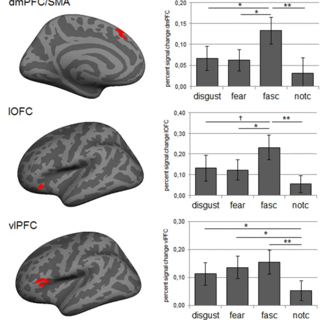
We tested the hypothesis that a negative image combined with morbid fascination feedback would be associated with stronger activation in the neural reference space for emotion than a negative image combined with fear, disgust or control feedback. First, based on an iterated leave-one-out Monte Carlo corrected cluster search, we identified significant clusters in the dmPFC/ SMA, lOFC and vlPFC that reflected general task activity. Figure 3contains a visual representation of the functionally defined ROIs by presenting the conjunction of clusters produced by the iterated leave-one-out cluster search.Table 1contains the peak coordinates and size of the clusters within this conjunction.
In the second step, we extracted percent signal change from each of the functionally defined ROIs for each of the feedback conditions separately. Figure 3presents the means and stand-ard errors for each feedback condition. As predicted, we found that the left dmPFC/SMA was significantly modulated by the feedback manipulation, F(3, 69)¼3.92; P¼0.036; g2¼0.15.
Consistent with our hypothesis, the dmPFC/SMA was more strongly engaged after morbid fascination feedback than fear feedback (P¼0.033), disgust feedback (P¼0.036) and control feedback (P¼0.003). Disgust and fear feedback did not differ sig-nificantly from the control condition (bothP’s>0.21). A similar pattern was present in the left lOFC,F(3, 69)¼4.52;P¼0.018;
g2¼0.16, with stronger activation after morbid fascination than after fear feedback (P¼0.022) and control feedback (P¼0.001). The comparison between morbid fascination feedback and dis-gust feedback was marginally significant (P¼0.074). Disgust and fear feedback did not differ significantly from the control condi-tion (bothP’s>0.12). The modulation of the left vlPFC by the feedback manipulation was marginally significant, F(3, 69)¼3.53;P¼0.057;g2¼0.13. Paired comparisons demonstrated that the vlPFC engaged significantly more after morbid fascin-ation feedback (P¼0.004), fear feedback (P¼0.014) and disgust feedback (P¼0.051) than after control feedback. Disgust and fear feedback did not differ significantly from morbid fascin-ation feedback (bothP’s>0.61). For each of the above functional ROIs, the comparison between morbid fascination and control feedback also passed a Bonferroni corrected level of significance.
Interaction with accuracy judgments
A possible alternative explanation for our findings concerns the individual variability in how people judged the accuracy of the
feedback. The patterns of neural activity that we found could have been driven solely by the participants with low accuracy reports, possibly because they were confused by the feedback. In order to test this, we split our sample into two groups. One group included participants that judged that ‘all’ feedback was correct in 70% or more of the trials (n¼11). The other group
included participants that thought that ‘one or more’ of the feedback types was correct in 60% or less of the trials (n¼13). It is important to note that this latter group included all partici-pants that showed any suspicion about the feedback manipula-tion. We then re-ran all ROI analyses with group membership as a between-subjects factor. None of analyses showed a signifi-cant interaction effect (allP>0.23, uncorrected).
Whole brain contrasts feedback
In addition to the ROI analyses, we also calculated whole brain contrasts comparing the different categories of feedback dir-ectly (see Table 2for an overview). Consistent with the ROI ana-lyses, we found that morbid fascination feedback activated the vlPFC, dmPFC and lOFC more than control feedback. Furthermore, the contrast between disgust feedback and control feedback revealed a cluster in the left vlPFC. Comparing the con-trol feedback to the emotion feedback conditions, we consist-ently found clusters in the right temporoparietal junction (TPJ)
Fig. 3.Left: visual representation of functional ROIs (conjunction) in dmPFC/SMA, lOFC and vlPFC activated by the task. Right: mean percent signal change (error bars represent standard errors) for images paired with disgust, fear, morbid fascination (fasc) and could not be calculated (notc) feedback, extracted from functional ROIs produced by the iterated leave-one-out cluster search.†P
<0.1; *P0.05; **P<0.01.
Table 1.Representation of functional ROIs within neural reference space for emotion
Region hemi max Size mm2 x y z k
dmPFC/SMA lh 2.81 140 7.3 28.5 42.4 241 lOFC lh 3.48 105 42.5 26.2 11.5 216 vlPFC lh 3.43 286 51.6 27.6 5.2 520
and the frontal pole. The contrasts that compared emotion feedback conditions with one another demonstrated clusters in the superior parietal lobule and fusiform gyrus for morbid fas-cinationvsfear feedback; clusters in the precuneus for morbid fascinationvsdisgust feedback; and clusters in the precentral gyrus for fearvsdisgust feedback.
Whole brain contrasts with passive viewing
As a final examination of the effect of feedback, we compared acti-vation patterns after each feedback condition with actiacti-vation pat-terns while people ‘passively’ viewed the same set of images (i.e. during the ‘collection’ runs) (see Table 3 for an overview). Consistent with the ROI analyses, we found that both disgust feed-back and morbid fascination feedfeed-back showed increased activation within the left vlPFC, compared with passively viewing the same
images. Morbid fascination feedback further demonstrated clusters in the left dmPFC, dlPFC, lOFC, TPJ and precuneus (seeFigure 4). When comparing control feedback with passive viewing, we found clusters of activation in the TPJ and precuneus. For passive view-ing, we consistently found clusters in bilateral occipital lobe (e.g. cuneus, pericalcarine and lingual gyrus) and superior parietal lob-ule across all comparisons. This may reflect stronger visual pro-cessing and spatial orientation when viewing images for the first time in the scanner. In addition, for all conditions except for mor-bid fascination, we found that passive viewing was associated with increased activation in the right lOFC.
Discussion
This study documents novel evidence for the neural mechan-ism that supports morbid fascination in reaction to negative
Table 2.Contrasts comparing emotion and control feedback
Contrast hemi max Size mm2 x Y z k Region
fear feedbackvscontrol feedback lh no clusters
rh 2.94 64 4.5 35.3 30.5 145 Precentral disgust feedbackvscontrol feedback lh 3.59 78 49.6 0.7 39.1 135 Precentral
3.48 89 36.2 31.2 5.2 198 vlPFC
rh 3.28 87 57.8 18.5 4.1 167 Superior temporal 2.99 52 46.1 30.6 3.5 83 vlPFC
Fascination feedbackvscontrol feedback lh 5.25 55 6.9 25.3 48.9 106 dmPC 4.08 59 36.4 30 5.8 112 vlPFC
3.98 176 34.9 48.5 56.2 443 Superior parietal 3.59 102 44.9 30.1 7.8 116 vlPFC
3.04 85 47.8 21.8 5.7 150 vlPFC 2.73 155 36.2 36.6 10.3 301 lOFC rh 3.5 80 31.8 6.1 43.9 180 Precentral
3.35 169 26.9 50.5 46.9 376 Superior parietal
2.58 50 37.6 5 3 94 Insula
Control feedback vs. fear feedback lh no clusters
rh 4.2 60 4.5 35.3 30.5 145 Posterior cingulate 3.08 55 56 43.5 7.4 87 Middle temporal 3.07 76 23.8 56.4 3.8 94 Frontal pole 2.94 295 35 67.8 41.7 554 IPL/TPJ 2.62 59 48.5 49.9 30.9 117 IPL/TPJ Control feedbackvsdisgust feedback lh no clusters
rh 3.89 72 49.1 49.8 31.1 145 IPL/TPJ 3.4 105 56.8 42.6 7 160 Middle temporal 2.95 54 12.1 53.8 20.6 109 Precuneus 2.75 50 23.5 56.7 3.1 61 Frontal pole control feedbackvsfascination feedback lh no clusters
rh 3.21 61 24 57 5.1 70 Frontal pole
2.77 86 37.3 62.9 46.5 164 IPL/TPJ fear feedbackvsdisgust feedback lh 3.41 68 35.2 19.1 47.2 153 Precentral
rh no clusters fear feedbackvsfascination feedback lh no clusters rh no clusters Disgust feedbackvsfear feedback lh no clusters rh no clusters Disgust feedback vs. fascination feedback lh no clusters rh no clusters Fascination feedbackvsfear feedback lh no clusters
rh 4.31 97 16.7 62.2 51.8 168 Superior parietal 2.97 71 28.3 58.9 7.6 132 Fusiform Fascination feedbackvsdisgust feedback lh no clusters
rh 2.38 52 8.7 55.8 21.4 85 Precuneus
Note: Clusters are significant atP<0.005 with a minimum cluster size of 50 mm2; k is size in vertices; coordinates are in Talairach space.
images. To manipulate the way participants conceptualized a negative image, we used a novel false feedback paradigm in which we claimed to ‘decode’ participants’ psychological state from their brain activity. In reality, we used the feedback as a form of suggestion to direct how participants ‘might’ conceptu-alize their experience of negative images in the moment. This method allowed us to directly compare the relative involvement of neural regions following morbid fascination, fear and disgust feedback.
Behavioral results suggest that our manipulation was success-ful: participants reported that the feedback was correct on the ma-jority of trials. Moreover, an analysis of ratings after scanning indicated that false feedback influenced participants’ subjective experience of the negative stimuli. This difference was subtle, but given that these ratings occurred well after the manipulation, they may be a conservative estimate of the shift in participants’ experi-ence caused by the feedback manipulation. It should be noted that we deliberately decided not to include trial-by-trial ratings of emotional experience or feedback accuracy during our task, as this would have confounded the feedback manipulation with the explicit evaluation of a stimulus or a label. These limitations not-withstanding, we believe that the subtle change in participants’ world-focused emotional experiences, along with participants’ general agreement about the accuracy of the feedback, are sug-gestive that our false feedback influenced participants’ emotional reactions, just as priming emotion concepts shapes emotional ex-periences in the behavioral literature (Schachter and Singer, 1962; Lindquist and Barrett, 2008; Oosterwijket al., 2010; Crumet al., 2013; Kassam and Mendes, 2013).
Our results reflected different patterns of activation in the neural reference space for discrete emotions, notably within re-gions associated with conceptualization (i.e. the dmPFC/SMA, lOFC and vlPFC). Consistent with our first hypothesis, the vlPFC showed a trend towards increased activation following emotion label feedback (i.e. fear, disgust, morbid fascination) as com-pared with control feedback. This finding is consistent with re-search observing vlPFC activation during labeling of emotional feelings (i.e. ‘affect labeling;’ Lieberman et al., 2007; Satpute
et al., 2013), semantic retrieval more generally (Wagner et al., 2001) and with constructionist predictions that language plays a role in emotion production (Barrett, 2006; Lindquistet al., 2015). Consistent with our second hypothesis, the dmPFC/SMA showed increased activation following morbid fascination feed-back, as compared with all other conditions. The dmPFC has been implicated when individuals show increased attention to their internal state (Satputeet al., 2013) and is also part of a dis-tributed network of brain regions involved in representing se-mantic knowledge (Binderet al., 2009). We thus interpret this finding in line with the proposal of the CAT (Barrett, 2006, 2009)
that ‘atypical’ emotions prompt a relatively greater reliance on conceptualization (Wilson-Mendenhallet al., 2014).
As a potential limitation, we should note that only the com-parison between morbid fascination feedback and control feed-back was significant at the most stringent level in our ROI analysis. Whole brain contrasts comparing morbid fascination feedback to control feedback and to passive viewing forwarded highly similar results. This suggests that our strongest effects result from comparing an atypical emotion label with the con-trol condition, which did not explicitly require participants to draw on knowledge about emotion categories. Nevertheless, we chose to present and discuss all findings since we worked with a novel paradigm that likely produced a subtle effect, both be-haviorally and neurally. Participants were in no way instructed to apply any feedback label or judge its accuracy during scan-ning, which may have resulted in less robust patterns of activa-tion than a more explicit instruction. These points notwithstanding, the brain activity we observed in the func-tional ROI analyses largely replicated the brain areas that are consistently observed in emotion meta-analyses (Vytal and Hamann, 2010; Lindquistet al., 2012) and in studies on affect labeling (Liebermanet al., 2007; Satputeet al., 2013), suggesting that our findings are reliable.
Consistent with our interpretation that morbid fascination, as an atypical emotional state, involved increased conceptual-ization, the dmPFC/SMA, vlPFC and lOFC clusters that demon-strated strong involvement for morbid fascination feedback fell within the boundaries of a so-called ‘default mode network’ (DMN) (i.e. Yeoet al., 2011; see also Oosterwijket al., 2012). The DMN is thought to support conceptualization by using represen-tations of prior experience to make meaning of sensations in the moment (Bar, 2009; Lindquistet al., 2012; Oosterwijket al., 2015). In line with this hypothesis, the DMN has been shown to have increased task-related functional activity across a number of different psychological processes, such as self-referential processing (e.g. Kelleyet al., 2001; Gusnardet al., 2001), semantic judgments (e.g. Binderet al., 2009), and emotional experience (Wilson-Mendenhall et al., 2011; Lindquist et al., 2012; Oosterwijket al., 2012). The suggestion that the default network may be important in the construction of complex and atypical states such as morbid fascination, is consistent with work that demonstrates DMN activation for other types of information that reflect atypicality, such as ambiguous or ambivalent stim-uli (Jenkins and Mitchell, 2010; Nohlenet al., 2013). The possible link between the DMN and the experience of fascination in gen-eral is a topic of investigation that we aim to address in future research.
Table 3.Contrasts comparing activation after feedback with activation during passive viewing
Contrast hemi max Size mm2 x y z k Region
fear feedbackvspassive viewing lh No clusters
rh 3.17 54 35.8 17.9 31 115 Caudal middle frontal 2.54 52 47.6 53.7 44.1 110 Inferior parietal
passive viewingvsfear feedback lh 6.36 2973 26.6 62.8 7.2 5233 Cuneus/pericalcarine/lingual 4.72 143 19.9 60.3 53.7 322 Superior parietal
-2.73 66 24.1 43.7 3.5 151 Lingual 2.31 57 29.2 65 25.5 140 Inferior parietal rh 7.62 2835 20.1 69.8 9.2 4815 Pericalcarine
3.34 97 44.8 32.5 4.7 166 Pars triangularis 3.31 77 20.7 56.1 49.8 151 Superior parietal
3.21 66 30.9 25.8 16.6 112 lOFC
3.06 56 22.6 7.9 56.9 102 Superior frontal 2.79 70 26.7 62 29.3 119 Superior parietal disgust feedbackvspassive viewing lh 2.92 63 39 38 2 86 dlPFC/vlPFC
rh 3.55 109 12 62 35.9 217 Precuneus
2.84 58 8.4 39.3 1.8 107 Rostral anterior cingulate passive viewingvsdisgust feedback lh 7.73 3765 3.2 72.4 17.2 6475 Cuneus/pericalcarine/lingual
3.54 182 17 61.9 56.8 409 Superior parietal 2.99 148 33.1 74 24 221 Inferior parietal 2.53 90 19.7 59.8 37.7 189 Superior parietal rh 6.57 3575 18.7 34.1 6.1 6141 Parahippocampal
4.21 384 25.9 52.3 46.4 932 Superior parietal 3.04 106 34.5 75 21.8 169 Inferior parietal
3.01 80 32.8 23.9 17.4 140 lOFC
2.98 86 13 75.6 0.5 95 Lingual 2.87 203 43.2 70.8 19.8 318 Inferior parietal 2.85 132 21.4 80.1 24.7 195 Superior parietal 2.75 102 37 36.8 9.3 215 Parahippocampal 2.64 97 32.5 64.7 26.7 183 Inferior parietal fascination feedback vs. passive viewing lh 4.71 249 5.7 64.4 40.4 488 Precuneus
4.23 251 31.6 55.9 37.4 531 IPL/TPJ 3.5 209 48.4 47.5 33.5 479 TPJ 3.5 178 40.1 42.7 8.2 240 lOFC
3.35 61 61.1 35.9 6.9 106 Middle temporal 3.26 60 46.9 58 41.7 151 IPL/TPJ 3.23 109 51.6 27.6 5.2 186 vlPFC 3.2 128 38.4 14.9 46.6 201 dlPFC 3.15 74 6.7 26.8 39.9 123 dmPFC rh 4.7 249 7.2 65.8 38.5 510 Precuneus
3.75 200 39.8 53.4 40.8 443 Inferior parietal 2.42 63 14 90.3 16.2 80 Lateral occipital passive viewing vs. fascination feedback lh 7.37 3124 26.4 60.1 7.5 5542 Precuneus
3.64 142 22.2 59 52 320 Superior parietal rh 7.84 2722 16.7 65.1 12.2 4494 Pericalcarine
2.53 83 21.5 77.6 41.7 123 Superior parietal notc feedback vs. passive viewing lh 3.8 142 37.8 63.8 45.6 293 IPL/TPJ
3.59 294 48.1 55.5 40.1 692 IPL/TPJ 3.2 66 40.6 13.7 38.6 86 dlPFC 3.17 116 46.4 54.6 26.7 257 IPL/TPJ 3.06 70 11.3 49.3 37.7 134 Precuneus 2.85 129 5.7 63.3 39.2 256 Precuneus rh 3.93 731 42.7 59.1 41.5 1524 Inferior parietal
3.46 60 38.6 23 33.1 120 Caudal middle frontal 3.45 82 9.1 66.4 39.3 170 Precuneus
3.41 73 17.1 96.5 13.4 98 Lateral occipital passive viewing vs. notc feedback lh 7.61 5305 14.3 78.4 12.7 9298 Pericalcarine
6.05 139 30.7 47.3 49.4 330 Superior parietal 5.16 62 44.3 0.8 18.3 155 Superior temporal/ATL 4.13 58 17 58.8 59.6 135 Superior parietal 3.93 63 32.6 50.3 5.8 131 Fusiform
3.39 83 36.4 36 10.1 145 lOFC
2.97 84 43.8 69.4 14.2 158 Inferior parietal
fascination, our findings are also consistent with an interpret-ation in terms of an increased role for ‘top-down’ interpretative processes (e.g. reappraisal). For example, previous research has observed patterns of activation in the dmPFC/SMA, lOFC and vlPFC when individuals regulate their emotional state by reap-praising the meaning of a stimulus (Wageret al., 2008; Diekhof
et al., 2011; Buhle et al., 2013) or interpret neutral stimuli as negative (Ochsneret al., 2009). Thus, the relatively stronger acti-vation of the dmPFC/SMA and lOFC for morbid fascination feed-back may suggest a link between the experience of fascination and reappraisal specifically. This link may have been empha-sized by our instruction, which mentioned that morbid fascin-ation could be interpreted as a state in which people ‘get drawn to a stimulus to examine the details’. Although this systematic way of viewing the stimulus is similar to the instructions that people receive in a reappraisal paradigm, it is also a characteris-tic of the mental state of fascination itself. Note, furthermore, that our instruction did not ask people to reappraise the nega-tivity of the stimulus, nor to down-regulate their emotions.
It is important to note that conceptualization and re-appraisal account are not mutually exclusive. In fact, conceptu-alization has been offered as a mechanism for achieving reappraisal (Barrettet al., 2014). We argue that paradigms in which participants are instructed to reinterpret a stimulus ‘top-down’ are in fact evoking a re-conceptualization of the sensory information present in that particular situation. Moreover, re-gions involved in emotion regulation (Buhleet al., 2013; Diekhof
et al., 2011) are also commonly involved during emotion experi-ence (Vytal and Hamann, 2010; Lindquistet al., 2012). Thus, the patterns of activation found in the present study may also re-flect the production of emotional reactions afresh. Ultimately, there may not be a strict division between the mechanisms involved in creating an emotion in the first place or modifying it after the fact (cf., Gross and Barrett, 2011; Lindquistet al., 2012; Ochsneret al., 2012).
Limitations
As the first study to use false mental state feedback, our study has several limitations. A first possible limitation concerns our choice of control condition. Although we deliberately chose to include a condition with a non-specific label, this could have led participants to give trials with this type of feedback reflect-ive scrutiny (e.g. ‘What am I actually feeling?’), resulting in increased activity in regions associated with conceptualization. Thus, the comparison between emotion feedback and control feedback is likely conservative, which may explain why we only found a significant difference between fear, disgust and control feedback in the VLPFC. Nevertheless, morbid fascination
feedback could also have resulted in self-reflective thoughts (‘Why am I fascinated by this image?’), thus matching morbid fascination and control feedback in this regard. In short, al-though there may be psychological implications of the control condition, these differences cannot explain the different pat-terns of neural activation following control feedback and mor-bid fascination feedback.
Second, there may be possible psychological consequences of giving people (false) morbid fascination feedback that could have influenced the patterns of neural activation in this study (e.g. by causing doubt, surprise). In this context, it is important to note that additional analyses suggested that our results did not differ between participants who reported that the labels were accurate descriptions of their feelings and those who re-ported low accuracy of the feedback. Although null findings are hard to interpret, this provides some support for the assump-tion that the patterns forwarded by our study were not merely driven by participants who were confused or in doubt about the veracity of the feedback labels. Nevertheless, as a future direc-tion, it is important to replicate the current findings by targeting the non-manipulated experience of morbid fascination.
Finally, although our experimental design presented fear, disgust and morbid fascination as mutually exclusive states, it is likely that these experiences often co-occur. The subjective ratings collected at Time 1 indeed demonstrated that the negative images were rated equally in terms of threat, repul-sion and interest, suggesting that experiencing a stimulus as disgusting does not preclude experiencing that stimulus as interesting. The integration of multiple experiences towards negative images, and an understanding of how these experi-ences develop over time is an important avenue for future re-search. Another important side note is that the present findings do not speak to the possible differences or similarities in how the brain represents fascination for negative stimuli, and for positive or non-emotional stimuli. Incorporating such a comparison was beyond the scope of the present project, but we believe that this is another important topic for future research.
Conclusion
To summarize, using an innovative paradigm, this study docu-ments the neural mechanisms underlying atypical and typical experiences towards negative images. Our findings provide an important starting point for future research into the experience of morbid fascination and emphasize that it is relevant to take this state into consideration when investigating reactions to negative events.
Note: Clusters are significant atP<0.005 with a minimum cluster size of 50 mm2; k is size in vertices; coordinates are in Talairach space.
Acknowledgements
This research was supported by a National Institutes of Health Director’s Pioneer Award to Lisa Feldman Barrett (DP1OD003312), a Marie Curie International Outgoing Fellowship (275214) awarded by the European Commission’s Seventh Framework Programme and Netherlands Organization for Scientific Research VENI grant (451-13-008) to Suzanne Oosterwijk and a Harvard University Mind/ Brain/Behavior Postdoctoral Fellowship to Kristen Lindquist. The authors would like to thank Evangeline Barnard, Mara Koerner and Jamie Nichols for their assistance with stimu-lus selection and development.
Supplementary data
Supplementarydata are available atSCANonline.
Conflict of interest. None declared.
References
Bar, M. (2009). The proactive brain: memory for predictions. Philosophical Transactions of the Royal Society B,364, 1235–43. Barrett, L.F. (2006). Solving the emotion paradox: categorization
and the experience of emotion.Personality and Social Psychology Review,10, 20–46.
Barrett, L.F. (2009). Variety is the spice of life: a psychological constructionist approach to understanding variability in emo-tion.Cognition and Emotion,23, 1284–306.
Barrett, L.F. (2012). Emotions are real.Emotion,12, 413–29. Barrett, L.F. (2013). Psychological construction: a Darwinian
ap-proach to the science of emotion.Emotion Review,5, 379–89. Barrett, L.F., Satpute, A.B. (2013). Large-scale brain networks in
affective and social neuroscience: towards and integrative functional architecture of the brain. Current Opinion in Neurobiology,23, 1–12.
Barrett, L.F., Wilson-Mendenhall, C.D., Barsalou, L.W. (2014). A psychological construction account of emotion regulation and dysregulation: the role of situated conceptualizations. In: Gross, J.J., editor.The Handbook of Emotion Regulation, 2nd edn, pp. 447–465, New York: Guilford.
Barsalou, L.W. (1985). Ideals, central tendency, and frequency of instantiation as determinants of graded structure in catego-ries. Journal of Experimental Psychology: Learning, Memory, and Cognition,11, 629–54.
Binder, J.R., Desai, R.H., Graves, W.W., Conant, L.L. (2009). Where is the semantic system? A critical review and meta-analysis of 120 functional neuroimaging studies.Cerebral Cortex, 19(12), 2767–96.
Borg, C., de Jong, P.J., Renken, R., Georgiadis, J.R. (2013). Disgust trait modulates frontal-posterior coupling as a function of dis-gust domain.Social Cognitive and Affective Neuroscience,8, 351–8. Buhle, J.T., Silvers, J.A., Wager, T.D.,et al. (2013). Cognitive
re-appraisal of emotion: a meta-analysis of human neuroimaging studies.Cerebral Cortex,24(11), 2981–90.
Crum, A., Salovey, P., Achor, S. (2013). Rethinking stress: the role of mindsets in determining the stress response. Journal of Personality and Social Psychology,104, 716–33.
Dale, A.M., Fischl, B., Sereno, M.I., (1999). Cortical surface-based analysis. I. Segmentation and surface reconstruction. Neuroimage,9, 179–94.
Desikan, R.S., Segonne, F., Fischl, B.,et al. (2006). An automated labeling system for subdividing the human cerebral cortex on
MRI scans into gyral based regions of interest.Neuroimage,31, 968–80.
Diekhof, E.K., Geier, K., Falkai, P., Gruber, O. (2011). Fear is only as deep as the mind allows: a coordinate-based meta-analysis of neuroimaging studies on the regulation of negative affect. Neuroimage,58, 275–85.
Esterman, M., Tamber-Rosenau, B.J., Chiu, Y., Yantis, S. (2010). Avoiding non-independence in fMRI data analysis: Leave one subject out.Neuroimage,50, 572–6.
Fischl, B., Sereno, M.I., Dale, A.M., (1999a). Cortical surface-based analysis. II: inflation, flattening, and a surface-based coordin-ate system.Neuroimage,9, 195–207.
Fischl, B., Sereno, M.I., Tootell, R.B., Dale, A.M., (1999b). High-resolution intersubject averaging and a coordinate system for the cortical surface.Human Brain Mapping,8, 272–84.
Gray, M.A., Harrison, N.A., Wiens, S., Critchley, H.D. (2007). Modulation of emotional appraisal by false physiological feed-back during fMRI. PLoS One2(6): e546.
Gross, J.J., Barrett, L.F. (2011). Emotion generation and emotion regulation: one or two depends on your point of view.Emotion Review,3, 8–16.
Gusnard, D.A., Akbudak, E., Shulman, G.L., Raichle, M.E. (2001). Medial prefrontal cortex and self-referential mental activity: relation to a default mode of brain function.Proceedings of the National Academy of Sciences,98, 4259–64.
Hariri, A.R., Tessitore, A., Mattay, V.S., Fera, F., Weinberger, D.R. (2002). The amygdala response to emotional stimuli: a com-parison of faces and scenes.Neuroimage,17, 317–23.
Haynes, J., Rees, G. (2006). Decoding mental states from brain ac-tivity in humans.Nature Reviews Neuroscience7, 523–34. Jenkins, A., Mitchell, J.P. (2010). Mentalizing under
uncer-tainty: dissociated neural responses to ambiguous and unambiguous mental state inferences. Cerebral Cortex, 20, 404–10.
Kassam, K.S., Mendes, W.B. (2013). The effects of measuring emotion: physiological reactions to emotional situations de-pend on whether someone is asking.PLoS One,8(7),e64959. Kelley, W.M., Macrae, C.N., Wyland, C.L., Caglar, S., Inati, S.,
Heatherton, T.F.(2002).Finding the self? an event-related fMRI study.Journal of Cognitive Neuroscience,14, 785–94.
Kober, H., Barrett, L.F., Joseph, J., Bliss-Moreau, E., Lindquist, K.A., Wager, T.D. (2008). Functional networks and cortical-subcortical interactions in emotion: a meta-analysis of neuroi-maging studies.Neuroimage,42, 998–1031.
Kriegeskorte, N., Simmons, W.K., Bellgowan, P.S.F., Baker, C. (2009). Circular analysis in systems neuroscience – the dangers of double dipping.Nature Neuroscience,12, 535–40.
Lang, P.J., Bradley, M.M., Cuthbert, B.N. (2008). International Affective Pictures System (IAPS): Technical manual and affective rat-ings (Tech. Rep. A-8). Gainesville: University of Florida, Research Center for Research in Psychophysiology.
Lieberman, M.D., Cunningham, W.A. (2009). Type I and Type II error concerns in fMRI research: re-balancing the scale.Social Cognitive and Affective Neuroscience,4, 423–8.
Lieberman, M.D., Eisenberger, N.I., Crockett, M.J., Tom, S.M., Pfeifer, J.H., Way, B.M. (2007). Putting feelings into words: affect labeling disrupts amygdala activity in response to affective stimuli.Psychological Science,18, 421–8.
Lindquist, K.A. (2013). Emotions emerge from more basic psy-chological ingredients: a modern psypsy-chological construction-ist approach.Emotion Review,5, 356–68.
Crone, E.A. (2013). Evaluating ambivalence: social-cognitive and affective brain regions associated with ambivalent decision-making.Social Cognitive and Affective Neuroscience,9(7), 924–31.
Ochsner, K.N., Ray, R.R., Hughes, B.,et al. (2009). Bottom-up and top-down processes in emotion generation: common and dis-tinct neural mechanisms.Psychological Science,20, 1322–31. Ochsner, K.N., Silvers, J.A., Buhle, J.T. (2012). Functional imaging
studies of emotion regulation: a synthetic review and evolving model of the cognitive control of emotion.Annals of the New York Academy of Sciences,1251, E1–24.
Oosterwijk, S., Lindquist, K.A., Anderson, E., Dautoff, R., Moriguchi, Y., Barrett, L.F. (2012). States of mind: emotions, body feelings and thoughts share distributed neural networks. Neuroimage,62, 2110–28.
Oosterwijk, S., Topper, M., Rotteveel, M., Fischer, A. H. (2010). When the mind forms fear: embodied fear knowledge potenti-ates bodily reactions to fearful stimuli.Social Psychological and Personality Science,1, 65–72.
Oosterwijk, S., Touroutoglou, A., Lindquist, K.A. (2015). The neuroscience of construction: what neuroimaging approaches can tell us about how the brain creates the mind. In: Barrett, L.F., Russell, J.A., editors. The Psychological Construction of Emotion. New York: Guilford.
Rime´, B., Delfosse, C., Corsini, S. (2005). Emotional fascination: responses to viewing pictures of September 11 attacks. Cognition and Emotion,19, 923–32.
Satpute, A.B., Shu, J., Weber, J., Roy, M., Ochsner, K.N. (2013). The functional neural architecture of self-reports of affective ex-perience.Biological Psychiatry,73, 631–8.
Schachter, S., Singer, J. (1962). Cognitive, social, and physio-logical determinants of emotional state.Psychological Review,
69, 379–99.
Shirer, W.R., Ryali, S., Rykhlevskaia, E., Menon, V., Greicius, M.D. (2012). Decoding subject-driven cognitive states with whole-brain connectivity patterns.Cerebral Cortex,22(1), 158–65.
Valins, S. (1966). Cognitive effects of false heart-rate feedback. Journal of Personality and Social psychology,4, 400–8.
Van der Kouwe, A.J., Benner, T., Salat, D.H., Fischl, B. (2008). Brain morphometry with multiecho MPRAGE.Neuroimage,40, 559–69.
Vytal, K., Hamann, S. (2010). Neuroimaging support for discrete neural correlates of basic emotions: a voxel-based meta-analysis.Journal of Cognitive Neuroscience,22, 2864–85.
Wager, T.D., Davidson, M.L., Hughes, B.L., Lindquist, M.A., Ochsner, K.N. (2008). Prefrontal-subcortical pathways media-ting successful emotion regulation.Neuron,59, 1037–50. Wagner, A.D., Pare´-Blagoev, E.J., Clark, J., Poldrack, R.A. (2001).
Recovering meaning: left prefrontal cortex guides controlled semantic retrieval.Neuron,31(2), 329–38.
Wild, J., Clark, D.M., Ehlers, A., McManus, F. (2008). Perception of arousal in social anxiety: effects of false feedback during a so-cial interaction.Journal of Behavioral Therapy and Experimental Psychiatry,39, 102–16.
Wilson-Mendenhall, C.D., Barrett, L.F., Barsalou, L.W. (2014). Variety in emotional life: within-category typicality of emo-tional experiences is associated with neural activity in large-scale brain networks.Social Cognitive and Affective Neuroscience,
10(1), 67–71.
Wilson-Mendenhall, C.D., Barrett, L.F., Simmons, W.K., Barsalou, L.W. (2011). Grounding emotion in situated conceptualization. Neuropsychologia,49, 1105–27.
Wright, P., He, G., Shapira, N.A., Goodman, W.K., Liu, Y. (2004). Disgust and the insula: fMRI responses to pictures of mutila-tion and contaminamutila-tion.NeuroReport,15, 2347–51.
Yeo, B.T.T, Krienen, F.M., Sepulcre, J., et al. (2011). The or-ganization of the human cerebral cortex estimated by functional connectivity. Journal of Neurophysiology, 106, 1125–65.
Zuckerman, M., Litle, P. (1986). Personality and curiosity about morbid and sexual events.Personality and Individual Differences,