1
THE ROLE OF MUTUALISTS IN PLANT RESPONSE TO PATHOGEN INFECTION
Megan Arlene Rúa
A dissertation submitted to the faculty of the University of North Carolina at Chapel Hill in
partial fulfillment of the requirements for the degree of Doctor of Philosophy in the Curriculum for the Environment and Ecology.
Chapel Hill 2012
Approved by: John Bruno Lauren Buckley Shuijin Hu
iii ABSTRACT
MEGAN ARLENE RÚA: The Role of Mutualists in Plant Response to Pathogen Infection
(Under the direction of Charles E. Mitchell)
Plants interact with a diversity of microorganisms including enemies and
mutualists. Plant pathogens and mutualistic fungi are two classes of microorganisms that directly impact the plant and may in turn alter each other’s success. While their roles have often been considered independently, few researchers have considered their
concurrent role. Dynamics of these two groups of widespread microbes may modify plant nutrient allocation in response to abiotic environmental changes. Furthermore, early
models suggest that mutualists and pathogens may profoundly impact not only their shared host plant, but each other. In one of the first thorough explorations of three-species interactions, I use both experimental and theoretical approaches to investigate the
interaction between plants, their pathogenic enemies and fungal mutualists in the context of changing abiotic conditions.
In two separate greenhouse experiments I show that mutualistic strategy is important for determining the direction of change by which mutualists alter pathogen dynamics. In additional work, I also confrim the reverse can also be true in that pathogen
infection influences mutualists. Both mathematical theory and an experiment indicate that a pathogen can alter host-mutualist dynamics and consequently alter long-term
iv
environmental conditions can modify the relationships between hosts, mutualists and pathogens. Using greenhouse and field experiments, I demonstrate that increases in
atmospheric CO2, temperature and precipitation all modify mutualist-host-pathogen relationships. Overall, my thesis demonstrates that mutualists and pathogens can have
important impacts on not only the host but also on the success of each other. Such dynamics can be further modified by changes in the abiotic environment. Precipitation, temperature, and atmospheric CO2 are all expected to continue to change for the foreseeable future. Thus, in order to make accurate projections about ecosystem, community or population dynamics, changes in microorganisms and their interactions
v
To my family and my friends
whose encouragement and patience have given me the inspiration
vi
Acknowledgments
The road to my dissertation has been winding with lots of starts, stops, bumps and
detours but throughout I have had the support of many people. First, I would like to thank the members of my committee for all of their support, insights and feedback throughout
this process. My advisor, Charles Mitchell, has been nothing but supportive in everything I’ve tried to do. From harebrained experiments to programs in Africa, Charles has never wavered in his commitment to helping me improve as a scientist. James Umbanhowar has
always been available for a cup of tea and provided a crucial balance to my Type A personality. I would like to thank Rebecca McCulley and Shuijin Hu for fostering my
intellectual development in the mutualists used in this project and hosting me at various points in their lab. I would also like to thank John Bruno and Lauren Buckley for their helpful comments and insights throughout the completion of this dissertation. Finally, I
would like to thank Jack Weiss for giving me an ever expanding statistical toolbox. A variety of organizations and people have supported me throughout my time at
UNC. I have had the support of four chairs of the Curriculum for the Environment and Ecology (and its predecessor the Ecology Curriculum) including: Bob Peet, Seth Reice, David Moreau and Jaye Cable. I have also had the support of a number of administrative
assistants in the Curriculum including: Cottie Pasternak, Denise Kenney, Mary Beth Robbins and especially Kathleen McNeil. Finally, Valerie Ashby and Kathy Wood with
vii
there with a well-timed email, free food and general moral support. During my dissertation I have been supported by fellowships from AGEP as well as an NSF
Graduate Research Fellowship. I have received additional support via teaching
assistantships from the Ecology Curriculum and the Biology Department. AGEP has also
provided important travel support throughout my dissertation. Finally, my two writing groups have provided important honest and critical feedback on everything I’ve written this year. My fellow graduating PhD students Alicia Frame and Josie Reinhardt have
helped me navigate this final year with grace, dignity and laughter that I would not have thought possible.
Members of the Mitchell Lab, both past and present, have provided invaluable logistical and moral support throughout this process. Miranda Welsh has been there with me from the start and has been very important to me in many different roles throughout
my dissertation. Robert Heckman and Fletcher Halliday have been important in providing support, laughter and motivation when my gas tank was running empty. Our lab
technician, Marty Dekkers, has helped me manage all of the different arms of my projects and is always available for a good conversation. Additionally we have been blessed with a stellar crew of undergrads, without whom I would never have been able to get anything
accomplished. Gabe Hobsen is a root washer extraordinaire and washed many of the roots for the work presented in Chapter 3. Additionally, Alexandra Chang, Jin Bae, Dan
Chen, Lee Dunn, Sarah Hatcher, Drea Paulk, K. Amanda Saunders, Juan Caldron, Bianca White, Angela Ancheta, Carmen Kuan, Tyler Fletcher, Kristine Nguyen, Courtney
Whitaker and Xin Wu have all helped with various components of my dissertation.
viii
pleasure of working with. Her hard work and humor never cease to amaze me. She has pushed me to a better mentor and re-invigorated the scientist in me.
Volunteer work has always been important to me, but while at UNC I found it a crucial reminder as to why I became an ecologist in the first place. My fellow volunteers
with the Sierra Club Inner City Outings Program have all taught me many things and introduced me to many features of North Carolina during my five years here. Fellow Chapel Hillian Amanda Damweber has always been available for drinks and a bite to eat.
Sara Felsen’s leadership, laughter and amazing ability to turn camp food into a gourmet meal are nothing short of an inspiration. Rachel Harden-Banyal was the first person to
take the time to teach me the flora and fauna of North Carolina. Sarah Johnson and Zach Wilcox are both wonderful with kids and bring imagination back into my life. Last of all, I want to thank the kids who never cease to make me laugh and inspire me with all
they’ve overcome. Together we have conquered the darkness and bugs at Hanging Rock, biked the Raleigh greenway and Tobacco Trail, flipped canoes in Merchant’s Millpond,
waffed the Eno, learned to swim at Bear Island, Falls Lake and Lake Wheeler, hiked Umstead and attended numerous exhibits and festivals throughout the Triangle.
Fleet Feet Sports in Carrboro has provided an outlet for my energy I didn’t even
know I needed. Leaving my Southern California roots behind to come to North Carolina not only meant joining the true South (sorry Texas) and initiation into the BBQ wars, but
also leaving behind a long history of swimming. Running with Fleet Feet has not only provided a source of exercise but introduced me to a wonderful group of people. Natalie Reder, our fearless leader, is an amazing source of energy at 7am on a Saturday. Katie
ix
there to pick me up when I’m feeling drained and push me that extra mile. Together we completed my first half marathon in March and the Tarheel Ten Miler in April.
Finally, I need to thank my closest friends and family for giving me perspective and keeping me balanced. Vanessa Mendoza has been my best friend since high school
JV tennis. Even though she’s 5000 miles away, she’s always had my back. Hugham Chan has always been a one man cheering squad. Most of all, this dissertation is made possible by the love, support and sacrifices of my family. My mom has always been an inspiration
and a strong believer that you’re never too old for care packages with gold fish and brownies. My dad’s unique humor is a constant reminder to not sweat the small things.
Both of my parents have an uncompromising faith in my ability to succeed at whatever I put my mind to and this has helped me through many a tough moment. My brother Colin, like my dad, can always make me laugh and is always there for me when I need him.
Even if it means a panicked phone call at 5am Pacific Time (oops). My Aunt Sue has provided the quiet and constant support that only a Minnesotan aunt can provide. During
my dissertation I have lost the loving support of my Grandpa whose mind has fully succumbed to dementia and the support of my Abuela who passed from a stroke. It is with their spirit, my grandparents who worked so hard to give their children a different
x
TABLE OF CONTENTS
Contents
LIST OF TABLES ... 13
LIST OF FIGURES ... 15
CHAPTER I: INTRODUCTION ... 17
CHAPTER II: ELEVATED CO2 SPURS RECIPROCAL POSITIVE EFFECTS BETWEEN A PLANT VIRUS AND AN ARBUSCULAR MYCORRHIZAL FUNGUS ... 25
Abstract... 25
Introduction ... 26
Materials and Methods ... 29
Results ... 35
Discussion... 37
Acknowledgements ... 41
CHAPTER III: FUNGAL ENDOPHYTE INFECTION AND HOST CULTIVAR JOINTLY MODULATE HOST RESPONSE TO AN APHID-TRANSMITTED VIRAL PATHOGEN ... 47
Abstract... 47
Introduction ... 48
Materials and Methods ... 52
Results ... 57
xi
Acknowledgements ... 62
CHAPTER IV: IMPACTS OF CLIMATE DRIVERS, HOST SPECIES IDENTITY, AND FUNGAL ENDOPHYTE INFECTION ON THE PREVALENCE OF THREE VIRUS SPECIES IN A GRASSLAND ECOSYSTEM ... 69
Abstract... 69
Introduction ... 70
Materials and Methods ... 74
Results ... 80
Discussion... 81
CHAPTER V: THE EFFECT OF MUTUALISTS ON PATHOGEN-HOST DYNAMICS ... 90
Abstract... 90
Introduction ... 91
Model Description and Analyses ... 94
Numerical analysis ... 96
Discussion... 98
Acknowledgements ... 102
CHAPTER VI: CONCLUSIONS ... 110
APPENDICES ... 114
APPENDIX A SUPPLEMENTARY MATERIAL FOR CHAPTER 2 ... 114
A1. Supplementary Figures ... 114
A2. Full Statistical Models. ... 118
xii
B1. Supplementary Figures ... 122
B2. Full Statistical Models ... 123
APPENDIX C SUPPLEMENTARY MATERIAL FOR CHAPTER 4 ... 127
C1. Supplementary Figures ... 127
C2. Supplementary Tables ... 129
C3. Full Statistical Models ... 132
APPENDIX D SUPPLEMENTARY MATERIAL FOR CHAPTER 5 ... 135
Dimensionalized Equations ... 135
Parameterization ... 135
Greenhouse experimental design and treatments ... 137
13
LIST OF TABLES
Table 4.1. Parameter estimates from the binomial model describing
virus infection in 2009. ... 88
Table 4.2. Parameter estimates from the binomial model describing Schedonorus phoenix virus infection 2009-2011. ... 89
Table 5.1a. Nondimensionalized parameters and their dimensionalized components, as well as their biological meaning... 108
Table 5.1b. Dimensionalized parameters and their biological meaning and source. ... 109
Table A2.1. Model of Optical Density values, estimating relative virus titer. ... 118
Table A2.2. Model of percent hyphal colonization. ... 118
Table A2.3. Model of Total Plant Biomass. ... 119
Table A2.4. Model of Root Fraction. ... 120
Table A2.5. Model of Percent Leaf Tissue Phosphorus ... 121
B2.1. Model of Total Plant Biomass ... 123
B2.2. Model of Total Plant Biomass: within the PDF cultivar ... 123
B2.4. Model of Root Biomass: within the PDF cultivar ... 123
B2.5. Model of Shoot Biomass ... 124
B2.6. Model of Shoot Biomass: within the PDF cultivar ... 124
B2.7. Model of Root Fraction ... 124
B2.8. Model of Root Fraction: within the PDF cultivar ... 124
B2.9. Model of Tiller Number ... 124
B2.10. Model of Tiller Number: within the PDF cultivar ... 124
14
B2.12. Model of adult aphid abundance for the aphid Rhopalosiphum
padi ... 125 B2.13 Model of total aphid abundance for the aphid Rhopalsiphum
padi ... 125 B2.14. Model of nymph production by the aphid Rhopalosiphum padi:
within the PDF cultivar ... 125 B2.15. Model of adult aphid abundance for the aphid Rhopalosiphum
padi: within the PDF cultivar ... 125 B2.16 Model of total aphid abundance for the aphid Rhopalsiphum
padi: within the PDF cultivar ... 125 B2.17. Model of Model of Optical Density values, estimating relative
virus titer. ... 126 B2.18a. Model of Model of Optical Density values, estimating relative
virus titer: within the KY cultivar ... 126 B2.18b. Model of Model of Optical Density values, estimating relative
virus titer: within the PDF cultivar ... 126 Table C2.1. Frequencies of climate change treatment × species
combinations for aphid counts. ... 129 Table C3.1. Summary for model of virus infection described by heat,
precipitation or host species ... 132 Table C3.2. Summary for model of Tall Fescue (Schedonorus phoenix)
virus infection described by heat, precipitation, endophyte-infection
status or year ... 133 Table C3.3. Summary for model of aphid presence described by heat,
15
LIST OF FIGURES
Figure 2.1. Effect of mycorrhizal colonization on viral titer ... 42
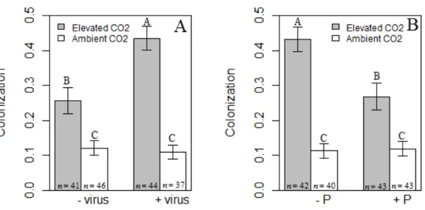
Figure 2.2. Results for root colonization by AMF. ... 43
Figure 2.3. The role of phosphorus, viral infection and host species for root colonization by AMF. ... 44
Figure 2.4. Plant response to virus infection... 45
Figure 2.5. Leaf phosphorus concentration. ... 46
Figure 3.1. Biomass response to endophyte infection. ... 64
Figure 3.2. Root Fraction by virus and endophyte infection. ... 65
Figure 3.3. Average tiller number by endophyte and virus infection status. ... 66
Figure 3.4. Viral titer by genotype. ... 67
Figure 3.5. Aphid response to endophyte infection. ... 68
Figure 4.1. Bayesian credible intervals for the log odd ratio of virus infection. ... 86
Figure 4.2. Bayesian credible intervals for the log odds ratio of aphid presence... 87
Figure 5.1. Isocline analysis for our model... 103
Figure 5.2. Numerical integration across the plant-mutualist nullcline space, constant k... 104
Figure 5.3. Nullclines for the enemy graphed on the plant-mutualist nullcline space, varying k. ... 105
Figure 5.4. Nullclines for the enemy graphed on the plant-mutualist nullcline, varying c... 106
Figure 5.5. Numerical integration across the plant-mutualist nullcline space, constant c... 107
16
A1.2. Role of elevated CO2 and host species on root colonization by
AMF. ... 115 A1.3.Effect of CO2 and host species on total biomass. ... 116 A1.4. Role of phosphorus and host species in altering leaf phosphorus
concentration. ... 117
B1.1. Tiller number by endophyte infection status for PDF. ... 122 C1.1. The proportion of plants infected by a virus varied among host
17
CHAPTER I: INTRODUCTION
Plants interact with a diversity of microorganisms, including both enemies and
mutualists. Plant pathogens and mutualistic fungi are two classes of microorganisms that directly impact the plant and may in turn alter each other’s success. Despite their
ubiquitous nature, little research attention has been given to the interaction of multiple microorganisms as they alter host growth and the success of each other. Early three-species models have shown that the third player can alter the intensity, outcome and even
the symbiotic state (mutualistic or parasitic) of an association (Bronstein and Barbosa 2002). Furthermore, dynamics of these two groups of widespread microbes may modify
plant nutrient allocation in response to abiotic environmental changes. In one of the first thorough explorations of three-species interactions, I used both experimental and
theoretical approaches to investigate the interaction between plants, pathogens and
mutualists under changing abiotic conditions.
Plant hosts provide an important ecological arena in which to examine
multispecies interactions. Specifically, plant phenotypes can be fundamentally altered by microbes, which may provide novel nutritional and defense pathways via their influence on plant biochemical pathways (Friesen et al. 2011). Plant pathogens are one type of
enemy which may capitalize on such phenotypic changes (Rúa et al. 2011); however, not all pathogen-plant interactions are created equal. Plants vary in the severity of disease
18
infection), susceptibility (probability of infection when exposed to the pathogen) or resistance (ability of the plant to defend against infection). Often plant tolerance traits are
positively associated with traits involved with resource acquisition such as root biomass, the ability to shunt carbon from roots to shoots after foliar damage, leaf area, and
photosynthetic rate (Strauss and Agrawal 1999, Stowe et al. 2000). Conversely, tolerance may also be negatively associated with plant resistance traits like concentrations of secondary compounds (Strauss and Agrawal 1999, Stowe et al. 2000).
Variation is common within different enemy-host-mutualist interactions. Few theoretical studies have examined such three-way interactions (but see (Bennett et al.
2006)), and a unifying framework is lacking. Further, most studies consider the plant as the key component for determining the outcome of such three species interactions. Realigning theoretical models to instead utilize the mutualist as the center of focus may
prove a more efficient way to explain the large amount of variation surrounding these interactions. Mutualism classes can be grouped based on the nature of the benefits they
exchange with their partners (Bronstein and Barbosa 2002): nutritional mutualisms (partners provide each other with essential limiting nutrients) and protection mutualisms (one partner provides protection from biotic or abiotic environmental stresses for the
other partner) are two examples of traditionally recognized mutualisms (Bronstein and Barbosa 2002). The fundamental characteristics describing the differences between each
of these types of mutualistic relationships may explain some of the variation by which mutualists have been shown to influence plant-enemy interactions.
When exploring the interaction of mutualists and pathogens it is important to study
19
environments. The extent to which plant-microbe interactions are mutalistic or parasitic may often be a function of resource availability, which is currently being altered by
global change. Few studies have directly investigated how such interactions are altered in the context of global change. For example, since viral pathogens and fungal mutualists
can be integral players in plant allocation of carbon, the growth, fecundity and population dynamics of these two groups of widespread microbes may modify plant performance in response to elevated CO2 (Malmstrom and Field 1997, Johnson et al. 2005). Additionally, association with one particular class of mutualist - foliar endophytic fungi - may bolster a plant’s ability to withstand changes to temperature and precipitation regimes
(Stuedemann and Hoveland 1988, Arachevaleta et al. 1989). In order to advance our understanding of host-mutualist-enemy interactions, I investigated the relationship between a viral pathogen and two different types of mutualists (a nutrition mutualist and
a protection mutualist) under varying abiotic conditions. I explored multiple aspects of this relationship by combining, greenhouse/lab work, field studies, and a new theoretical
model of enemy-mutualist-host interactions. Using greenhouse experiments, we tested the roles mutualists play in altering plant response to viral infection (Chapter 2 and 3). First we considered whether a nutritional mutualist, arbuscular mycorrhizal fungi (AMF),
alters host response to pathogen pressure when soil nutrients and atmospheric concentrations of carbon dioxide (CO2) are altered (Chapter 2). We then explored whether a protective mutualist, foliar endophytic fungi, alters host response to pathogen pressure and vector abundance (Chapter 3). The nature of these three-way interactions was further pursued through the use of field studies. We explored the role that
20
temperature regimes within a managed grassland (Chapter 4). Finally, theoretical work exploring multispecies interactions has only recently attempted to dissect
enemy-mutualist interactions (Bennett et al. 2006), but previous models have neglected to include pathogens. Since pathogen-mutualist affiliations are characterized by a more
intimate connection (primarily they both exist internally in the host), their interactions may exhibit different dynamics then generic plant-enemy relationships. Therefore, we created and analyzed a model of the interaction of a fungal mutualist, a viral pathogen
and their shared host in order to explore pathogen-specific influences on three way species interactions (Chapter 5).
CHAPTER SUMMARIES
Chapter 2: Elevated CO2 spurs reciprocal positive effects between a plant virus and an arbuscular mycorrhizal fungus
While many studies have considered the individual effects of pathogens and mutualists on their hosts, few studies have investigated interactions among microbial
mutualists and pathogens in the context of global change. Together with Dr. Kent Burkey at the USDA, Dr. Shuijin Hu at North Carolina State University, Dr. James Umbanhowar at the University of North Carolina at Chapel Hill and my advisor, Dr. Charles Mitchell, I
experimentally tested the interactive effects of increased atmospheric CO2 concentration, soil phosphorus supply, an ecologically important nutrition mutualist (AMF), and a
widespread viral pathogen. Under elevated CO2, mycorrhizal association increased viral titer, and virus infection reciprocally increased the colonization of roots by mycorrhizal hyphae. This indicates that when carbon was abundant, the mycorrhizal fungus and the
21
decreased plant allocation to root biomass, increased the accumulation of phosphorus in leaves, as well as modulated the effects of elevated CO2 and (for one plant species) of phosphorus addition on mycorrhizal colonization of roots. These results emphasize the importance of interactions among multiple microorganisms for plant performance in the
context of global change. Overall, our research indicates that these mutualist and
pathogenic organisms interact to alter each other’s success, and predicts these interactions will respond to changes in resource availability under global change.
Chapter 3: Fungal endophyte infection and host cultivar jointly modulate host response to an aphid-transmitted viral pathogen
With Drs. Rebecca McCulley (University of Kentucky) and Charles Mitchell, we investigated how an aphid-transmitted viral pathogen and a protection mutualist (an endophytic fungus) alter host growth and allocation for two different genotypes of the
same grass host. While endophyte infection reduced the negative impact of virus infection on root allocation, it also rendered one host genotype more sensitive to the
negative impacts of virus infection on tillering. Further, endophyte infection decreased vector production, abundance of adult aphids and total number of aphids on the host, but this did not interact with virus infection status. These results indicate that many of the
beneficial effects provided by endophytic infection arise not from the alteration of host interactions with the vectors (aphids), but rather by changing host responses to viral
infection. These results highlight the importance of exploring multi-species
22
Chapter 4: Impacts of climate drivers, host species identity, and fungal endophyte infection on the prevalence of three virus species in a grassland ecosystem
Under climate change, shifts in precipitation and temperature regimes are expected to impact ecosystem structure and function. These impacts may be determined
by feedbacks between plants and their microbes, including both endophytic fungal symbionts and viral pathogens. In collaboration with Drs. McCulley and Mitchell, I considered the role of biotic and abiotic factors in shaping disease dynamics within a
managed grassland. After one growing season, all species were tested for infection with three species of barley and cereal yellow dwarf viruses (B/CYDVs). B/CYDVs are
aphid-vectored, host-generalist plant viruses that are widespread in natural and agricultural grasslands. Since endophytes produce alkaloids which can deter aphids from feeding, B/CYDV prevalence should be lower in endophyte-infected plants. However, endophyte
infection can also confer drought resistance to its host by increasing host water uptake and storage while reducing transpiration loss. This may increase aphid feeding under
drought conditions which could increase the prevalence of B/CYDVs despite endophyte induced alkaloids. Thus, how alterations to temperature and precipitation regimes will alter these interactions remains unclear.
Plant species identity influenced risk of pathogen infection, as the odds of infection with one or more B/CYDV viral species were higher for Bluegrass and
Dallisgrass compared to Tall Fescue (endophyte-infected or endophyte-free) or Goosegrass. The environmental context provided by abiotic factors also had a strong impact on viral disease dynamics in grasslands. Precipitation decreased overall viral
23
had a relative positive effect for endophyte-infected plants. Also in 2011, but regardless of endophyte infection, elevated heat by itself increased virus prevalence for tall fescue.
This effect may have been driven by vector population size, as aphid presence was greater in high temperature plots.
Overall, our investigation suggests that disease dynamics in managed grasslands are complex, with both biotic and abiotic factors having important roles. Thus, changes in climate which alter temperature and precipitation regimes are likely to have strong
impacts for disease dynamics by not only altering vector presence but also by changing the prevalence of individual viral species; which can scale up to changes in overall
disease dynamics.
Chapter 5: The effect of mutualists on pathogen-host dynamics
In order to examine the interaction of a fungal mutualist, viral pathogen and their
shared host, Dr. Umbanhowar and I created and analyzed a dynamic systems model based on classic Lotka-Volterra model of predation. Both microbes were assumed to
alter the uptake and use of soil nutrients by the plant. Qualitative analysis of nullclines demonstrated the presence of threshold dynamics that depend on both the productivity of the system and the strength of the plant-fungal mutualism. In particular, at very low
resource availability, plants are obligately dependent on their mutualist to forage for soil resources. Further, we identified complex equilibria states such that the enemy depends
on mutualist for persistence, but could also cause the extinction of the mutualist. In order to more accurately quantify these dynamics, we derived our parameter values from a greenhouse experiment and from the literature, and used them to
24
that the microbes may enhance the abundance of one another or hinder the success of one another. Further parameter exploration demonstrated that if the pathogen is too
exploitative it drives the host and fungus extinct. On the other hand, if the fungus is not effective enough as a mutualistic partner, the pathogen can drive the host extinct before
25
CHAPTER II: ELEVATED CO2 SPURS RECIPROCAL POSITIVE EFFECTS BETWEEN A PLANT VIRUS AND AN ARBUSCULAR MYCORRHIZAL FUNGUS
Abstract
Plants form ubiquitous associations with diverse microbes. These interactions range from parasitism to mutualism, depending partly on resource supplies that are being altered by
global change. While many studies have considered the separate effects of pathogens and mutualists on their hosts, few studies have investigated interactions among microbial mutualists and pathogens in the context of global change. Here we experimentally test the
interactive effects of increased atmospheric CO2 concentration, soil phosphorus supply, mycorrhizal association and virus infection on the performance of a widespread,
ecologically important mutualist and pathogen infecting two wild grass species. Under elevated CO2, mycorrhizal association increased the titer of virus infections, and virus infection reciprocally increased the colonization of roots by mycorrhizal hyphae. Thus,
when carbon supply was increased, the mycorrhizal fungus and the virus stimulated one another’s performance. These results indicate that plant mutualists and pathogens can
alter each other’s success, and predict that these interactions will respond to increased resource availability under global change. Additionally, virus infection decreased plant allocation to root biomass, increased the accumulation of phosphorus in leaves, and
modulated effects of elevated CO2 and phosphorus addition on mycorrhizal colonization of roots. Overall, this study emphasizes the importance of interactions among multiple
26 Introduction
Effects of increased atmospheric CO2 on plant growth and productivity are expected to occur both directly via plant physiological responses (Lee et al. 2001) and indirectly via impacts on microbes that associate with plants (Malmstrom and Field 1997,
Johnson et al. 2005). Plant pathogens and arbuscular mycorrhizal (AM) fungi are two ubiquitous classes of microorganisms that can directly impact plant allocation of carbon, and may in turn indirectly alter each other’s success (Bennett et al. 2006, Smith and Read
2008). Elevated CO2 generally increases the positive impact of AM fungi on plant growth (Treseder 2004). Additionally, elevated CO2 can reduce the negative impacts of pathogen infection on plant growth, increasing disease tolerance (Malmstrom and Field 1997). Together, these studies suggest the potential for interactive effects of plant pathogens and AM fungi on plant performance under elevated CO2. Yet, there have been no studies considering their joint impact on plant performance under elevated CO2. Thus, the goals of this experiment were to explore the independent and interactive effects of a viral plant
pathogens, fungal mutualists and changing resource levels as they impact plant performance.
Plants often simultaneously support mutualists and are attacked by natural
enemies, creating the potential for interactions that impact plant performance. A meta-analysis of plant-enemy-mutualist interactions concluded that, on average, the presence
of mutualists lessens the negative effect of enemies on plant performance (Morris et al. 2007). However, the impact of AM fungi on the performance of plants exposed to natural enemies depended upon the identity of the enemy examined (Borowicz 2001). In
27
the plant mutualist (Bennett and Bever 2007). The context dependency and variability highlighted by these studies demonstrates that we cannot safely extrapolate from one type
of interaction for another, so understanding interactions between mycorrhizal fungi and plant viruses will require direct study of those systems.
Mutualists and natural enemy interactions are often mediated by their shared host. Theoretical models predict that, by improving plant nutrition and tolerance, AM fungi will also increase enemy populations (Bennett et al. 2006). This prediction may be
relevant to effects of AM fungi on plant viruses, with plant phosphorus as the
mechanism. Among natural enemies, viruses may be particularly limited by phosphorus
availability within hosts because they are comprised chiefly of nucleic acids, which have a relatively high concentration of phosphorus (Clasen and Elser 2007). AM fungi
generally increase plant phosphorus concentration under both ambient and elevated CO2 conditions (Smith and Read 2008). Therefore, host plants associating with mycorrhizal fungi may have higher viral titer due to their higher shoot phosphorus content. There is
some evidence in agricultural systems which suggest an increase in viral titer as a result of association with mycorrhizae (Daft and Okusanya 1973, Schonbeck 1979), but such reports are limited.
The impact of global change on plant communities may be mediated through indirect effects, including via pathogens (Burdon et al. 2006). Because pathogens do not
28
potentially increasing the resources available to pathogens infecting the host plant (Clasen and Elser 2007, Alexander 2010). Alternatively, elevated CO2 may alter plant-pathogen interactions by changing plant defense traits, including those traits associated with tolerance and resistance (Burdon et al. 2006). Elevated CO2 can alter traits that are associated with pathogen tolerance, the capacity to vegetatively or reproductively compensate for damage by enemies (Strauss and Agrawal 1999). Specifically, elevated CO2 can enhance traits associated with tolerance such as photosynthetic capacity, root biomass, and carbon stores (Strauss and Agrawal 1999, Ainsworth and Long 2005), leading to an increase in plant tolerance of infection (Malmstrom and Field 1997).
Overall, changes in plant performance and physiology in response to elevated CO2 may change the growth, fecundity and population dynamics of pathogens (Alexander 2010).
Just as with plant pathogens, alterations of plant physiology due to elevated CO2 may in turn impact mycorrhizal fungi (Johnson et al. 2003, Treseder 2004, Klironomos et al. 2005). The carbon limitation hypothesis suggests that when carbon is limiting, such as
can occur under ambient CO2 or under foliar herbivory, AM fungal growth will be reduced because carbon will be preferentially allocated to parts of the plant or soil pool other than AM fungi (Gehring and Whitham 2002). Therefore we expect that elevated
CO2 will alter plant physiology to increase the available carbon to AM fungi, thereby strengthening the mutualism by increasing one currency of the mutualism. A
meta-analysis of atmospheric CO2 studies found that mycorrhizal fungi consistently and significantly increased their growth in response to elevated CO2 (Treseder 2004).
However, mycorrhizae have also been reported to reduce their beneficial effects on plant
29
In total, association with mutualists can alter plant-enemy interactions, and enemies can also alter plant-mutualist interactions. In addition, elevated CO2 can alter both plant-pathogen and plant-mutualist interactions. Together, this suggests that elevated CO2 will alter interactions between plant pathogens and mutualists. Yet to date, no experimental studies have examined the effects of elevated CO2 on plants associating with both mutualists and pathogens.
Materials and Methods Study System
Barley and cereal yellow dwarf viruses (B/CYDVs) are a group of
aphid-transmitted generalist viral pathogens that infect over 150 crop and noncrop grasses (D'Arcy 1995, Halbert and Voegtlin 1995). Infection is systemic and localized to the phloem where it causes necrosis and disruption of carbohydrate translocation (Irwin and
Thresh 1990, D'Arcy 1995). BYDV infection stunts plant growth (Malmstrom et al. 2005a), reduces root/shoot ratio (Kolb et al. 1991) and reduces longevity. B/CYDVs are
obligately transmitted by aphids, including the globally common aphid species Rhopalosiphum padi (L.).
AM fungi are ubiquitous plant symbionts that play an important role in the
acquisition of less mobile mineral nutrients, particularly phosphorus (Smith and Read 2008). In return, AM fungi receive carbohydrates from the plant. In addition to altering
leaf level photosynthesis (Smith and Read 2008), AM fungi can increase plant root growth (Bryla and Eissenstat 2005).
For this experiment we used two C3 Eurasian annual host plants, Bromus
30
al. 2005b). These host plants were chosen because they are both colonized by AM fungi (Hu et al. 2005, Rillig 2006) and are hosts for B/CYDVs (Malmstrom et al. 2005b). To
ensure genetically diverse hosts, experimental seed from multiple wild plants was hand-collected in Oregon and germinated in the experimental pots. When multiple germinates
were observed, plants were thinned down to one plant. Plants were watered every three days.
Experimental Conditions
The experiment was conducted in the CO2 exposure facility at the USDA-ARS Air-Quality greenhouse at North Carolina State University in Raleigh, NC. The CO2 facility consists of a 9m x 12m greenhouse bay containing 20 continuously stirred tank reactor (CSTR) chambers, each measuring 1.2m in diameter by 1.4 m tall (Chen et al. 2007). Gasses were dispensed and monitored in a laboratory adjacent to the greenhouse.
A blower system provided a constant flow of charcoal-filtered air through each CSTR. For those chambers assigned to an elevated CO2 treatment, compressed CO2 was added to the air entering the CSTR. To maintain CO2 at a constant concentration, a rotameter was used to control flow. The potential heating effect of the chambers was alleviated by air which was continuously moved through the CSTR. Monitoring of CO2 concentration was accomplished using computer-activated solenoid valves to direct gas exiting the CSTR into infrared analyzers (model 6252, LiCor Inc., Lincoln, NE, USA).
Experimental design and treatments
31
concentration (ambient + 200 ppm) were randomly assigned to each chamber within a block. The CO2 concentration in elevated chambers is within the range of concentrations predicted by the IPCC for the end of this century (IPCC 2001). Measured values for ambient and elevated CO2 treatments during the study were 387±11 and 581±11 ppm, respectively. AM fungi (mycorrhizal and non-mycorrhizal), virus (infected and
uninfected) and phosphorus (addition and ambient) were manipulated as subplot factors at the individual plant level in a full-factorial design.
Individual plants were grown in D60 Deepots (Steuwe and Sons Inc., Oregon, USA). We were interested in the effects of phosphorus and mycorrhizae on plants
growing under very nutrient-poor conditions. Each plant received 800 g of steam-sterilized field soil in a mixture of one part sandy loam with two parts of pure sand (by mass). Field soil was collected from a site adjacent to the CSTR facility and steam
sterilized to remove any existing soil microbes. The very nutrient-poor soil resulted in slow plant growth, which allowed the plants to grow for an extended period without
producing enough biomass to become either light-limited or root-bound. To inoculate plants with AM fungi, we added 50 g of active mycorrhizal spore inoculum per pot. We used commercially available inoculum AM120 from Reforestation Technologies
International (Salinas, CA, USA) which consists of the AM fungal species Glomus intraradices. Control plants received 50 g of autoclave sterilized inoculum to control for potential changes in nutrient content due to the inoculum. To ensure that, aside from the AM fungus, the same soil microbial community was added to all treatments. All pots received 100 mL of microbial filtrate solution filtrated by Whatman No. 1 filter paper
32
possible differences in the microbial community and mineral content between
mycorrhizal and no mycorrhizal treatments. Plants in the phosphorus addition treatment
received 1.42 g of triple super phosphate [Ca(H2PO4)2] per pot, mixed into the soil before planting.
To infect plants with virus, we used an isolate of Barley yellow dwarf virus – PAV (hereafter referred to as BYDV for brevity) that has previously been used in inoculation experiments (Cronin et al. 2010). This isolate was obtained in August 2007 from a
naturally infected Bromus vulgaris individual in Oregon; since collection, it has been maintained (approximately three transmission cycles per year) in laboratory plants of the
Avena sativa cultivar Coast Black Oats. The virus isolate has yet to be sequenced and is not currently in GenBank .Virus inoculations occurred approximately two weeks after germination when plants were at the two leaf stage. Uninfected aphids of the species R.
padi were fed in petri dishes for 72 hours on infected plant tissue. Five infected aphids were then transferred to each experimental plant, at which time a plastic / nylon mesh cap
was placed on plants to prevent the spread of aphids. Aphids were allowed to feed on each experimental plant for 48 hours and then uncapped. Plants were then sprayed with a horticultural oil solution (SAF-T-SIDE, ClawEl Specialty Products, Pleasant Plains, IL)
to kill the aphids. Mock-inoculated plants received the same treatment but uninfected aphids were fed on uninfected tissue prior to being transferred to experimental plants. To
test the plants for BYDV infection and to quantify relative viral titer concentration, a compound indirect double-antibody sandwich Enzyme-linked Immunosorbent Assay (ELISA; Agdia Inc., Elkhart, IN, USA) was used on 0.1-0.3g wet aboveground tissue
33
Plants were allowed to grow for five and a half months and then harvested. At harvest, plants were separated into above- and belowground portions. Both above- and
belowground biomass was placed in a drying oven. Plants were dried at 60˚C for a minimum of 72 hours to obtain dry biomass values. Soils were frozen and stored at -20˚C
until they could be processed. The belowground fraction was washed to separate roots from soil. A subset of the roots from each individual were collected before drying,
stained with trypan blue following the methods outlined in (Koske and Gemma 1989) and
scored for intraradical AM fungal colonization using the magnified gridline intersect method (McGonigle et al. 1990). Using this method, the percentage of root length
colonized by intraradical hyphae was measured using a compound microscope (200-400x).
Plant phosphorous concentration was determined using the dry ash/acid extraction
method (Stable Isotope/Soil Biology Laboratory of the University of Georgia’s Odum School of Ecology).
Statistical Analysis
Plants that did not survive or did not become inoculated with the appropriate treatment were eliminated from analyses, resulting in 339 total plants (162 A. fatua and
177 B. hordeaceus). We used several response variables to assess experimentally induced changes in plant performance. To assess changes in allocation we used root fraction. Root
fraction (root biomass divided by total plant biomass) quantifies the portion of the plant’s total biomass allocated to roots. We measured root mass and fraction because BYDV is known to have strong negative effects on root biomass of crop plants (Irwin and Thresh
34
plant. Total plant biomass was the sum of all above-and below-ground biomass. To account for the portion of aboveground tissue removed for ELISA, we used a wet/dry
conversion factor. To calculate this conversion factor, aboveground material minus ELISA tissue was weighed immediately after harvest and divided by the weight of the
same aboveground material after drying. The dry weight for tissue removed from ELISA was then added to complete the total biomass metric. After removing material for ELISA, 91 of the 339 total plants did not have enough plant material for phosphorus
analysis and were removed from analyses for this response variable. Thus, analyses considering percent leaf phosphorus as a response variable used 248 plants.
To evaluate specific microbe responses subsets of the dataset were used for analyses. To assess AM fungal response, hyphal colonization can be used as a measure of fungal performance (Smith and Read 2008). Higher percent colonization values can also
indicate a greater proportion of plant resource allocation to mycorrhizae (Smith et al. 2009, Smith 2009). To assess treatment-induced changes in hyphal colonization, analyses
were limited to only those plants that received active mycorrhizal inoculum and had greater than 5% colonization, resulting in 168 total plants for this response variable. In order to assess viral responses, we used relative viral titer. Viral titer is the concentration
of virus present in plant tissue. ELISAs generate optical density (OD) values that can be used as a measure of relative viral titer (Cronin et al. 2010). While compounds in healthy
plant sap can influence OD values, comparison of OD values from infected plants with OD values from healthy control plants of the same species indicated that the variation among treatments in OD values from infected plants was several times greater than could
35
optical density of infected plants was caused by titer, and therefore our observed optical density data can be used to indicate viral titer. To assess treatment effects on viral titer,
analyses were limited to plants indicated infected with BYDV based on ELISA, resulting in 161 total plants for this response variable.
All data were analyzed using R (v.2.13.1, R Foundation for Statistical Computing, Vienna Austria) with the ‘lme4’ package and the ‘lmer’ function (Bates and Maechler 2009). Data from the experiment was subjected to analysis of variance using general
linear models with appropriate error terms to a split plot design. Response variables were log transformed to fit model assumptions of homogeneity of variances when necessary.
Differences within in a treatment were determined using Tukey’s HSD with the ‘glht’ function of the ‘multcomp’ package (Hothorn et al. 2010). When interactions included plant species, Tukey’s tests were performed within each species since main effects
already indicated differences between species. Appendix A2 includes full statistical model tables for all response variables.
Results Viral Titer
As an indicator of relative viral titer (concentration) in leaf tissue, we analyzed
optical density (OD) values from ELISAs. Plant association with mycorrhizal fungi increased the OD of virus infections under elevated CO2, but not under ambient CO2 (AM fungi × CO2 interaction: F1,141=4.622, p=0.033; Fig. 2.1). Phosphorus addition decreased OD for A. fatua, but not for B. hordeaceus (phosphorus × plant species interaction: F1,141=4.26, p=0.0409; Appendix A1.1).
36
Mirroring the effect of mycorrhizal fungi on relative viral titer, virus infection increased hyphal colonization of roots by mycorrhizal fungi 69% under elevated CO2, but not under ambient CO2 (CO2 × virus interaction: F1,148=11.4, p=0.0009; Fig. 2.2A). Looking at the same interaction another way, elevated CO2 increased hyphal colonization of virus-infected plants more than virus-uninfected plants. Further, phosphorus addition decreased hyphal colonization 37% under elevated CO2, but not under ambient CO2 (CO2 × phosphorus interaction: F1,148=10.5, p=0.0015; Fig. 2.2B). Phosphorus addition also decreased mycorrhizal colonization of virus-infected B. hordeaceus, but not A. fatua or virus-uninfected B. hordeaceus (virus × phosphorus × plant species interaction:
F1,148=4.62, p=0.033; Fig. 2.3). Finally, elevated CO2 increased hyphal colonization of both plant species, and more for A. fatua than B. hordeaceus (CO2 × plant species interaction: F1,148=13.6, p=0.0003; Appendix A1.2).
Plant Biomass and Root Fraction
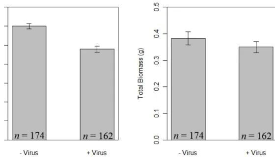
Across all treatments and both plant species, virus infection reduced root fraction
by 20% (F1,300=45.2, p<0.0001; Fig. 2.4A) and tended to decrease total plant biomass by 8.6% (F1,300=2.92, p=0.088; Fig. 2.4B). Elevated CO2 increased total plant biomass of non-mycorrhizal Avena fatua, but not mycorrhizal A. fatua or Bromus hordeaceus (AM
fungi × CO2 × plant species interaction: F1,300=4.4, p=0.037, Appendix A1.3). Across all treatments, B. hordeaceus individuals had 52% less total biomass (F1,300=79.1, p<0.0001) and 43% smaller root fraction than A. fatua ( (F1,300=126.2, p<0.0001).
Leaf Phosphorus Concentration
Phosphorus addition to the soil increased phosphorus concentration in leaves of
37
interaction: F1,257=61.9, p<0.0001; Appendix A1.4). Virus infection also increased leaf phosphorus concentration for both species, and more for A. fatua than B. hordeaceus
(virus × plant species interaction: F1,257=5.15, p=0.0241; Fig. 2.5). Mycorrhizal fungi did not increase leaf phosphorus concentration (F1,257=0.21, p=0.644).
Discussion
Changes in abiotic resource supply have been hypothesized to alter plant
interactions with microbes (Suding et al. 2008). Our results support this concept, showing
that alterations in resource supply can influence performance of both pathogenic and mutualistic plant-associated microbes. In turn, effects on these microbes can influence
not only their host but also the performance of each other.
General ecological theory has predicted that host associations with mutualists may increase enemy populations and thus the severity of enemy damage (Bennett et al. 2006).
The stoichiometric hypothesis for virus production (Clasen and Elser 2007) leads to a more specific prediction: the association of plants with arbuscular mycorrhizal fungi may
increase the titer of virus infections, because AM fungi typically increase host phosphorus concentration (Smith et al. 2009). Our experimental results partially
supported this prediction in that virus infections of plants with AM fungi had 20% higher
relative titers than did infections of plants without AM fungi (Fig. 2.1). However, AM fungi did not significantly increase host tissue phosphorus (Table A2.5), which suggests
that the viral response did not result from the transfer of phosphorous to the plant from AM fungi. This does not completely rule out a role for phosphorus because our
phosphorus data was collected at the leaf level, rather than at the level most relevant to
38
1995). However, physiological mechanisms other than phosphorus transfer may be important. Our finding that AM fungi increased viral titer under elevated CO2, but not under ambient CO2 (Figure 1) suggests that the flow of carbon may also be important in viral production (Malmstrom and Field 1997).
Additionally, the viral pathogen stimulated fungal performance as measured by hyphal colonization (Fig. 2.2A). Specifically, virus infection increased hyphal
colonization of roots under elevated CO2. By the same token, elevated CO2 increased hyphal colonization of virus-infected plants more than virus-uninfected plants. Also, virus infection interacted with phosphorus addition to alter fungal performance for one
plant species. Phosphorus addition decreased fungal colonization for virus-infected B. hordeaceus, but not for virus-free B. hordeaceus or for A. fatua (Fig. 2.3). Together, these results suggest the possibility that the virus derives a fitness benefit under elevated
CO2 by stimulating its host to invest more in a mutualism. While the possible selective pressures behind this are unclear, one possible physiological mechanism involves sucrose
conductance via phloem. Typically, B/CYDVs disrupt the flow of carbohydrates, including sucrose flow through the plant (Irwin and Thresh 1990, Jensen and D'Arcy 1995, Malmstrom and Field 1997), which may interfere with or induce the signaling
39
starvation response, thereby stimulating greater colonization of roots by AM fungi (Smith et al. 2011).
Within host individuals, pathogen populations can be limited by nutrient supplies (Smith et al. 2005, Smith 2007). For example, in an algal-viral system where
post-infection viral production was reduced in low-phosphorus host cultures, presumably as a result of insufficient intracellular phosphorus for production of phosphorus-rich viral particles (Clasen and Elser 2007). The universally high phosphorus concentration of
nucleic acids, the main component of viruses, suggests that low phosphorus concentration may similarly constrain production and titer of viruses infecting terrestrial plants. This
stoichiometric hypothesis predicts that soil phosphorus amendments will increase viral titer in experimental plants. Effects of phosphorus amendment on the prevalence of virus infection in a field experiment were consistent with this hypothesis, although relative
virus titer and leaf phosphorus concentration were not analyzed (Borer et al. 2010). In the first experiment to consider the role of BYDV and leaf phosphorus concentration in
wild grasses, we demonstrated the reverse in that soil phosphorus amendment
significantly decreased relative viral titer for A. fatua and had no effect on titer for B. hordeaceus (Appendix A1.1). This result indicates that effects of phosphorous supply on viral titer can vary among host species, perhaps depending on their physiological uptake rate or allocation of phosphorus.
In addition to altering microbe performance, changes in resources can also have direct effects on plant biomass and allocation. In a previous study, elevated CO2
increased the biomass of BYDV-infected Avena sativa more than uninfected plants
40
in plant carbon uptake caused by BYDV infection. We did not see such a
counterbalancing effect in our experiment. This may be because we used two wild host
species which have not been selected for agronomic yield, whereas Malmstrom and Field (1997) used A. sativa, an agricultural species which could react differently to changes in
CO2 availability due to differences in evolutionary history.
Elevated CO2 and mycorrhizal fungal colonization often jointly stimulate plant growth,but such responses can vary with host-fungal species identity (Johnson et al.
2003, Klironomos et al. 2005). In our experiment, elevated CO2 increased total biomass of non-AM-fungal A. fatua, but not of AM-fungal A. fatua, or of B. hordeaceus
(Appendix A3). AM fungi had no net impact on total biomass of B. hordeaceus or of A. fatua plants under elevated CO2, even though elevated CO2 stimulated AM fungal colonization of both plant species (Appendix A1.2). This result suggests that AM fungi
did not stimulate plant biomass despite increased activity as measured by hyphal colonization of roots.
Our two study plant species were similar in life history and growth form, as well as in serving as common hosts for mycorrhizal fungi, aphids, and viruses, yet they often differed in their responses to our experimental manipulations. While such differences
may be idiosyncratic, study of a larger number of host species may reveal these to be part of a broader pattern. For instance, both A. fatua and B. hordeaceus fall along a
phenotypic continuum in leaf ecophysiological traits which may influence not only the way they respond to biotic factors such as mycorrhizae or pathogen infection, but also to abiotic factors (Wright et al. 2004, Cronin et al. 2010). Further study of the combined
41
to understanding and predicting large-scale changes to ecosystems (Treseder 2004, Suding et al. 2008).
Acknowledgements
We are thankful to J. Barton at the CSTR facility for technical help. We would
like to thank J. Bever and K. Vogelsang for mycorrhizal inoculum. We are grateful to the Borer-Seabloom lab for collecting and providing all plant seed, and to Jack Weiss for statistical advice. We would also like to thank the Mitchell lab for assistance. This
research was partially supported by the joint NSF-NIH Ecology of Infectious Disease program through NSF Grants EF-05-25641 and DEB-10-15909 to C.E.M. and an NSF
42
Figure 2.1. Effect of mycorrhizal colonization on viral titer. Across plant species and phosphorus treatment, mycorrhizal colonizaton (+AMF vs. –AMF) increased relative viral titer as measured by Optical Density (OD) value for plants under elevated CO2 but had no effect under ambient CO2. Data shown are means ± SEM; letters indicate
43
Figure 2.2. Results for root colonization by AMF. Across plant species, virus infection increased root colonization by mycorrhizal fungi under elevated CO2 but not under ambient CO2 (A). Phosphorus addition (+P vs. –P) decreased root colonization by
44
Figure 2.3. The role of phosphorus, viral infection and host species for root colonization by AMF. Phosphorus addition (+P vs. –P) did not alter root hyphal
45
46
47
CHAPTER III: FUNGAL ENDOPHYTE INFECTION AND HOST CULTIVAR JOINTLY MODULATE HOST RESPONSE TO AN APHID-TRANSMITTED VIRAL PATHOGEN Abstract
1). Despite their ubiquitous nature, interactions between multiple microorganisms and their effect on not only host growth but also one another’s success have received
limited scientific attention. In this study, we investigated how an aphid-transmitted viral pathogen and a mutualistic endophytic fungus altered host growth and allocation.
2). In a greenhouse experiment, we manipulated endophyte status and virus infection (Barley Yellow Dwarf Virus - PAV) of two tall fescue cultivars. We assessed host, virus and vector responses.
3). Endophyte infection mitigated the negative impact of the virus on root allocation but also allowed the virus to decrease host tillering. Both of these effects had
either host or endophyte genotype dependent responses. Endophyte infection universally decreased reproduction and abundance of aphid vectors, and this did not interact with host plant virus infection status.
4). These results indicate that some of the beneficial effects provided by endophyte infection do not arise strictly from altering host interactions with the vector
(aphids), but also occur by changing host responses to viral infection. Furthermore, these results emphasize the importance of exploring multi-species microbial interactions and genotype controls on these interactions in order to more fully understand their role in
48 Introduction
Plant hosts are often confronted simultaneously with a diverse array of
microorganisms, including both pathogens and mutualists (Arnold 2007, Pieterse and Dicke 2007, Friesen et al. 2011). The close relationships between hosts and their
microbes are characterized by a high degree of recognition and signaling between the plant and the associated microbe at molecular, morphological and physiological levels (Harrison 2005). Furthermore, association with microbes can alter plant phenotypes by
supplying novel nutritional and defense pathways for the plant as well as influencing plant biochemical pathways (Friesen et al. 2011). Such alterations in plant phenotypes
due to association with one microbe may in turn alter plant relationships with other microbes. These relationships may be altered either directly via the shared host or indirectly via a third player such as an arthropod vector. For example, mutualistic
microbes can help protect plants against pathogens either by increasing plant defense against pathogens themselves, or by increasing plant defense against herbivores,
including arthropods that transmit pathogens (Clay and Schardl 2002, Hartley and Gange 2009). Thus, a broad community context may be important for understanding at least some of these microbial interactions (Saunders et al. 2010). Despite this recognition, few
studies examine the impact of interactions among multiple microorganisms on host growth and allocation, or the impact of different microorganisms on each other’s success.
49
A majority of plant-infecting viruses are dependent upon arthropod vectors for transmission between hosts (Nault 1997, Hogenhout et al. 2008). Therefore, virus
ecology is often dependent on the population dynamics, host preference, and movement of vectors (Power and Flecker 2008). Barley and cereal yellow dwarf viruses (B/CYDVs)
are a widespread group of aphid-transmitted, generalist viral pathogens that have
provided a model system for plant-virus-vector interactions (Gray and Gildow 2003). For example, consumption of B/CYDV-infected host tissue often increases aphid fecundity,
with some variation among host, vector and virus species (Power and Gray 1995). Additionally, increased abundance of aphid vectors generally increases the rate at which
B/CYDVs are transmitted to healthy plants (Burnett and Gill 1976, Jensen and D'Arcy 1995, Power and Gray 1995). Thus, plant characteristics that alter vector population dynamics are likely to alter their transmission of viruses.
Many agronomic and wild grass species host endophytic fungi in the Ascomycete family Clavicipitaceae. These endophytes receive nutrients, protection, reproduction and
dissemination via seeds from the plant (Schardl et al. 2004). In return, the host receives a variety of services from the symbiont including increased soil nutrient uptake
(Malinowski et al. 2000) and increased drought resistance (Arachevaleta et al. 1989,
Malinowski and Belesky 2000). In addition, many of these endophytes are thought to provide herbivore deterrence via the production in planta of several distinct classes of
biologically active alkaloids that can reduce arthropod feeding, population size, and consequent damage for the host plant (Clay 1990, Schardl et al. 2004). However, benefits to the host provided by fungal alkaloid production can vary among herbivore species,
50
Endophyte-produced alkaloids may influence aphid-transmitted plant pathogens because, among insect herbivores, aphids are some of the most negatively affected by endophyte
infection (Hartley and Gange 2009). Endophytes commonly deter aphid consumption and reduce aphid fecundity (Schardl and Phillips 1997, Hartley and Gange 2009).
For viruses transmitted by aphids and other arthropods, the arthropod deterrence that results from endophyte infection may in turn decrease the severity of virus infection for the host plant. Transmission of B/CYDVs to the plant from the aphid typically
requires several hours of aphid feeding (Power and Gray 1995), so decreased aphid feeding duration as a result of endophyte infection may decrease transmission of
B/CYDVs to the plant. Furthermore, a decreased number of feeding aphids can decrease the titer of resulting virus infections (Power and Gray 1995), so impacts of endophytes on both aphid population size and feeding duration may reduce the titer of resulting virus
infections in endophyte-infected hosts. In turn, reduced virus titer can both decrease the negative impacts of infection on the host plant, and increase the amount of feeding time
necessary for uninfected aphids to acquire the virus from the plants (Power and Gray 1995).
Another way in which endophytes may influence B/CYDV infections is through
the alteration of biochemical pathways related to pathogen defense. Infection by endophytes may result in mismatches between plant and pathogen signaling, including
51
recognition pathway, they broaden the potential for mismatches and result in changes in pathogen protection of the host via endophyte infection.
Within grass-fungal endophyte associations, such as that of tall fescue (Schedonorus phoenix = Festuca arundinacea) and Neotyphodium coenophialum,
endophytes can produce a suite of alkaloid compounds that deters both mammalian and insect herbivory (Schardl et al. 2004). So called ‘common toxic’ genotypes of these endophytes have been demonstrated to consistently deter arthropods in agroecosystems
(Breen 1994), but such deterrence may change with time and abiotic conditions (Hunt and Newman 2005, Rasmussen et al. 2007). ‘Novel’ forms of some of these endophytes
exist and generally lack the ability to produce the mammalian active compounds but retain the compounds important in deterring arthropod herbivores (Malinowski and Belesky 2006). It is possible that host plants infected with novel endophytes may
respond differently to virus infection than those infected with the common toxic strain of the endophyte. The limited previous research suggests that novel endophyte infected
hosts may be at a competitive disadvantage compared to common toxic endophyte infected individuals when exposed to biotic stresses, such as herbivory, and abiotic stresses, such as variation in growing conditions (Malinowski and Belesky 2006).
Additionally, there is evidence to suggest that novel endophytes do not provide the same degree of protection from aphids as common toxic endophytes (Hunt and Newman 2005).
Specifically, intrinsic rates of growth for enclosed populations of aphids were greatest for those aphids fed on endophyte-free plants, slower on novel endophyte-infected plants and slowest (or no growth at all) on the plants infected with the common toxic strain of
52
infection will provide less aphid deterrence, and consequently less protection for the host from virus infection, then common toxic endophytes.
Much of the previous research on virus-endophyte-aphid interactions has centered on community-level studies. These studies have generally focused on the impacts of such
interactions on agriculturally important host species, with conflicting results. For example, studies that attempted to correlate B/CYDV prevalence and the incidence of endophyte infection for Lolium perenne (perennial ryegrass) found no correlation (Guy
1992), while studies considering tall fescue have found that endophyte-infected plants were less likely to be infected by B/CYDVs (Mahmood et al. 1993, Guy and Davis
2002). On the other hand, most plant populations are genetically diverse, and the benefits of endophyte infection can vary among host genotypes (Cheplick 1998). Yet, this
previous research has not considered potential impacts of host genotypic differences
within the same species. Therefore our research, which examines both host and fungal endophyte genotypic effects of endophyte-host-B/CYDV interactions, can serve to
inform both future and past community level explorations of these interactions.
Here, we present the first experiment evaluating the interaction of virus infection and endophyte infection as they relate to impacts on the host. Specifically, we explore
how endophyte and host cultivar interact with virus infection to alter vector abundance, host biomass, allocation and tillering. Such impacts are likely to play a crucial role not
only in agroecosystems but in natural ecosystems, where fungal endophytes and B/CYDV are also common (Mitchell and Power 2006).
53
Barley and cereal yellow dwarf viruses (B/CYDVs) are a group of aphid-transmitted generalist viral pathogens that infect over 150 crop and noncrop grasses
(D'Arcy 1995, Halbert and Voegtlin 1995). B/CYDV infection is systemic and localized to the phloem where it causes necrosis and disruption of carbohydrate translocation
(Irwin and Thresh 1990, D'Arcy 1995). Impacts of infection include stunted plant growth, reduced root/shoot ratio and reduced longevity (Kolb et al. 1991, Malmstrom et al.
2005a). B/CYDVs are obligately transmitted by aphids, including the globally common
aphid species Rhopalosiphum padi (L.).
Tall fescue (Schedonorus phoenix = Lolium arundinaceum = Festuca
arundinacea) is a cool-season grass that has been widely planted for forage in the United States due to its ability to tolerate high temperatures, drought conditions and grazing (Stuedemann and Hoveland 1988). Many of the properties that make S. phoenix attractive
for use as a forage species can be attributed to the symbiotic fungal endophyte
Neotyphodium coenophialum (Clay and Schardl 2002). It is estimated that between 75 and 85% of S. phoenix in the US is infected with the common toxic form of N.
coenophialum (Ball et al. 1993, Clay and Schardl 2002). Tall fescue provides a valuable model system to investigate microbe-microbe interactions because pair-wise host–fungus
interactions and mechanisms for microbe-microbe competition have been well-described in this system (Saunders et al. 2010).
Experimental design, treatments and conditions
We used two S. phoenix cultivars, KY 31 and PDF. Experimental seed for the KY 31 cultivar was either endophyte free (E-) or contained the common toxic strain of N.