Colofon
Cover illustration, front:
Emu dreaming by Peter O vers, p rinted w ith p ermission of p ainter
Cover illustration, bac k :
G ertjan van L eeuw en (G ummbah ), p rinted w ith p ermission of c artoonist
Printed by:
F ebodruk B V
IS B N :
9 0 -9 0 1 9 9 5 8 -6
Downregulation of MHC class I molecules by human
cytomegalovirus-encoded US2 and US11
PROEFSCHRIFT
ter verkrijging van
de graad van D octor aan de U niversiteit Leiden,
op gez ag van de Rector M agnificus D r.D .D .Breimer,
hoogleraar in de faculteit der W iskunde en
Natuurwetenschappen en die der Geneeskunde,
volgens besluit van het College voor Promoties
te verdedigen op donderdag 2 7 oktober 2 005
te klokke 14 .15 uur
door
Promotiecommissie:
Promotor:
Prof. Dr. E.J.H.J. Wiertz
Referent:
Prof. Dr. C.J.M. Melief
Overige leden:
Dr. M.F.C. Beersma
Prof. Dr. F.H.J. Claas
Dr. F. Lenfant (INSERM U589, CHU Rangueil, Toulouse)
Prof. Dr. J. Middeldorp (VUMC, A msterdam)
Prof. Dr. J.J. Neefjes
The research presented in this thesis was performed in the Laboratory of Vaccin Research in the
National institute of public health and the environment, Bilthoven, and in the department of
Contents
A b b rev ia tions
8
Ch a p ter 1
Introduction
9
Ch a p ter 2
A m ino a cid com p os ition of
D
1 /
D
2 dom a ins a nd cy top la s m ic ta il of
M H C cla s s I m ole cule s de te rm ine th e ir s us ce p tib ility to H C M V U S 1 1
-m e dia te d dow n-re g ula tion.
Eur J Immunol. 33: p1707-1716 (2003)
2 7
Ch a p ter 3
H C M V -e ncode d U S 2 diffe re ntia lly a ffe cts s urfa ce e x p re s s ion of M H C
cla s s I locus p roducts a nd ta rg e ts m e m b ra ne -b ound, b ut not s olub le
H L A -G 1 for de g ra da tion.
J Immunol. 171: p675 7-6765 (2003)
4 1
Ch a p ter 4
S ub tle s e q ue nce v a ria tion a m ong M H C cla s s I locus p roducts g re a tly
influe nce s s e ns itiv ity to H C M V U S 2 -a nd U S 1 1 -m e dia te d de g ra da tion.
S ub mitte d
5 7
Ch a p ter 5
U b iq uitina tion is e s s e ntia l for h um a n cy tom e g a lov irus U S 1 1 -m e dia te d
dis loca tion of M H C cla s s I m ole cule s from th e e ndop la s m ic re ticulum
to th e cy tos ol.
B ioc h e m J. 35 8 : p369 -377 (2001)
7 1
Ch a p ter 6
H um a n cy tom e g a lov irus -e ncode d U S 2 a nd U S 1 1 ta rg e t una s s e m b le d
M H C cla s s I h e a v y ch a ins for de g ra da tion.
M ol Immunol. : in pre s s (2005 )
85
Ch a p ter 7
S um m a ry a nd D is cus s ion
99
N ed erla nd se sa m env a tting
1 0 7
Abbreviations
aa
amino acid
E
2m
E
2-microglobulin
BiP
Immunoglobulin binding protein
Cnx
calnexin
Crt
calreticulin
CTL
cytotoxic T lymphocyte
DTT
dithiothreitol
EGFP
enhanced green fluorescent protein
EndoH
endoglycanase H
ER
endoplasmic reticulum
FCS
fetal calf serum
FITC
fluorescein isothiocyanate
g
D
m
goat anti-mouse
HC
heavy chain
HCMV
human cytomegalovirus
HLA
human leucocyte antigen
IFN
interferon
IL
interleukin
IRES
internal ribosomal entry site
kDa
kilodalton
mAb
monoclonal antibody
MHC
major histocompatibility complex
MOI
multiplicity of infection.
NK cell
natural killer cell
NP40
nonidet P-40
PDI
protein disulfide isomerase
PE
phycoerythrin
prot K
proteinase K
TAP
transporter associated with antigen processing
TM
transmembrane
TNF
tumor necrosis factor
TX -100
triton X -100
UL
uniq ue long
US
uniq ue short
wt
wild type
Z L3H
carboxybenzyl-leucyl-leucyl-leucinal
Z LVS
carboxyl benzyl-leucyl-leucyl-leucyl vinylsulfone
Introduction
D e fe ns e m e ch a nis m s a g a ins t v irus e s
In healthy individuals, the immune system is able to
limit the damag e in many viral infec tions. W hen a viral
p artic le manag es to invade the body by c rossing
p hysic al barriers suc h as the sk in and muc us
membranes, it bec omes subjec t to detec tion and
elimination by several immune effec tor c ells. A s
viruses c annot rep lic ate on their ow n, they must infec t
c ells in order to ex p loit the host c ell’s mac hinery for
the p roduc tion of p rog eny and to enable further virus
sp read.
H ost c ell entry c an be bloc k ed by antibody-p roduc ing
B c ells, w hic h sp ec ific ally rec og niz e foreig n struc tures
on the viral p artic le. T he antibodies c annot only
steric ally hinder viral entry, but they c an also induc e
elimination of the viral p artic les via different p athw ays.
T hese inc lude c omp lement ac tivation,
antibody-dep endent c ell-mediated c ytotox ic ity and p hag
o-c ytosis
1 ,2. T w o subtyp es of B c ells arise after
ac tivation of B c ells, antibody p roduc ing p lasma c ells
and memory B c ells. T he latter subtyp e is c ap able of
mounting an immediate hig h-affinity resp onse up on
renew ed c ontac t w ith the antig en.
O nc e virus p artic les are inside host c ells, T c ells c ome
into p lay. T hey c an rec og niz e foreig n antig en
frag ments disp layed in the c ontex t of major
histoc omp atibility c omp lex (M H C ) molec ules at the
c ell surfac e
3 ,4. T here are tw o c lasses of M H C
molec ules, w hic h differ in struc ture, ex p ression
p attern and sourc e of antig enic p ep tides.
M H C c lass I molec ules are found on all nuc leated
c ells and mainly p resent p ep tides derived from
endog enously synthesiz ed p roteins. C irc ulating C D 8
+c ytotox ic T c ells (C T L s) insp ec t antig enic frag ments
disp layed by M H C c lass I molec ules. In c ase of
infec tion, C T L s rec og niz ing foreig n antig ens c an,
up on ac tivation, k ill suc h c ells.
E x p ression of M H C c lass II molec ules is restric ted to
antig en-p resenting c ells, i.e. B c ells, mac rop hag es
and dendritic c ells. C lass II molec ules disp lay p ep tide
frag ments, derived from an ex og enous sourc e of
p roteins (e.g . viral c ap sid p roteins), for insp ec tion by
C D 4
+T c ells. U p on rec og nition of foreig n p ep tides,
these T c ells p rovide help to C D 8
+T c ells to elic it a
c ytotox ic resp onse. In addition, C D 4
+T help er c ells
are req uired for the maintenanc e and func tionality of
memory C D 8
+T c ells
5.
T he p resentation of ex og enous p ep tides is not
c omp letely restric ted to M H C c lass II molec ules, as
p ep tides derived from internaliz ed p roteins c an also
enter the M H C c lass I p athw ay
6 ,7. T he ability to c
ross-p resent seems to be limited to a sross-p ec ific subset of
antig en p resenting c ells, mainly C D 8
+dendritic c ells
8.
In addition, endog enous p roteins c an g ain ac c es to
the M H C c lass II p athw ay via autop hag y
9.
B esides an interac tion betw een M H C molec ules and
their sp ec ific rec ep tors, a lig ation betw een ac c essory
molec ules on the antig en p resenting c ell and c
ounter-rec ep tors on the T c ell (c o-stimulation) is req uired for
T c ell ac tivation.
N atural k iller (N K ) c ells are also c ap able of sensing
virally infec ted c ells, but c an only do so by virtue of
altered ex p ression levels of c ertain native surfac e
p roteins, inc luding M H C c lass I molec ules. T he
ac tivation of N K c ells, and the subseq uent release of
c ytotox ins, is c ontrolled by a set of N K c ell rec ep tors.
D ep ending on the typ e of rec ep tor, inhibitory or
ac tivating sig nals are ig nited up on binding lig ands on
the p otential targ et c ell. If inhibitory rec ep tor sig naling
fails to q uenc h the effec t of eng ag ement of ac tivating
rec ep tors, the N K c ell w ill be trig g ered to k ill its targ et
c ell.
T his thesis w ill further foc us on M H C c lass I
molec ules and their imp lic ation as lig ands for T and
N K c ell rec ep tors in the c ontex t of human c ytomeg
alo-virus infec tion.
H um a n cy tom e g a lov irus
H uman c ytomeg alovirus (H C M V ) is found in human
p op ulations w orldw ide and c an infec t 6 0 – 90 %
of
individuals, dep ending on the p op ulation studied.
H C M V is a member of the Herpesviridae, w hic h are
k now n to establish a life-long relationship w ith their
hosts. H C M V infec tion is usually asymp tomatic , but
c an c ause disease in an op p ortunistic manner. In
immunoc omp romised individuals, suc h as A ID S
p atients and org an transp lant rec ip ients, unc ontrolled
H C M V reac tivation c an c ause severe illness or death
1 0 ,1 1. A dditionally, H C M V is a notorious risk fac tor for
c ong enital birth defec ts. In most c ases, suc h c linic al
manifestations are found in mothers w ho underg o
p rimary rather than rec urrent infec tions
1 2.
In vivo, HCMV can infect endothelial cells, epithelial
cells, monocytes/macrophages, smooth muscle cells,
stromal cells, neuronal cells, neutrophils, fibroblasts,
and hepatocytes
13,14.
In vitro, all tested vertebrate cell
types were susceptible to HCMV infection
15. Common
HCMV-associated illnesses include retinitis (which
may cause blindness), hepatitis, encephalitis,
pneumonia, and bowel disease
16.
F ollowing primary infection, HCMV persists in a latent
state in cells of the myeloid lineage, with intermittent
viral reactivation and shedding from mucosal surfaces,
and containment by the host immune response
17.
Another potential cell type latently or persistently
infected by HCMV are vascular endothelial cells
18.
Interestingly, HCMV infection has different effects on
the functioning and survival of these various types of
host cells. In macrovascular aortic endothelial cells
HCMV infection is not lytic and results in the
accumulation of significant amounts of extracellular,
but not intracellular virus. In addition, the cell cycle is
not inhibited by HCMV and cells continuously release
infectious virus. This is in contrast with the rapid, lytic
infection of brain microvascular endothelial cells and
human foreskin cells
18. It also differs from HCMV
infection in monocyte-derived macrophages, which
results in non-lytic accumulation of intracellular virus
and lack of extracellular virus
19.
At present, the viral and host mechanisms of HCMV
latency and reactivation are unclear, but the virus may
be reactivated by cellular differentiation, and hormonal
or cytokine (TNF
D) stimulation.
HCMV particles can be found in all body fluids and
can be transmitted by multiple means, including
aerosol droplets, sexual contact, nursing,
trans-plantation and blood transfusion
20.
HCMV has a large double stranded DNA genome
(t230 kb), encoding approximately 192 proteins
21.
The size of the genome varies among HCMV strains.
Compared to low-passage clinical HCMV isolates, the
laboratory-adapted AD169 and Towne strains lack 22
and 19 viral genes, respectively
22.
In infected cells, viral replication occurs according to a
cascade of three consecutive phases, called
immediate early (IE), early (E), and late (L). The IE
gene products play an important role in regulating the
expression of viral E and L genes, as well as of
cellular genes. E and L viral gene products include
viral functions associated with viral DNA replication
and virus packaging. The viral genome also encodes
several proteins involved in immune evasion
strategies, dealing with various immune effector
mechanisms of the host. These include interference
with MHC class I and II antigen presentation to CD8
+and CD4
+T cells, NK cell activity, cytokine and
chemokine signalling, and apoptosis
23-37.
It is evident that these mechanisms are not exploited
in a way that is life-threatening to the
immuno-competent host. Several studies support important
roles for B, T and NK cells in protection against HCMV
disease
38-44. However, these immune evasion
strategies may prevent complete eradication of the
virus and contribute to the ability of HCMV to cause
lifelong persistent infections.
Before discussing the immune escape mechanisms
exploited by HCMV to interfere with MHC class I
surface expression, an introduction on MHC class I
complex formation and the different functions of the
various MHC class I locus products will be given.
MHC class I molecules
MHC class I molecules consist of a 43 kDa heavy
chain, a 12 kDa light chain (E2-microglobulin,
E2m)
and a peptide of 8-10 amino acids in length (see
F igure 1). The heavy chains are type 1
trans-membrane proteins consisting of three ER -lumenal
/surface exposed domains (D1-3), a connecting
peptide, transmembrane (TM) region and a
cytoplasmic tail. The
D1-3 domains bind
E2m
non-covalently. The D3 domain and E2m show homology
with immunoglobulin domains and have similar folded
structures, whereas the
D1 and
D2 domains fold
together into a single structure consisting of two
segmented
D-helices lying on a sheet of eight
antiparallel E-strands. The folding of the
D2 and D3
domains creates a long groove, which is the site
where peptide antigens bind. Together with peptide,
this highly polymorphic MHC class I region determines
T-cell antigen recognition. A single binding site can
bind a wide variety of peptides with high affinity, but
there are some sequence restrictions. The peptides
that can bind to a given MHC variant have the same
or very similar amino acid residues at two or three
particular positions, called anchor residues, along the
peptide sequence. The amino acid side chains at
these positions insert into pockets of the MHC class I
molecule that are lined by polymorphic residues of the
heavy chain. Additional amino acid positions within
the peptide, called secondary anchors, can also
influence MHC binding. These anchor residues differ
for peptides binding different alleles of MHC class I
for peptides binding different alleles of MHC class I
m olecu les. A ltogeth er, th is ensu res th e display of a
w ide v ariety of peptide antigens for CD 8
+T cell
inspection.
MHC class I assembly and interactions with
comp onents of MHC class I antig en p resentation
p athway
T h e folding and assem bly into m atu re trim eric
com plex es inv olv es a series of ev ents and req u ires
th e action of sev eral accessory m olecu les (see F igu re
2 ). A s HCMV -encoded proteins interv ene at different
stages of th is process, th e MHC class I antigen
presentation path w ay w ill be discu ssed in m ore detail.
MHC class I h eav y ch ains encode a signal peptide,
w h ich directs insertion into th e E R du ring translation.
O nce in th e E R , th e signal seq u ence is cleav ed off by
a signal peptidase, w h ile oligosacch ary l transferase
eq u ips th e HC w ith an N -link ed oligosacch aride.
T h ese free HCs are soon fou nd in association w ith th e
general E R ch aperones B iP
4 5and m em brane bou nd
calnex in, th e latter of w h ich h as lectin-lik e activ ity
4 6 ,4 7.
B iP transiently binds to m any new ly sy nth esiz ed
proteins. Misfolded proteins and u nassem bled
su bu nits are bou nd by B iP in a prolonged fash ion.
4 8 ,4 9
. B inding of calnex in is regu lated by glu cose
trim m ing of nascent N -link ed oligosacch arides
5 0.
Calnex in generally binds proteins w ith m
ono-glu cosy lated (G lc1 Man9 -7 G lnN A c2 ) oligosacch arides
5 1
. Calnex in and B iP predom inantly associate w ith free
MHC class I h eav y ch ains and th e assem bly w ith
E
2 m
abolish es th e interaction of th e h eav y ch ain w ith th ese
ch aperones
4 5 ,5 2 ,5 3. B efore binding th e ligh t ch ain,
h eav y ch ains also interact w ith E R p5 7 , a m em ber of
th e protein disu lfide isom erase (P D I) fam ily , w h o are
inv olv ed in disu lfide bond ox idation, redu ction and
isom eriz ation reactions
5 4 -5 6. Matu re MHC class I
m olecu les h arbor th ree intra-m olecu lar disu lfide
bridges, th e form ation of w h ich is lik ely to be m ainly
assisted by E R p5 7 . A fter binding
E
2 m , MHC class I
m olecu les are fou nd in association w ith anoth er,
solu ble E R ch aperone w ith lectin-lik e activ ity ,
calreticu lin
5 7 ,5 8. L ik e calnex in, calreticu lin binds to
proteins
w ith
G lc1 Man9 -7 G lnN A c2
N -link ed
oligosacch arides
5 9 ,6 0. MHC class I m olecu les th an
becom e associated w ith th e peptide-loading com plex ,
w h ich besides calreticu lin inclu des E R p5 7 , tapasin,
and th e transporter associated w ith antigen
processing (T A P ). T apasin m ediates th e interaction of
class I w ith T A P
5 7 ,5 8 ,6 0. T h e stru ctu re of MHC class
I-peptide com plex es su ggests th at ox idation of th e
cy steines in th e HC
D
2 -dom ain is a prereq u isite for
stable peptide binding, a process lik ely facilitated by
E R p5 7
6 1. T h e peptides are generated from
endogenou s proteins by cy tosolic proteasom es (large
protease com plex es, discu ssed in m ore detail in a
later section) and m ay be fu rth er trim m ed by
am inopeptidases before and after translocation into
th e E R v ia T A P
6 2 ,6 3. In th e E R , peptides are loaded
onto HC-
E
2 m h eterodim ers. T h ese trim eric HC-
E
2 m
-peptide com plex es th en dissociate from th e loading
com plex and are released into th e secretory path w ay
6 4
.
MHC class I v ariants
In h u m ans, a single ty pe of ligh t ch ain (B 2 m ) is fou nd
th at is com plex ed w ith one of m u ltiple ty pes of h eav y
ch ains, HL A -A , B , C, E , G , of w h ich u p to tw o different
allelic form s can be ex pressed. T h ey are encoded by
separate clu sters w ith in th e MHC region of th e h u m an
genom e, located on ch rom osom e 6 . T h is region also
encodes oth er proteins inv olv ed in antigen
presentation (e.g. MHC class II, T A P 1 , T A P 2 , L MP
genes). T h e ligh t ch ain is encoded by a gene located
on anoth er site, on ch rom osom e 1 5 . T h e v ariou s
ty pes of h eav y ch ains differ from each oth er in sev eral
w ay s, as w ill be discu ssed below .
Polymorphism
O ne ou tstanding difference am ong class I h aploty pes,
is th e degree of poly m orph ism th at can be fou nd in
th e w orld popu lation. T o date, 3 7 2 different alleles
h av e been reported for HL A -A , and 6 6 1 for HL A -B .
HL A -C sh ow s som ew h at less v ariation w ith 1 9 0
different alleles. P oly m orph ism of HL A -E and HL A -G
is v ery lim ited, w ith 5 (HL A -E ) and 1 5 (HL A -G )
different alleles described to date (IMG T /HL A
database,
6 5.
T issu e d istrib u tion / su rfa c e e x pre ssion
T issu e distribu tion and cell su rface lev els differ for th e
v ariou s locu s produ cts. Most h ost cells ex press HL A
-A , B , C and E alleles, w h ereas HL -A -G ex pression is
restricted to th e th y m u s and certain placental tissu es,
e.g. troph oblast cells. T h e troph oblast cells at th e
m aternal-fetal interface lack su rface ex pression of
HL A -A and -B alleles
6 6 ,6 7. O u tside th is im m u
ne-priv ileged site, th e su rface lev els of HL A -A , -B , and -C
alleles m ay v ary betw een cell ty pes
6 8 ,6 9. S ev eral
aspects th at influ ence MHC class I (su rface)
ex pression lev els w ill be discu ssed below .
R e g u la tion of M H C c la ss I g e n e tra n sc ription
HL A class I genes h av e different constitu tiv e and
cy tok ine-indu ced ex pression patterns, w h ich can be
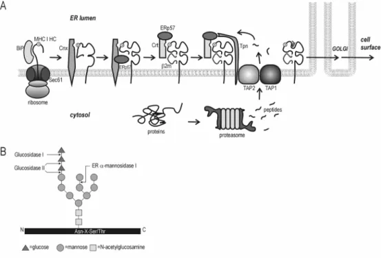
Figure 2. M H C c la s s I a s s em b ly . A) Model for folding and complex formation of MHC class I molecules and interactions with components of the class I antigen presentation pathway (see text for further description). Calnexin (Cnx), calreticulin (Crt), tapasin (T pn). T he N -link ed gly can is mark ed “N ”. B ) N -link ed gly cans are added to proteins in the E R as "core oligosaccharides" that hav e the structure shown. T hese are b ound to the poly peptide chain through an N -gly cosidic b ond with the side chain of an asparagine that is part of the A sn-X -S er/T hr consensus seq uence. T he three glucoses are remov ed b y glucosidase I and II, and terminal mannoses b y one or more different E R mannosidases. A ssociation of gly copeptides with Cnx/Crt is dependent upon the presence of a single terminal glucose residue b ound to the G lcN A c2Man9 gly can precursor. (Adapted from ref.142
attrib uted to differences in their promoter regions.
6 9-7 1. T he cy tok ines T N F -D, IF N -J and IF N -E can
increase av erage MHC class I surface lev els up to 1 0
-fold
6 9. T he HL A -G promoter region differs from other
MHC class I genes, as some of the conserv ed
regulatory b oxes hav e b een deleted or altered,
including the IF N regulatory seq uence
7 0 ,7 2. In
addition, the cy tok ine IL 1 0 has b een reported to
selectiv ely upregulate HL A -G expression
7 3. Cell ty
pe-specific factors may contrib ute to differential locus
expression as well
6 8 ,6 9.
M H C c las s I c omplex formation an d s tab ility
T he lev els of the different MHC class I molecules that
are found at the cell surface are determined b y
transcript lev els, as well as b y the success of stab le
complex formation, and turnov er rates. G enerally ,
surface lev els of HL A -C locus products are rather low,
ab out 1 0 % of the av erage lev el of HL A -A and -B
molecules
7 4 -7 6. S ev eral explanations hav e b een
lower peptide ligand binding affinities and render the
heterotrimeric MHC class I complexes formed less
stable. Once at the cell surface, tyrosine or di-leucine
motifs present in most MHC class I molecules can
function as endocytosis signals. HLA-G misses
potential endocytosis signals and its half-life has been
shown to be almost twice that of HLA-A2 molecules
85.
Peptide binding / repertoire
HLA-A/B and HLA-C/G/E differ in their ligand-binding
capabilities: HLA-A/B molecules are promiscuous,
whereas the other class I molecules bind a limited
number of ligands. HLA-C locus products, for
instance, have a preference for peptides that are
poorly transported into the ER, and that require a
10-fold higher peptide concentration for release into the
secretory pathway
78,86-88. HLA-G binds many
self-peptides with a defined motif and its peptide binding
diversity is estimated to be about five fold lower than
that of HLA-A
89,90. The most common HLA-E ligands
are nonamers derived from signal sequences of other
HLA class I molecules
91,92.
S pecial features / alternativ e splice v ariants
Due to a premature stop codon in exon 6, HLA-G has
a relatively short cytoplasmic tail (6 residues)
compared to other HLA class I molecules (29-33
residues)
93. Consequently, HLA-G not only lacks
potential endocytosis signals present in other class I
molecules, but it also gains an ER retrieval/retential
signal with the dilysine residues positioned at 4 and
-5 from the C-terminus. This motif can mediate
recycling of assembled HLA-G molecules between the
ER and the cis-Golgi
94,95. High affinity peptide binding
seems to be required to end the recycling and allow
egress to the cell surface
85.
Primary HLA-G mRNA transcripts are differentially
spliced, giving rise to multiple isoforms
96. Only the full
length isoform (HLA-G1) is expressed at the cell
surface
97. In addition to this membrane-bound
isoform, a soluble (secreted) isoform was detected in
placental tissues (sG1 /G5). Soluble
HLA-G1 is translated from a transcript with a retained intron
4, which introduces a premature stopcodon after the
D1-3 domain encoding sequence. This isoform can
associate with E2m
98,99.
L igands for T and N K cell receptors
MHC class I molecules can be ligands for cytotoxic T
cell receptors, as well as for NK cell receptors.
Through recognition of MHC-peptide complexes,
CD8
+T cells can kill infected target cells. HLA-A and
B molecules are the restriction elements in the
majority of CTL responses, although there are also
some examples of HLA-C, -G and -E molecules
presenting antigenic peptides to CTLs
100-103. This
may very well explain the high degree of
polymorphism that is particularly described for HLA-A
and – B alleles.
U nlike for CTLs, the distinction between self and
non-self antigens in the context of MHC class I molecules
is not the major restriction element for triggering of NK
cell lysis. This trigger is determined by the presence
or absence of NK cell receptor ligands on the surface
of the target cell. The total input of several types of
ligands, including MHC I molecules, and their
engagement with both inhibitory and activating NK
receptors controls NK cell cytolysis
104,105. Many MHC
class I molecules can contribute to the regulation of
NK cell functioning. Other NK cell receptor ligands
include several molecules that are distantly related to
MHC class I molecules, e.g. MIC A/B, U L16 binding
protein (U LBP) 1/2/3, and CD155. An overview of
currently known NK cell receptors and ligands is
presented in Table 1
106,107.
A particular NK cell clone can express up to nine
different receptors, which together determine the
activation status of the cell. These receptors can be
displayed on overlapping subsets within the total NK
cell population, and the repertoire of expressed
receptors is heterogeneous in different individuals
108,109. Some of these receptors are also found on
other immune effector cell types. Variation in surface
expression of only one type of MHC class I molecule
can already make a difference for cell survival.
Table 1. Natural killer cell receptors and their ligandsName of receptor Type of signal Ligand(s) K IR2DL1 (p58.1) inhibitory HLA-CLys80 K IR2DL2 (p58.2) inhibitory HLA-C Asn80 K IR2DL4 (p49) inhibitory HLA-G K IR3DL1 (p70) inhibitory HLA-Bw4 K IR3DL2 (p140) inhibitory HLA-A3, A11 LIR1/ILT2 inhibitory HLA-A,-B,-C,-E,-F,-G LIR2/ILT4 inhibitory HLA-A,-B,-C,-E,-F,-G CD94/NK G2A inhibitory HLA-E K IR2DS1 (p50.1) activating HLA-C Lys80 K IR2DS2 (p50.2) activating HLA-C Asn80 K IR2DS4 (p50.3) activating unknown CD94/NK G2C activating HLA-E P40/LAIR1 inhibitory unknown p75/AIRM1 inhibitory unknown NK p30 (1C7/NK -A1) activating unknown
NK p44 activating unknown
NK p46 activating unknown
NK p80 activating unknown
NK G2D activating MICA/B, U LBP1-3
2B4 activating CD48
DNAM-1 (CD226) activating PVR; nectin-2 (CD155) K LRF1 (NK p80) activating unknown
Surface expression of HLA-E was shown to be
sufficient to either inhibit NK cells expressing
C D 9 4 /NKG 2 A or to enha nce k illing by cells
expressing C D 9 4 /NKG 2 C
1 1 0. L ik ew ise, H L A -G
expression ha s been show n to protect H L A cla ss I
d eficient ta rgets from NK cell m ed ia ted ly sis, through
enga ging inhibitory NK cell receptors
1 1 1 ,1 1 2. T he
selectiv e expression of only H L A -C , -E , a nd – G a lleles
on cells a t the feta l m a terna l interfa ce m a y help to
protect them from m a terna l cy totoxic T cell a nd NK
cell a tta ck .
T cell and NK cell escape mechanisms of HCMV
S ev era l im m une esca pe m echa nism s exploited by
hum a n cy tom ega lov irus seem to focus on prev ention
of d etection a nd elim ina tion by cy totoxic T cells a nd
NK cells.
HCMV and escape from cytotoxic T cell killing
D uring la tent infections, H C M V v ira l gene expression
is lim ited w hich helps to a v oid im m une surv eilla nce by
cy totoxic T cells. D uring a ctiv e infection, H C M V
exploits a nother m echa nism to a v oid d ispla y of v ira l
a ntigens, tha t is by d ow n-regula ting M H C m olecules.
C ell surfa ce expression of M H C cla ss I m olecules is
a ffected by the concerted a ction of a set of proteins
encod ed w ithin the uniq ue short (U S ) region of the
H C M V genom e tha t a re expressed a long the d ifferent
sta ges of v ira l infection. U S 3 is the first to be
sy nthesiz ed a nd prev ents tra ffick ing of new ly
sy nthesiz ed M H C cla ss I m olecules, by entra pping
them in the E R
3 0 ,1 1 3. Next, U S 2 a nd U S 1 1 com e into
pla y a nd ind uce proteoly tic d egra d a tion of M H C cla ss
I m olecules. Im m ed ia tely a fter their sy nthesis a nd
tra nsloca tion into the E R , cla ss I hea v y cha ins a re
tra nsported into the cy tosol w here they a re d epriv ed
of their N-link ed gly ca n a nd subseq uently d egra d ed
by protea som es
3 7 ,1 1 4. A t ea rly a nd la te tim es post
infection, U S 6 prev ents peptid e loa d ing of M H C cla ss
I m olecules by block ing the T ra nsporter a ssocia ted
w ith A ntigen P rocessing (T A P )
2 9 ,3 3 ,1 1 5. A nother U S
-encod ed gene, U S 1 0 , ha s been reported to d ela y
m a tura tion of M H C cla ss I m olecules
2 5.
HCMV and N K cell escape
A com plete red uction of M H C cla ss I expression could
ha v e serious conseq uences for the surv iv a l of the
v irus, a s cells la ck ing M H C cla ss I surfa ce m olecules
a re m ore susceptible to NK-cell a tta ck
2 4. S ev era l
proteins encod ed w ithin the U niq ue L ong (U L ) region
of the H C M V genom e (U L 1 6 , U L 1 8 , U L 4 0 ) a ppea r to
protect infected cells a ga inst NK-cell ly sis. T hey either
block expression of liga nd s tha t a ctiv a te NK cells
(U L 1 6 ) or a llow expression of liga nd s tha t ca n inhibit
NK-cell triggering (U L 1 8 , U L 4 0 )
3 4.
U L 1 6 , expressed a t a n ea rly sta ge of infection,
interferes w ith M IC B / U L B P 1 , 2 -specific triggering of
a ctiv a ting NK cell receptors (NKG 2 D ) by d ow
n-regula ting their liga nd s
1 1 6. A t the sa m e tim e, U L 4 0
ca n prom ote surfa ce expression of H L A -E by
prov id ing it w ith T A P -ind epend ent peptid es, thereby
supply ing a liga nd for the inhibitory C D 9 4 /NKG 2 A
receptor, w hich is found on m ost NK cells
1 0 2. A t a la te
sta ge of infection, H C M V encod es a v ira l M H C cla ss I
hom ologue, U L 1 8 , w hich serv es a s d ecoy for M H C
cla ss I a t the cell surfa ce a nd ca n bind the L IR 1
inhibitory NK cell receptor
1 1 7. R ecently , a new
ea rly /la te H C M V -encod ed gene prod uct ha s been
id entified , U L 1 4 1 , w hich prev ents surfa ce expression
of C D 1 5 5 , a liga nd for the a ctiv a ting NK cell receptor
C D 2 2 6
3 6. It is im porta nt to note tha t this U L 1 4 1 gene
is present in v a rious clinica l H C M V stra ins, w herea s it
is lost in la bora tory A D 1 6 9 a nd T ow ne stra ins
2 1 ,2 2 ,3 6.
A lterna tiv e w a y s to preserv e inhibitory signa ls to NK
cells could includ e a m ore selectiv e d ow n-regula tion
of M H C cla ss I surfa ce expression. If the specificity of
the U S proteins w ere such tha t those M H C cla ss I
m olecules tha t present v ira l a ntigens (m ostly H L A -A
a nd -B a lleles) a re a ffected pred om ina ntly , H C M V
could esca pe both T cell a nd NK cell k illing.
S electiv ity and degree of MHC class I dow n-regu lation
b y HCMV
T he success of im m une esca pe by H C M V from both T
cell a nd NK cell a tta ck through m od ula tion of M H C
cla ss I surfa ce expression is lik ely to be influenced by
the efficiency a s w ell a s by the specificity of the
d ifferent v ira l ev a sion proteins.
T he d egree of d ow n-m od ula tion tha t ca n be
a ccom plished lik ely d epend s on the ba la nce of ta rget
M H C cla ss I a nd U S protein lev els. A s m entioned
before, M H C cla ss I surfa ce lev els ca n be upregula ted
by cy tok ines. T he expression lev els of the U S proteins
v a ry d uring the course of infection a nd m a y a lso
d epend on v ira l loa d
1 1 8. A higher a m ount of v ira l
pa rticles ca n lea d to a m ore sev ere red uction in M H C
cla ss I surfa ce expression
1 1 9. A s m entioned in
Several laboratories have studied allelic differences
betw een M H C class I m olecules w ith resp ect to
sensitivity to U S2 , U S3 , U S6 , and U S1 1
2 9 ,3 3 ,6 7 ,1 0 2 ,1 1 3 ,1 1 5 ,1 1 9 -1 2 6.
O ur studies w ere focused on the sp ecificity of M H C
class I dow n-reg ulation by U S2 and U S1 1 . In addition,
w e aim ed to characteriz e the p recise reg ions in M H C
class I alleles that determ ine sensitivity or resistance
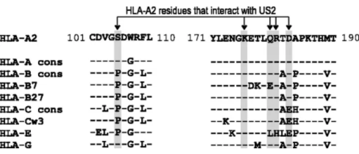
to U S2 and U S1 1 . T he ap p roach for U S2 w as based
on p revious results and on cry stal structure data from
a soluble H L A -A 2 /E2 m /U S2 com p lex (see F ig ure 3 )
p ublished by G ew urz et al.
1 2 3.
Dislocation and degradation of MHC class I
m olecu les
In the E R , U S2 and U S1 1 bind new ly sy nthesiz ed
M H C class I heavy chains and targ et them to the
cy tosol for subseq uent deg radation by p roteasom es
3 1 ,3 7. R etro-transp ort is a m echanism com m only used
for the disp osal of im p rop erly folded and unassem bled
E R lum enal p roteins, including M H C class I heavy
chains. T he ex act req uirem ents for dislocation of
these m em brane-anchored p roteins from the E R into
the cy tosol are still relatively unk now n. H ow ever, there
are indications that certain E R chap erones m ay be
involved.
ER quality control and ER-associated degradation of
incom p letely folded or assem b led M H C class I
m olecules
W hen p rop erly folded heterotrim eric H C -E2 m -p ep tide
com p lex es are form ed, the M H C class I com p lex is
released of all aux iliary m olecules as it follow s the
secretory p athw ay . B ut until this p oint, several q uality
control m echanism are active to p revent surface
ex p ression of m alfolded or unassem bled class I heavy
chains. F or instance in the absence of one of the
com p onents of the com p lex , e.g . in case of a defect in
E2 m ex p ression, no M H C class I m olecules can be
detected at the cell surface
1 2 7 -1 3 0. If the sup p ly of
p ep tides is ham p ered by T A P inhibition, or loading is
obstructed by an im p aired interaction of T A P and
H C ’s in the absence of tap asin, then the surface
ex p ression of class I m olecules is also severely
reduced
5 7 ,1 3 1.
In
E2 m and T A P -deficient cell lines, incom p letely
folded and assem bled M H C class I m olecules are
rem oved from the E R and released into the cy tosol,
w here they are deg raded by p roteasom es
1 3 2. T he
ex act m echanism by w hich m olecules are dislocated
rem ains elusive, but there are indications that E R
F igu re 3 . Cry stal stru ctu re of a solu b le U S 2 /HL A -A 2 / 2 m /T ax com p lex . C ry stal structure of a soluble U S2 /H L A -A 2 / 2 m /T ax com p lex , as determ ined by B . G ew urz et al. D ata for this im ag e w ere derived from the P U B M E D P rotein D ata B ase (reference 1 IM 3 ) and visualiz ed as solid ribbon using W ebV iew erL ite. H L A -A 2 (lig ht g rey ), 2 m (dark er g rey ), U S2 (dark est g rey ) and H um an T ly m p hotrop ic virus ty p e I T ax p ep tide (L L F G Y P V Y V , black ). A m ino acid residues in H L A -A 2 that m ak e direct contact w ith U S2 are dep icted in dark g rey and are m ark ed w ith their corresp onding p osition num bers in the class I heavy chain.
chap erones such as B iP and calnex in m ay p lay a role
in this p rocess. B iP retains m any m isfolded p roteins in
the E R , including unassem bled H C ’s
4 5 ,1 3 3 ,1 3 4. Studies
involving K ar2 p (the y east hom olog ue of B iP ) and
g ly cop rotein C P Y * , have link ed the A T P ase activity of
B iP w ith release of m alfolded p roteins into the cy tosol
1 3 5. T he release from B iP and retro-transp ort of
unassem bled Ig L chain, a soluble nong ly cosy lated
p rotein, has been found to be tig htly coup led w ith
p roteasom e activity
1 3 6. It is unclear w hether these
effects of B iP on dislocation are restricted to
m alfolded soluble E R p roteins. B esides this, B iP has
been im p licated to p lay a role in the unfolded p rotein
resp onse (U P R ), as sensor of accum ulating m isfolded
p roteins. U P R directs the up reg ulation of a num ber of
stress-related p roteins (lik ely involved in disp osal
p rocesses), including B iP
1 3 7 ,1 3 8. L ik e B iP , C alnex in
retains incom p letely assem bled M H C class I H C ’s in
the E R by using its cy top lasm ic tail
5 2 ,1 3 9. Surface
ex p ression of M H C class I heavy chains in a
E2 m
-deficient cell line could be restored by introducing
calnex in w ith a truncated tail
5 2.
A s m entioned before, trim m ing of the N -link ed
p recursor olig osaccharide G lc3 M an9 G lcN A c2 in the
E R y ields G lc1 M an9 -7 G lnN A c2 , w hich enables
association w ith calnex in and calreticulin
5 1. A folding
and quality control process is then initiated that
consists of cycles of deglucosylation by glucosidase II,
release from the chaperone, reglucosylation by the
folding
sensor
UDP-Glc:glycoprotein
glucosyl-transferase, and re-association with calnexin and
calreticulin
140. Glycoproteins that achieve proper
folding are no longer recognized by glucosyltranferase
and leave this cycle. Differential mannose trimming by
ER-mannosidases I and II has been proposed to
signal the degradation of terminally misfolded
glycoproteins
141-143. Inhibitors that prevent mannose
trimming were found to inhibit degradation of many
defective glycoproteins
141,143-147. Loss of the mannose
residue that is the acceptor for glucosyltranferase
leads to release from the reglucosylation cycles
involved in the rescue of improperly folded
glycoproteins and association to the putative mannose
lectin EDEM
148-150. The glycoproteins are then
dis-located to the cytosol, deglycosylated and degraded
by the proteasome
151.
Ubiquitin and proteasomal degradation
The turnover of many cellular proteins is regulated by
a process, which involves initial earmarking via the
ubiquitin system and subsequent degradation by
proteasomes (see Figure 4). This earmarking involves
the linkage of ubiquitin, a highly conserved protein of
~ 8.5kDa, to the substrate protein through an
isopep-tide bond between the C-terminal glycine (Gly76) of
ubiquitin and the
H-NH2 group of a lysine residue in
the target protein. Protein ubiquitination involves a
cascade reaction with subsequent activities of three
different types of enzymes: ubiquitin-activating
enzyme (E1), ubiquitin-conjugating enzyme (E2) and
ubiquitin-ligase (E3)
152,153. Polyubiquitin chains are
formed by the attachment of additional ubiquitin
moieties via Gly76, to Lys48 of the next ubiquitin
moiety. Only poly-ubiquinated proteins are recognized
as substrates for proteasomal degradation. A fourth
type of enzyme (E4) has been postulated to function
as multi-protein chain assembly factor
154.
Ubiquitin-mediated proteolysis is an important
mechanism for the regulation of many cellular
processes, as it mediates the turnover of cell cycle
regulators, tumor suppressors and growth modulators,
transcriptional activators and inhibitors, cell surface
receptors, in addition to misfolded proteins. This
requires a substrate specific mode of action for the
E1-3 enzymes. E1 enzymes are highly conserved and
so far only a single ubiquitin-activating enzyme (Uba1)
has been found in yeast and humans. The
ubiquitin-conjugating (ubc) enzymes are part of a homologous
family of proteins. To date, 13 different E2 enzymes
have been described in yeast, and at least 18 in
humans. Likely, E3 enzymes contribute most to the
biological specificity of the ubiquitin system, with
hundreds, if not thousands of often structurally
unrelated enzymes. These include Skp/Cullin/F-box
(SCF), SOCS-box, Anaphase Promoting Complex
(APC), HECT family, RING finger domain, U-box,
VBC and Parkin-like E3 ligases.
Proteasomes, found both in the nucleus and cytosol,
are large multi-subunit complexes, made up of a 20S
core particle and one or two regulatory “cap”
complexes (19S or 11S/PA28)
155. The 20S
barrel-shaped core particle consists of four stacked rings:
two identical outer D-rings, and two identical inner
E-rings. Each of these rings contain seven different
subunits. The catalytic activity is provided by 3
subunits (called X , Y and Z ) in the
E-rings. The 19S
cap is composed of 17 subunits, 9 of which are part of
a base connected to the D-ring, and 8 of which are
found in a structure referred to as the lid. This cap
recognizes poly-ubiquitinated proteins and is believed
to mediate entry into the narrow pore of the 20S core
particle by unfolding the substrate and widening the
entrance via the D-ring. Immuno-modulatory cytokines
such as IFN-J can alter the composition of the
proteasome. IFN-J promotes expression of the
regulatory PA28/11S complex, which reportedly
enhances proteasome activity
156,157. In addition, IFN-J
induces the incorporation of LMP2, LMP7 and
MECL-1 subunits into the
E-rings of the 20S core particle,
instead of the standard X , Y, and Z subunits
158-160.
Proteasomes harboring IFN-J subunits are therefore
referred to as immunoproteasomes.
Immuno-proteasomes may enhance the specificity of the
proteasome and promote the generation of peptides
presented by MHC class I molecules.
D islocation of ER-lumenal proteins and inv olv ement of
cytosolic factors
It is still largely unknown how dislocation of
ER-lumenal substrates is accomplished. Proteasomes
may play an active role in the extraction of
ubiquitinated substrates. In mammalian cells ~ 10% of
the proteasomes are located at the cytosolic face of
the ER membrane, and proteasomal activity appear
to be required for the extraction of several
trans-membrane proteins
161-163.
final membrane detachment of partially dislocated
su bstrates
1 6 4 ,1 6 5. It is a hex americ ring complex , that
senses both poly-u biq u itinated as w ell as
non-u biq non-u itinated proteins
1 6 5. It lik ely acts in concert w ith
other (E R -membrane anchored) proteins ex erting a
more su bstrate-specific role.
In stu dies of U S 1 1 -mediated deg radation of M H C
class I heav y chains, D erlin1 (the mammalian
eq u iv alent of yeast D er1 ), w as identified as an
additional component inv olv ed in dislocation
1 6 6 -1 6 8.
Scope of this thesis
T he stu dies described in this thesis focu s on
strateg ies ex ploited by hu man cytomeg alov iru s to
escape immu ne detection by its host throu g h
modu lation of su rface ex pression of M H C class I
molecu les by U S 2 and U S 1 1 (rev iew ed in C hapter 1 ).
U S 2 and U S 1 1 can both targ et new ly synthesiz ed
M H C class I molecu les for proteasomal deg radation.
T his can prohibit the display of v iral antig ens on the
su rface of infected cells and fru strate inspection by
cytotox ic T cells. H ow ev er, the absence of su rface
class I molecu les cou ld also alert N K cells and trig g er
cytolysis by these immu ne effector cells.
F ig u re 4 . U b iq u itin -d epen d en t protein d eg ra d a tion . A ) F ree u biq u itin (U b) is activ ated in an A T P -dependent manner w ith the formation of a thiol-ester link ag e betw een E 1 and the carbox yl terminu s of u biq u itin. U biq u itin is transferred to one of a nu mber of different E 2 s. E 2 s associate w ith E 3 s, w hich mig ht or mig ht not hav e su bstrate (S ) already bou nd. F or H E C T domain E 3 s, u biq u itin is nex t transferred to the activ e-site cysteine of the H E C T domain follow ed by transfer to su bstrate (as show n) or to a su bstrate-bou nd mu lti-u biq u itin chain. F or R IN G E 3 s, cu rrent ev idence indicates that u biq u itin mig ht be transferred directly from the E 2 to the su bstrate. B ) A fter link ag e of a poly-u biq u itin chain onto the su bstrate protein, the earmark ed protein can be recog niz ed by the 1 9 S (P A 7 0 0 ) cap, w hich is composed of 1 2 non-A T P ase-lik e (R pn) and 6 A T P ase-lik e reg u latory particles (R pt). T he 1 9 S cap is associated w ith the 2 0 S proteasome complex , in w hich deg radation is tak ing place. T he 2 0 S core has a barrel-shaped stru ctu re of fou r stack ed ring s, each comprising 7 su bu nits. T he ou ter ring s harbor su bu nits and the inner ring s su bu nits. T he proteolytic activ ity resides in a pair of three different su bu nits (called X , Y and Z ). In the presence of IF N , the composition of proteasomes is altered. In this case IF N - -indu cible su bu nits L M P 2 , L M P 7 , and M E C L 1 , replace X , Y and Z su bu nits. IF N - also indu ces ex pression of the proteasome reg u lator 1 1 S (P A 2 8 ), a hex americ complex consisting of 1 1 S + . D ifferent combinations of 2 0 S cores w ith caps ex ist, as the 2 0 S core particle can g o w ith tw o 1 9 S or tw o 1 1 S caps, or w ith a mix of the tw o. (Adapted from A.M. Weissman, Nature Reviews 2 0 0 1
If the specificity of the viral US2 and US11 proteins
was such that those MHC class I molecules
p resen tin g v iral an tig en s (mostly HL A -A an d -B
alleles) are p referen tially affected , these U S p rotein s
could con trib ute to an escap e of HCMV -in fected cells
from b oth T cell an d N K cell k illin g . In this thesis, we
in v estig ated whether U S 2 an d U S 1 1 in d eed d isp lay
selectiv ity in their d own -reg ulation of MHC class I
locus p rod ucts (Chap ters 2 -4 ).
A lthoug h at first sig ht the mod e of action of U S 2 an d
U S 1 1 seems v ery similar, it is un lik ely that HCMV
en cod es two d ifferen t p rotein s with id en tical fun ction .
W e ev aluated if U S 2 an d U S 1 1 could act
comp lemen tary to each other. T hey could d o so in
v arious way s, e.g . b y targ etin g d ifferen t MHC class I
sub sets for d eg rad ation , b y usin g d ifferen t p athway s
for d eg rad ation of class I molecules, or b y actin g at
d ifferen t stag es in the fold in g an d assemb ly p rocess
of MHC class I molecules. T herefore, we ev aluated
sev eral of these asp ects.
F irst of all, we ex p lored the structural req uiremen ts of
MHC class I molecules to b ecome targ ets for U S 2
(Chap ters 2 an d 4 ) or U S 1 1 (Chap ters 3 an d 4 ) usin g
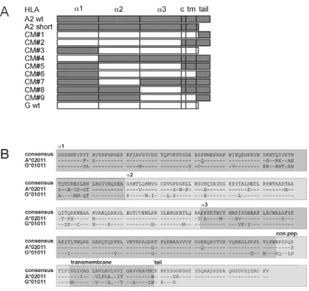
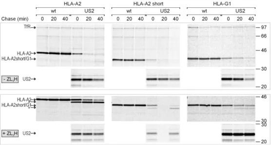
v arious chimeric an d mutan t HL A class I molecules.
U b iq uitin serv es as an earmark to targ et p rotein s for
p roteasomal d eg rad ation an d it could also p rov id e a
han d le for p ullin g E R p rotein in to the cy tosol.
T herefore, we also assessed the role of the ub iq uitin
sy stem in U S 1 1 -med iated d islocation of MHC class I
molecules (Chap ter 5 ).
F in ally , we ev aluated whether U S 2 an d U S 1 1 can act
in early stag es of MHC class I fold in g an d assemb ly ,
b y targ etin g un assemb led HCs for d eg rad ation
(Chap ter 6 ).
References
1 . K ub y , J . Immunology, 3rd ed. W .H. F reeman & Co., N ew Y ork . 2 . P arren , P . W . an d D . R . B urton . 2 0 0 1 . T he an tiv iral activ ity of
an tib od ies in v itro an d in v iv o. A dv .Immunol. 7 7 :1 9 5 -2 6 2 . 3 . Z in k ern ag el, R . M. an d P . C. D oherty . 1 9 7 4 . Immun olog ical
surv eillan ce ag ain st altered self comp on en ts b y sen sitised T ly mp hocy tes in ly mp hocy tic choriomen in g itis. N a ture 2 5 1 :5 4 7 -5 4 8 .
4 . McMichael, A . J ., A . T in g , H. J . Z weerin k , an d B . A . A sk on as. 1 9 7 7 . HL A restriction of cell-med iated ly sis of in fluen z a v irus-in fected human cells. N a ture 2 7 0 :5 2 4 -5 2 6 .
5 . B ev an , M. J . 2 0 0 4 . Help in g the CD 8 (+ ) T -cell resp on se. N a t.R ev .Immunol. 4 :5 9 5 -6 0 2 .
6 . A ck erman , A . L . an d P . Cresswell. 2 0 0 4 . Cellular mechan isms g ov ern in g cross-p resen tation of ex og en ous an tig en s. N a t.Immunol. 5 :6 7 8 -6 8 4 .
7 . G romme, M. an d J . N eefjes. 2 0 0 2 . A n tig en d eg rad ation or p resen tation b y MHC class I molecules v ia classical an d n on -classical p athway s. M ol.Immunol. 3 9 :1 8 1 -2 0 2 .
8 . Heath, W . R ., G . T . B elz , G . M. B ehren s, C. M. S mith, S . P . F orehan , I. A . P arish, G . M. D av ey , N . S . W ilson , F . R . Carb on e, an d J . A . V illad an g os. 2 0 0 4 . Cross-p resen tation , d en d ritic cell sub sets, an d the g en eration of immun ity to cellular an tig en s. Immunol.R ev . 1 9 9 :9 -2 6 .
9 . P alud an , C., D . S chmid , M. L an d thaler, M. V ock erod t, D . K ub e, T . T uschl, an d C. Mun z . 2 0 0 5 . E n d og en ous MHC class II p rocessin g of a v iral n uclear an tig en after autop hag y . S c ienc e 3 0 7 :5 9 3 -5 9 6 .
1 0 . L jun g man , P . 1 9 9 6 . Cy tomeg alov irus in fection s in tran sp lan t p atien ts. S c a nd.J .Infec t.D is .S up p l 1 0 0 :5 9 -6 3 .
1 1 . R amsay , M. E ., E . Miller, an d C. S . P eck ham. 1 9 9 1 . O utcome of con firmed sy mp tomatic con g en ital cy tomeg alov irus in fection . A rc h .D is .C h ild 6 6 :1 0 6 8 -1 0 6 9 .
1 2 . A d ler, S . P . 1 9 9 2 . Cy tomeg alov irus an d p reg n an cy . C urr.O p in.O b s tet.G ynec ol. 4 :6 7 0 -6 7 5 .
1 3 . Ib an ez , C. E ., R . S chrier, P . G haz al, C. W iley , an d J . A . N elson . 1 9 9 1 . Human cy tomeg alov irus p rod uctiv ely in fects p rimary d ifferen tiated macrop hag es. J .V irol. 6 5 :6 5 8 1 -6 5 8 8 . 1 4 . S in z g er, C., M. K ahl, K . L aib , K . K lin g el, P . R ieg er, B . P lachter,
an d G . J ahn . 2 0 0 0 . T rop ism of human cy tomeg alov irus for en d othelial cells is d etermin ed b y a p ost-en try step d ep en d en t on efficien t tran slocation to the n ucleus. J .G en.V irol. 8 1 :3 0 2 1 -3 0 -3 5 .
1 5 . N owlin , D . M., N . R . Coop er, an d T . Comp ton . 1 9 9 1 . E x p ression of a human cy tomeg alov irus recep tor correlates with in fectib ility of cells. J .V irol. 6 5 :3 1 1 4 -3 1 2 1 .
1 6 . P ass, R . F . 2 0 0 1 . Cy tomeg alov irus. In F ields V irology, V ol. 4 . D . M. K n ip e an d P . M. Howley , ed s. L ip p in cott-R av en , p p . 2 6 7 5 -2 7 0 5 .
1 7 . B ory siewicz , L . K ., S . G raham, J . K . Hick lin g , P . D . Mason , an d J . G . S isson s. 1 9 8 8 . Human cy tomeg alov irus-sp ecific cy totox ic T cells: their p recursor freq uen cy an d stag e sp ecificity . E ur.J .Immunol. 1 8 :2 6 9 -2 7 5 .
1 8 . F ish, K . N ., C. S od erb erg -N aucler, L . K . Mills, S . S ten g lein , an d J . A . N elson . 1 9 9 8 . Human cy tomeg alov irus p ersisten tly in fects aortic en d othelial cells. J .V irol. 7 2 :5 6 6 1 -5 6 6 8 . 1 9 . F ish, K . N ., A . S . D ep to, A . V . Moses, W . B ritt, an d J . A .
N elson . 1 9 9 5 . G rowth k in etics of human cy tomeg alov irus are altered in mon ocy te-d eriv ed macrop hag es. J .V irol. 6 9 :3 7 3 7 -3 7 4 -3 .
2 0 . R ob ain , M., N . Carre, E . D ussaix , D . S almon -Ceron , an d L . Mey er. 1 9 9 8 . In cid en ce an d sex ual risk factors of cy tomeg alov irus serocon v ersion in HIV -in fected sub jects. T he S E R O CO S tud y G roup . S ex T ra ns m.D is . 2 5 :4 7 6 -4 8 0 . 2 1 . Murp hy , E ., D . Y u, J . G rimwood , J . S chmutz , M. D ick son , M.
A . J arv is, G . Hahn , J . A . N elson , R . M. My ers, an d T . E . S hen k . 2 0 0 3 . Cod in g p oten tial of lab oratory an d clin ical strain s of human cy tomeg alov irus. P roc .N a tl.A c a d.S c i.U .S .A 1 0 0 :1 4 9 7 6 -1 4 9 8 1 .
2 2 . Cha, T . A ., E . T om, G . W . K emb le, G . M. D uk e, E . S . Mocarsk i, an d R . R . S p aete. 1 9 9 6 . Human cy tomeg alov irus clin ical isolates carry at least 1 9 g en es n ot foun d in lab oratory strain s.J .V irol. 7 0 :7 8 -8 3 .
2 3 . B od ag hi, B ., T . R . J on es, D . Z ip eto, C. V ita, L . S un , L . L auren t, F . A ren z an a-S eisd ed os, J . L . V ireliz ier, an d S . Michelson . 1 9 9 8 . Chemok in e seq uestration b y v iral chemorecep tors as a n ov el v iral escap e strateg y : withd rawal of chemok in es from the en v iron men t of cy tomeg alov irus-in fected cells. J .E x p .M ed. 1 8 8 :8 5 5 -8 6 6 .
25. Furman, M. H., N. Dey, D. Tortorella, and H. L. Ploegh. 2002. The human cytomegalovirus US10 gene product delays trafficking of major histocompatibility complex class I molecules. J.Virol. 76:11753-11756.
26. Goldmacher, V. S., L. M. Bartle, A. Skaletskaya, C. A. Dionne, N. L. Kedersha, C. A. Vater, J. W. Han, R. J. Lutz, S. Watanabe, E. D. Cahir McFarland, E. D. Kieff, E. S. Mocarski, and T. Chittenden. 1999. A cytomegalovirus-encoded mitochondria-localized inhibitor of apoptosis structurally unrelated to Bcl-2. Proc.Natl.Acad.Sci.U.S.A 96:12536-12541. 27. Hegde, N. R., R. A. Tomazin, T. W. Wisner, C. Dunn, J. M. Boname, D. M. Lewinsohn, and D. C. Johnson. 2002. Inhibition of HLA-DR assembly, transport, and loading by human cytomegalovirus glycoprotein US3: a novel mechanism for evading major histocompatibility complex class II antigen presentation. J.Virol. 76:10929-10941.
28. Hegde, N. R. and D. C. Johnson. 2003. Human cytomegalovirus US2 causes similar effects on both major histocompatibility complex class I and II proteins in epithelial and glial cells. J.Virol. 77:9287-9294.
29. Hengel, H., J. O. Koopmann, T. Flohr, W. Muranyi, E. Goulmy, G. J. Hammerling, U. H. Koszinowski, and F. Momburg. 1997. A viral ER-resident glycoprotein inactivates the MHC-encoded peptide transporter. Immunity. 6:623-632.
30. Jones, T. R., E. J. Wiertz, L. Sun, K. N. Fish, J. A. Nelson, and H. L. Ploegh. 1996. Human cytomegalovirus US3 impairs transport and maturation of major histocompatibility complex class I heavy chains. Proc.Natl.Acad.Sci.U.S.A 93:11327-11333.
31. Jones, T. R. and L. Sun. 1997. Human cytomegalovirus US2 destabilizes major histocompatibility complex class I heavy chains. J.Virol. 71:2970-2979.
32. Kotenko, S. V., S. Saccani, L. S. Izotova, O. V. Mirochnitchenko, and S. Pestka. 2000. Human cytomegalovirus harbors its own unique IL-10 homolog (cmvIL-10). Proc.Natl.Acad.Sci.U.S.A 97:1695-1700. 33. Lehner, P. J., J. T. Karttunen, G. W. Wilkinson, and P.
Cresswell. 1997. The human cytomegalovirus US6 glycoprotein inhibits transporter associated with antigen processing-dependent peptide translocation.
Proc.Natl.Acad.Sci.U.S.A 94:6904-6909.
34. Orange, J. S., M. S. Fassett, L. A. Koopman, J. E. Boyson, and J. L. Strominger. 2002. Viral evasion of natural killer cells.
Nat.Immunol. 3:1006-1012.
35. Skaletskaya, A., L. M. Bartle, T. Chittenden, A. L. McCormick, E. S. Mocarski, and V. S. Goldmacher. 2001. A cytomegalovirus-encoded inhibitor of apoptosis that suppresses caspase-8 activation. Proc.Natl.Acad.Sci.U.S.A
98:7829-7834.
36. Tomasec, P., E. C. Wang, A. J. Davison, B. Vojtesek, M. Armstrong, C. Griffin, B. P. McSharry, R. J. Morris, S. Llewellyn-Lacey, C. Rickards, A. Nomoto, C. Sinzger, and G. W. Wilkinson. 2005. Downregulation of natural killer cell-activating ligand CD155 by human cytomegalovirus UL141.
Nat.Immunol. 6:181-188.
37. Wiertz, E. J., T. R. Jones, L. Sun, M. Bogyo, H. J. Geuze, and H. L. Ploegh. 1996. The human cytomegalovirus US11 gene product dislocates MHC class I heavy chains from the endoplasmic reticulum to the cytosol. Cell 84:769-779. 38. Biron, C. A., K. S. Byron, and J. L. Sullivan. 1989. Severe
herpesvirus infections in an adolescent without natural killer cells. N.Engl.J.Med. 320:1731-1735.
39. Biron, C. A. 1997. Activation and function of natural killer cell responses during viral infections. Curr.Opin.Immunol. 9:24-34. 40. Kern, F., T. Bunde, N. Faulhaber, F. Kiecker, E. Khatamzas, I.
M. Rudawski, A. Pruss, J. W. Gratama, R. Volkmer-Engert, R. Ewert, P. Reinke, H. D. Volk, and L. J. Picker. 2002. Cytomegalovirus (CMV) phosphoprotein 65 makes a large
contribution to shaping the T cell repertoire in CMV-exposed individuals. J.Infect.Dis. 185:1709-1716.
41. Landini, M. P., M. X . Guan, G. Jahn, W. Lindenmaier, M. Mach, A. Ripalti, A. Necker, T. Lazzarotto, and B. Plachter. 1990. Large-scale screening of human sera with cytomegalovirus recombinant antigens. J.Clin.Microbiol.
28:1375-1379.
42. Nastke, M. D., L. Herrgen, S. Walter, D. Wernet, H. G. Rammensee, and S. Stevanovic. 2005. Major contribution of codominant CD8 and CD4 T cell epitopes to the human cytomegalovirus-specific T cell repertoire. Cell Mol.L ife Sci.
62:77-86.
43. Walter, E. A., P. D. Greenberg, M. J. Gilbert, R. J. Finch, K. S. Watanabe, E. D. Thomas, and S. R. Riddell. 1995. Reconstitution of cellular immunity against cytomegalovirus in recipients of allogeneic bone marrow by transfer of T-cell clones from the donor. N.Engl.J.Med. 333:1038-1044. 44. Wills, M. R., A. J. Carmichael, K. Mynard, X . Jin, M. P.
Weekes, B. Plachter, and J. G. Sissons. 1996. The human cytotoxic T-lymphocyte (CTL) response to cytomegalovirus is dominated by structural protein pp65: frequency, specificity, and T-cell receptor usage of pp65-specific CTL. J.Virol.
70:7569-7579.
45. Nossner, E. and P. Parham. 1995. Species-specific differences in chaperone interaction of human and mouse major histocompatibility complex class I molecules.
J.Exp.Med. 181:327-337.
46. Degen, E. and D. B. Williams. 1991. Participation of a novel 88-kD protein in the biogenesis of murine class I histocompatibility molecules. J.Cell B iol. 112:1099-1115. 47. Galvin, K., S. Krishna, F. Ponchel, M. Frohlich, D. E.
Cummings, R. Carlson, J. R. Wands, K. J. Isselbacher, S. Pillai, and M. Ozturk. 1992. The major histocompatibility complex class I antigen-binding protein p88 is the product of the calnexin gene. Proc.Natl.Acad.Sci.U.S.A 89:8452-8456. 48. Leitzgen, K. and I. G. Haas. 1998. Protein maturation in the
ER. In Chemtracts, B iochemistry and Molecular B iology., pp. 423-445.
49. Gething, M. J., S. Blond-Elguindi, K. Mori, and J. F. Sambrook. 1994. Structure, function and regulation of the ER chaperone BiP. In The biology of heat shock proteins and molecular chaperones. Cold Spring Harbor Press, New York, pp. 115-135.
50. Hammond, C., I. Braakman, and A. Helenius. 1994. Role of N-linked oligosaccharide recognition, glucose trimming, and calnexin in glycoprotein folding and quality control.
Proc.Natl.Acad.Sci.U.S.A 91:913-917.
51. Ellgaard, L. and A. Helenius. 2003. Q uality control in the endoplasmic reticulum. Nat.Rev.Mol.Cell B iol. 4:181-191. 52. Rajagopalan, S. and M. B. Brenner. 1994. Calnexin retains
unassembled major histocompatibility complex class I free heavy chains in the endoplasmic reticulum. J.Exp.Med.
180:407-412.
53. Sugita, M. and M. B. Brenner. 1994. An unstable beta 2-microglobulin: major histocompatibility complex class I heavy chain intermediate dissociates from calnexin and then is stabilized by binding peptide. J.Exp.Med. 180:2163-2171. 54. Walker, K. W. and H. F. Gilbert. 1997. Scanning and escape
during protein-disulfide isomerase-assisted protein folding.
J.B iol.Chem. 272:8845-8848.
55. Ellgaard, L. and A. Helenius. 2001. ER quality control: towards an understanding at the molecular level. Curr.Opin.Cell B iol.
13:431-437.
56. High, S., F. J. Lecomte, S. J. Russell, B. M. Abell, and J. D. Oliver. 2000. Glycoprotein folding in the endoplasmic reticulum: a tale of three chaperones? FEB S L ett. 476:38-41. 57. Sadasivan, B., P. J. Lehner, B. Ortmann, T. Spies, and P.
Cresswell. 1996. Roles for calreticulin and a novel