Original Article
Development of a Double Glass Mounting Method Using
Formaldehyde Alcohol Azocarmine Lactophenol (FAAL)
and its Evaluation for Permanent Mounting of Small
Nematodes
Farzaneh ZAHABIUN 1, *Seyed Mahmoud SADJJADI 1,2, Farideh ESFANDIARI 1
1. Department of Parasitology and Mycology, School of Medicine, Shiraz University of Medical Sciences, Shiraz, Iran 2. Basic Sciences in Infectious Diseases Research Center, Shiraz University of Medical Sciences, Shiraz, Iran
Received 13 Feb 2015
Accepted 11 Oct 2015 Abstract Background:: Permanent slide preparation of nematodes especially small ones is time consuming, difficult and they become scarious margins. Regarding this problem, a modified double glass mounting method was developed and com-pared with classic method.
Methods: A total of 209 nematode samples from human and animal origin were fixed and stained with Formaldehyde Alcohol Azocarmine Lactophenol (FAAL) followed by double glass mounting and classic dehydration method us-ing Canada balsam as their mountus-ing media. The slides were evaluated in differ-ent dates and times, more than four years. Differdiffer-ent photos were made with dif-ferent magnification during the evaluation time.
Results: The double glass mounting method was stable during this time and comparable with classic method. There were no changes in morphologic struc-tures of nematodes using double glass mounting method with well-defined and clear differentiation between different organs of nematodes in this method. Conclusion: Using this method is cost effective and fast for mounting of small nematodes comparing to classic method.
Keywords: Nematodes,
Double Glass Mount-ing,
Permanent Mounting, Formaldehyde Alcohol Azocarmine Lactophe-nol
*Correspondence Email:
Introduction
ematodes are one of the most im-portant human parasitic diseases agents. They are divided into three
types; parasitic, zoonotic and free living agents. Usually parasitic and zoonoses cause disease in human (1-3). However, in some
cir-N
Iranian Society of Parasitology http:// isp.tums.ac.ir
Iran J Parasitol
Open access Journal at http:// ijpa.tums.ac.ir Tehran University of Medical
cumstances free-living nematodes may also cause disease in human (4). Accordingly, study on all aspects of nematodes specially those related to their morphology is necessary for better understanding of these organisms. Basic knowledge for study of nematodes is based on their morphology and their related life cycle. In this regard, it is very important to have good specimens with fine details. Stu-dents learn about morphology of nematodes or their pathological effects in practical ses-sions along with theoretical courses. This causes better understanding of related dis-eases and subsequently better achievements in the control and treatment of nematode para-sites. It is obvious that without good quality specimens in teaching laboratories, students cannot be trained well.
Re-emerging of the eradicated helminthes makes us serious to collect and mount speci-mens as much as possible in high quality condition to be taught to the new students, who are not familiar with the eradicated parasites. Therefore, lack of these samples for teaching to medical and allied sciences will result misunderstanding and misdiagno-sis in their future career. For many years, the scientists have tried to prepare permanent slides (5-9). Recently, a few staining and mounting protocols including the applica-tion of red beet extract on the staining and differentiation of the helminth organs have been used for better differentiation between initial organs of worms (10). The application of different methods for staining different type of helminthes is necessary in order to laid in classification and identical key (10). Other chemicals such as silver staining have been used for elucidation of the synloph in Trichostrongyle nematodes (11). The classic way of mounting using Canada balsam with dehydration of the worms needs a critical work and special attention during worm processing, otherwise, it will destroy the worm (12).
It is very important to have good knowledge and information about fixatives
and chemicals that are used for preserving the samples; as the samples may be pre-served for several purposes. For example if we decide to determine both the morpho-logical and molecular aspects of nematode, high concentration of alcohol is not suitable and causes changes in organs measurements which itself could be used for numerical taxonomy (13).
Among parasite helminthes, nematodes (coelomates) especially the small ones are difficult to be dehydrated during staining and mounting. Sometimes the prepared slides will be black and destroyed; so, not suitable for teaching purposes.
Present study was carried out with the aim of preparing high quality permanent slides from small nematodes using Formaldehyde Alcohol Azocarmine Lactophenol (FAAL) as their fixing, staining and preserving media using a double glass method for better teaching parasitology specially taxonomy of nematodes which itself is very important to human related diseases. Classic mounting using dehydration of the specimens was used to evaluate and compare them in dif-ferent dates for a long time to find which one is superior for mounting of small nema-todes.
Material and Methods
(Merck, Germany). The specimens were transferred into FAAL, which clear, and stains the specimens (14-16).
Samples after recognition (17) were di-vided into two groups to be mounted in dif-ferent ways: the first group was mounted by Canada balsam (Sigma-Aldrich, USA) using double glass mounting while the specimens were transferred into a Glycerin Jelly (GJ) medium. In this regard, the stained samples with FAAL, were transferred carefully into a suitable amount of GJ on the middle of a slide and covered with a small coverslip. Af-ter 3-5 minutes when GJ was gelated, the first coverslip was covered with a bigger co-verslip while suitable amount of Canada bal-sam had been poured onto the first co-verslip. The second group, were stained with Azocarmine followed by dehydration and using Canada balsam in a classic way (18). Briefly, all samples were passed through gradient dilution of alcohols, 70%, 80%, 90%, 96% and 100%, respectively. This step was followed by clearing with xylene- alco-hol and pure xylene. Every step was repeat-ed 2 times each time 5-10 minutes followrepeat-ed by transferring the samples into Canada bal-sam and covered with a coverslip.
The mounted specimens were carefully observed for their changes in cuticle and other organs for several months and were photographed using an Olympus micro-scope (CH-2, Japan) equipped with camera. The specimens were also photographed us-ing Phase Contrast condenser.
Results
A total of 172 small nematodes including males, females and larvae were mounted us-ing double glass mountus-ing method in which FAAL was used as staining and clearing me-dia. The smallest one was 250 µm and the largest one was about 30 mm. A total of 37 samples were mounted with classic method and compared with the specimens of double
glass mounting method.
The color of the most specimens using double glass mounting method were nearly light brown to pink (Figs.1-4) and their qual-ity were high with very good differentiation between their organs especially reproductive organs (Figs. 5-7). No changes were ob-served during the time of observations.
Many slides, which prepared in classic method, have very good quality (Figs. 8-9). A number of these slides became black im-mediately after mounting (Fig. 10).
Fig. 1: The copulatory bursa of male Necator
americanus, double glass mounting
Fig. 2: The male of Necator americanus double glass
Fig. 3: The ovojector of Marshallagia marshali, double glass mounting after 4 years
So it was necessary to transfer back them to xylene or even serials of alcohols to re-peat dehydration again. In spite of doing this long time procedure, some of those samples remain black (Fig. 10).
Discussion
The impact of the methods for processing including killing, fixing, staining and mount-ing on some species of nematodes has been studied (18, 19).
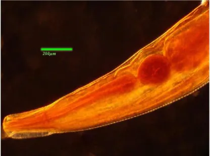
Fig. 4: The whole mount (WM) of male Strongyloides stercoralis in double glass mounting
Fig. 5: The head of Enterobius vermicularis mounted in
double glass method
Fig. 8: A slide including two Rhabditis axei male
Fig. 6: The vulva of a female Trichuris trichiura mounted in double glass method
Fig. 9: A single glass mounted male of Trichuris
trichiura after three years
Fig. 7: A female Rhabditis axei mounted in double glass mounting
Fig. 10: A slide including two Rhabditis axei male worm mounted with classic method, Canada balsam after 3
months
Since the process of preparing a slide, ex-poses a worm to many chemicals, pressures and temperature changes that has effect on their taxonomic characters, choosing a perfect method is very important. Grewal et al. com-pared the effect of several methods for killing, fixing and mounting of the Coenorhabditis ele-gans on its taxonomic characters. They found TAF fixative (7 ml 40% formaldehyde, 2 ml triethanolamine, 91 ml distilled water) at 95 °C and also slow transfer of the nematode from fixative to glycine as mountant has minimum effect on shrinkage of nematode (19). In addi-tion, they tried to prepare a special agar
mounting method. Taxonomic identification is very time consuming and might require the submission of specimens to an expert. In the survey, Sepulveda and Kinsella fixed the hel-minths which were obtained from the wild animals (either in 70% ethanol, 10% buffered formalin, or alcohol-formalin-acetic acid) and for species identification, they cleared nema-todes and small acanthocephalans in lacto-phenol and stained trematodes, cestodes, and large acanthocephalans, using Harris' hema-toxylin or Semichon's carmine. Then they rec-ognized the species by examining different structures (e.g. male spicules in nematodes or the rostellum in cestodes) (21). In all of the works differentiation of organs are very im-portant. Comparing to other works, our re-sults showed that the double glass mounting revealed well-defined and clear differentiation between different organs of nematodes in this method.
Ryss reported a technique to prepare per-manent slides of nematodes in which the liv-ing nematode was put in hot formaldehyde, and then it was transferred into glycerin to be mounted, during a definite process. A drop of glycerin containing nematode was occupied in the hole of slide. Two pieces of waxed paraffin were put on both sides of the hole. A cover glass was put on the top of hole and paraffin. Then it was fixed on the sample by means of heater. All of internal and external structures were fine (22). In our work, this kind of slide was also used. Lack of using the paraffin and heat causes a big bubble. Kumagai et al. had the experience like Ryss for helminth eggs. There were some dis-advantages with their slides. One of them was related to use of Glycerin-Jelly, which has low melting point, without Can-ada balsam, so they must be kept away from heat. In addition, an objective lens of 100× magnification cannot be used for them be-cause eggs are in the hole of the slide and are away from cover glass (23). It seems that the problem of Ryss and Kumagai slides are the same. Anyway, in our experience,
Glycerin-Jelly was used and protected by Canada bal-sam, in a double glass mounting method.
In spite of the traditional method that is difficult to mount the small worms, the dou-ble glass is easier. Moreover, the color of the slides in double glass yields a better differen-tiation between different organs in the worm especially when they are observed with Phase Contrast condenser (Figs. 3, 4 and 8-11). Re-production and digestive systems are set in the pseudo-coelom of nematodes. Every part of these two systems has especial histologic tissues. This kind of preparing and mounting slides is very useful for determining and comparing of these tissues in a nematode and/or between several nematodes.
Anyway, this experience showed that the classic method for preparation slides from small nematodes like, Strongyloides stercoralis or Rhabditis axei, face to a plenty of problems so that most of them are going to have shrink-age or black, while the mentioned problems for bigger nematodes are moderate.
The most important item for double glass method is recovering the nematodes in dif-ferent angles. They can be used for viewing their specific structure using specific con-densers such as Nomarski and Phase Con-trast (24, 25). Our results showed a very in-teresting photos using Phase Contrast con-denser for viewing double glass mounted nematodes. Therefore, double glass mount-ing could be one of the best methods to pre-pare permanent slides for small nematodes. Using this method is cost effective and fast for mounting of small nematodes comparing to classic method.
Conclusion
In comparison between two methods of single and double glass mounting the follow-ing are the advantages of double glass mounting:
2.Saving samples for small nematodes and larvae, since these fine nematodes are going to be missed during the process of dehydration.
3.Saving the money, because there is not any need to use Alcohols and xylene. 4.Three-dimensional investigation of samples, despite of being permanent slides, changing the coverslips is very simple and also hydrated samples are flexible, so that if it is necessary we can change the samples figure.
5. Solving the blackness problem dur-ing dehydration due to their pseudo-coe-lom.
Acknowledgements
This study was granted by grant no. 89-01-43-2120 by office of Vice Chancellor for Re-search at Shiraz University of Medical Sci-ences, Shiraz, Iran. The authors thank Mrs S. Kazemian for her help in preparation of ma-terials. The authors declare that there is no conflict of interests.
References
1. Ashford RW and Crewe W. The parasites of Homo sapiens – an annotated checklist of the protozoa, helminths and arthropods for which we are home. 2nd ed. London: Taylor and Francis; 2003.
2. Zibaei M, Sadjjadi SM, Jahad-Hosseini SH. Toxocara cati larvae in the eye of a child: a case report. Asian Pac J Trop Biomed. 2014; 4(1): 53-5.
3. Sadjjadi SM, Ardehali SM, Shojaei A. A case report of Linguatula serrata in human pharynx from Shiraz, Southern Iran. Med J Islam Re-pub Iran.1988; 12 (2):193-4.
4. Meamar AR, Kia EB, Zahabiun F, Jafari-Mehr A, Moghadam A, Sadjjadi SM. The occurrence of severe infections with Rhabditis axei in AIDS patients in Iran. J Helminthol. 2007; 81(4): 351-2.
5. Seinhorst JW. A rapid method for the transfer of nematodes from fixative to anhydrous
glyc-erin. Nematologica. 1959; 4(1): 67-9.
6. Baker AD. Rapid method for mounting nema-todes in glycerin. Can Ent.1953; 85(2): 77-8. 7. Hooper DJ. Handling, fixing, staining and
mounting nematodes. In: Southey, JF. (Ed.) Laboratory methods for work with plant and soil nematodes. London: MAFF; 1986. P. 59-80.
8. Sadjjadi SM. A polyvinyl alcohol (PVA)-containing medium for permanent mounting of helminth eggs. Trans Roy Soc Trop Med Hyg. 2002; 96(1):104.
9. Sabu L, Devada K, Sreekrishnan R. Single step processing of nematodes in FAAL solution. Vet Parasitol. 2012; 26(1): 91 – 2.
10. Al-Amura MFA, Hassen ZA, AL-Mhanawi BH. Staining technique for helminth parasites by use red beet (beta vulgaris l.) Extract. Bas J Vet Res. 2012;11(1):283-92.
11. Khrustalev AV, Hoberg EP. Silver staining for elucidation of the synloph in trichostrongyle nematodes. J Parasitol.1995; 81(6): 1016-18. 12. Khrustalev AV, Hoberg EP.
Carmine-propi-onic acid stain for elucidation of fine cellular structure in nematodes. J Parasitol. 1996; 82(1): 176--8.
13. Naem S, Pagan C, Nadler SA.Structural resto-ration of nematodes and acanthocephalans fixed in high percentage alcohol using DESS solution and rehydration. J Parasitol. 2010; 96(4): 809–11.
14. Sadjjadi SM, Massoud J. Helminth parasites of wild rodents in Khuzestan province, south west of Iran. J Vet Parasitol. 1999; 13:55-6. 15. Zibaei M, Sadjjadi SM, Sarkari B. Prevalence of
Toxocara cati and other intestinal helminths in stray cats in Shiraz, Iran. Trop Biomed. 2007; 24(2):39-43.
16. Tanideh N , Sadjjadi SM , Mohammadzadeh T. Helminthic infections of laboratory animals in animal house of Shiraz University of Medical Sciences and the potential risks of zoonotic in-fections for researchers. Iran Red Crescent Med J. 2010; 12(2):151-7.
17. Yamaguti S. Systema Helminthum. New York: Interscience Publishers; 1958-1963. Vol.I-IV. 18. May HG. Killing, staining and mounting
para-sitic nematodes. Trans Am Microsc Soc. 1922; 41(2): 103-5.
taxonomic characters of parthenogenetic adult female Caenorhabditis elegans (Nematoda: Rhab-ditidae). Revue Nématol. 1990; 13 (4): 437-44. 20. Grewal PS. The use of agar as a cover-glass
support for mounting nematodes. Revue Né-matol. 1990; 13 (1): 121-2.
21. Sepulveda MS, Kinsella JM. Helminth collec-tion and identificacollec-tion from wildlife. J Vis Exp. 2013;(82):e51000.
22. Ryss AY. Express technique to prepare perma-nent collection slides of nematodes. Zoosyst Rossica. 2003; 11: 257-60.
23. Kumagai M, Inaba T, Makioka A, Ishiwata K,
Onishi K, Watanabe N. An improved glycerin- jelly mounting procedure for permanent prepa-rations of helminth eggs. J Parasitol. 2010 ; 96(2):440-1.
24. Bird AF. Changes associated with parasitism in nematodes. I. Morphology and physiology of preparasitic and parasitic larvae of Meloidogyne ja-vanica. J Parasitol. 1967; 53(4): 768-76.
25. Bird AF. Further observations on the structure of nematode cuticle. Parasitol. 1958; 48(1-2): 32-7.