R E S E A R C H
Open Access
Microbiome-driven identification of
microbial indicators for postharvest
diseases of sugar beets
Peter Kusstatscher
1,2, Christin Zachow
1, Karsten Harms
3, Johann Maier
3, Herbert Eigner
4, Gabriele Berg
2and
Tomislav Cernava
2,5*Abstract
Background:Sugar loss due to storage rot has a substantial economic impact on the sugar industry. The gradual spread of saprophytic fungi such asFusariumandPenicilliumspp. during storage in beet clamps is an ongoing challenge for postharvest processing. Early detection of shifts in microbial communities in beet clamps is a promising approach for the initiation of targeted countermeasures during developing storage rot. In a combined approach, high-throughput sequencing of bacterial and fungal genetic markers was complemented with
cultivation-dependent methods and provided detailed insights into microbial communities colonizing stored roots. These data were used to develop a multi-target qPCR technique for early detection of postharvest diseases. Results:The comparison of beet microbiomes from six clamps in Austria and Germany highlighted regional differences; nevertheless, universal indicators of the health status were identified. Apart from a significant decrease in microbial diversity in decaying sugar beets (p≤0.01), a distinctive shift in the taxonomic composition of the overall microbiome was found. Fungal taxa such asCandidaandPenicilliumtogether with the gram-positive Lactobacilluswere the main disease indicators in the microbiome of decaying sugar beets. In contrast, the genera PlectosphaerellaandVishniacozymaas well as a higher microbial diversity in general were found to reflect the microbiome of healthy beets. Based on these findings, a qPCR-based early detection technique was developed and confirmed a twofold decrease of health indicators and an up to 10,000-fold increase of disease indicators in beet clamps. This was further verified with analyses of the sugar content in storage samples.
Conclusion:By conducting a detailed assessment of temporal microbiome changes during the storage of sugar beets, distinct indicator species were identified that reflect progressing rot and losses in sugar content. The insights generated in this study provide a novel basis to improve current or develop next-generation postharvest
management techniques by tracking disease indicators during storage.
Keywords:Beta vulgaris, Storage rot, Indicator species, Phytopathogens, Bacterial microbiome, Fungal microbiome
Background
Plant-colonizing microorganisms live in close relation-ship with their host and are a crucial factor for plant growth and health [1–3]. For various crop plants, this was observed along the entire value-chain including the postharvest period [4]. The exploration of plant-microbe interactions, plant-beneficial bacteria and fungi including
yeasts, their functions, and modes of action is a key for advanced developments related to biotechnological applications in agriculture [2,5]. However, the develop-ment of postharvest applications based on biologicals is challenging due to the great diversity of postharvest pathogens as well as the often highly challenging post-harvest treatments and storage conditions [6, 7]. The herbaceous dicotyledonous plant,Beta vulgarisL. (sugar beet) is the main crop for sugar production (sucrose content up to 18%) in temperate regions all over the world [8]. A number of plant pathogens such asPythium
© The Author(s). 2019Open AccessThis article is distributed under the terms of the Creative Commons Attribution 4.0 International License (http://creativecommons.org/licenses/by/4.0/), which permits unrestricted use, distribution, and reproduction in any medium, provided you give appropriate credit to the original author(s) and the source, provide a link to the Creative Commons license, and indicate if changes were made. The Creative Commons Public Domain Dedication waiver (http://creativecommons.org/publicdomain/zero/1.0/) applies to the data made available in this article, unless otherwise stated. * Correspondence:[email protected]
2Institute of Environmental Biotechnology, Graz University of Technology, Petersgasse 12, 8010 Graz, Austria
ultimum Trow [9], Rhizoctonia solani Kühn [10], and Cercospora beticola Sacc. [11] cause severe harvest shortfalls due to seedling rot or late root rot [12]. After harvest, starting from late October, sugar beets are stored in Europe directly on the fields for a maximum of 60 days due to limited process capacities and increased economic viability of sugar refineries. High water (76%) and sugar content (18%) in the unprocessed beets [13] provide perfect conditions for microbial colonization, es-pecially when cracks, root tip breakage, and fresh wounds on the surface provide easy entry points [14]. Microbial colonization, mainly by pathogenic or sapro-phytic fungi such as Fusarium,Penicillium, and Botrytis spp., leads to substantial sugar yield losses. A major ob-servation is microbial inversion of sucrose into un-wanted glucose and fructose molecules [15]. The combined occurrence of microbial degradation, respir-ation of the beet root, synthesis of raffinose, and other causes can yield sugar losses of up to 50–60% during storage [16,17].
Natural antagonists that are part of the indigenous beet microbiome, previously studied by Zachow and colleagues (2008) [18], carry the potential for alternative plant protection applications during growth and posthar-vest [19, 20]. In our previous study, we found correla-tions between the disease incidence in sugar beet fields and the antagonistic potential of the prevalent micro-biota [21]. These observations provide the basis for sustainable methods to prevent high sugar yield losses, caused by fungal infection with a targeted use of antag-onistic microorganisms that could also provide posthar-vest protection [22]. However, in order to develop targeted and sustainable countermeasures, it is crucial to identify key players in the rot onset and to improve early
detection strategies of rot-causing pathogens for beet clamps. Moreover, when biological control is employed, it is important to understand to which natural
counter-parts beneficial microorganisms will be exposed.
Although rot-causing fungal pathogens were previously identified [14], the health-related dynamics of bacteria and fungi in stored sugar beets remained unexplored.
The aim of this study was to analyze temporal community changes in the microbiome of stored roots, correlate them to sugar beet health, and finally integrate the generated knowledge into a novel disease detection technique. Therefore, we investigated the bacterial and fungal microbiome of stored sugar beets in different beet clamps located in important cultivation areas of Austria and Germany. By implementing a detailed assessment of the beet clamp microbiome, specific biological markers indicating disease development in stored beets were found. These observations were thereafter confirmed with sugar beets stored under controlled conditions to verify the applicability of the identified markers. The overall findings provide a basis for novel postharvest management techniques that implement microbial and molecular markers for targeted countermeasures.
Results
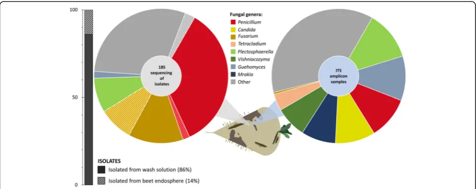
Identification of fungal taxa from decaying sugar beets In order to identify fungal taxa in infected sugar beets from clamps in Austria and Germany, two complemen-tary methods were applied. The community structure was reconstructed with Sanger sequencing of 18S rRNA gene fragments from fungal isolates and ITS Illumina amplicon sequencing of total community DNA (Fig. 1). The 18S rRNA gene sequencing-based community reconstruction with 120 fungal strains indicated a fungal
community structure with 11 different genera, which
was dominated by Penicillium (37%) and Fusarium
(22%) species, while ITS amplicon sequencing indicated a more diverse composition. A total of 80 amplicon datasets revealed more than 50 different fungal genera.
The most prominent genera were assigned to
Plecto-sphaerella (11%),Guehomyces (10%), Penicillium (10%), Candida (10%), Mrakia (8%), Vishniacozyma (8%), and Tetracladium (4%). While Penicillium was abundant in both approaches,Fusariumwas only predominant in the isolate-based community reconstruction. Moreover, the highest proportion of fungal strains (86%) was recovered from the beet surface; however, a substantial fraction of the identified Fusarium species (39%) originated from the sugar beet endosphere.
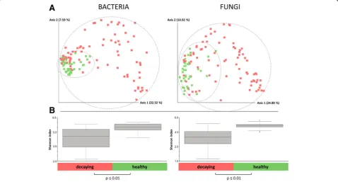
Microbial diversity was significantly decreased in decaying sugar beets
The comparison of amplicon data obtained from 120 samples of healthy and decaying sugar beets showed a significantly lower bacterial diversity in infected samples (Shannon index: 4.5 (16S) and 3.5 (ITS)) compared to the microbiome of healthy sugar beets (Shannon index 5.5 (16S) and 4.5 (ITS)) (Fig. 2b). The calculated Bray-Curtis distances showed significant differences in the composition of the microbiomes of the two groups. When a group-wise comparison was conducted, samples of decaying sugar beets (n= 80) clustered significantly
(p value ≤0.01) different from samples of healthy
sugar beets (n= 40). The variation within the infected group was found to be higher, compared to the healthy samples, which clustered more closely together (Fig.2a).
The core microbiome composition was altered in decaying sugar beets
Taxonomic assignments of the identified features
indicated a decay-specific microbiome of the analyzed sugar beets. The comparison of healthy and decaying samples showed a clearly distinguishable composition of taxa in both bacterial and fungal amplicon reads. Proteo-bacteria with an average relative abundance of 41% (healthy samples) and 51% (decaying samples) were the most abundant taxa on phylum level.Bacteriodetes(27% and 12.5%) andActinobacteria(28% and 11%) were also highly abundant in both groups. The main difference between both groups was due to the phylumFirmicutes (0.4% in healthy and 25% in decaying samples). A major fraction of Firmicutesin the decaying samples belonged to the order of Lactobacillales (24%). The predominant Proteobaceria in healthy samples were mainly members
of the orders Pseudomonadales (10%),
Sphingomona-dales(9%), Rhizobiales(8.5%),Xanthomonadales (6.5%), and Enterobacteriales (2.5%). In contrast, the 51% Proteobacteria found in decaying samples belonged to the orders Rhodospirillales (20%), Enterobacteriales
(8%), Pseudomonadales (8%), Xanthomonadales (5%),
Sphingomonadales (4%), and Rhizobiales (4%). At order level, the most abundant taxa of healthy sugar
beets were Flavobacteriales (21%), Micrococcales
(21%), and Pseudomonadales (10%), whereas the
predominant taxa of decaying sugar beets were Lacto-bacillales (24%), Rhodospirillales (20%), and Flavobac-teriales (9%). At genus level Lactobacillus (18.4%), Gluconobacter (16%), and Leuconostoc (11.3%) were the most abundant taxa in decaying samples, whereas Flavobacterium (20.6%), Pseudarthrobacter (13.5%), and Pseudomonas (9%) were the most abundant taxa in healthy samples. (Fig. 3a).
The ITS dataset showed diversified fungal micro-biomes in both healthy and decaying sugar beets. When the structure of the whole dataset was assessed, a total
of 60–62% Ascomycota and 33% Basidiomycota were
observed within the fungal community. At class level, an increased fraction of Saccharomycetes (+ 10% points; 12% total) and Eurotiomycetes (+ 9% points; 10% total) as well as a decreased fraction ofSordariomycetes(−16% points; 24% total) was found in the decaying samples. At
order level, an increased abundance ofCystofilobasidiales
(+ 11% points; 21% total), Saccharomycetales (+ 10%
points; 12% total), and Eurotiales (+ 9.5% points; 10% total) was observed. At genus level, this resulted in an increased number of Candida(+ 7.5%; 9.5 total), Penicil-lium (+ 9.5%; 10% total), Guehomyces (+ 5%; 10% total), andMrakia(+ 5%; 8% total). Healthy samples by contrast showed an increased amount of the genera Plectosphaer-ella (+ 10%; 21% total) as well asVishniacozyma (+ 12%; 18% in total). This was already shown in an increased abundance of the classesSordariomycetes (+ 16%; 40% in total) as well as Tremellomycetes(+ 2%; 30% in total). In comparison, at genus level, the most abundant genera in
decaying samples were Plectosphaerella, Guehomyces,
Candida, and Penicillium (all 10%), whereas in healthy samples the generaPlectosphaerella(21%) and Vishniaco-zyma(18%) dominated (Fig.3b).
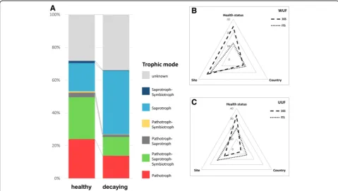
Trophic specialization in the fungal microbiome
Taxonomic differences between healthy and decaying sugar beets were found to be accompanied by changes
in the trophic modes of the identified core features. Healthy samples were mainly colonized by pathotrophic (24%) and pathotrophic-saprotrophic-symbiotrophic (26%) fungi. The trophic distribution in the decaying sam-ples, however, was dominated by saprotrophic fungi (39%) with a decreased fraction of pathotrophic (14%)
and pathotrophic-saprotrophic-symbiotrophic (12%)
fungi. Overall, a decrease in pathotrophic and symbio-trophic functions and an increase in saprosymbio-trophic functions from the microbiome in healthy to the micro-biome in decaying sugar beets was observed (Fig.4a).
The health status of beets was identified as the major driver for microbial community composition
The comparison of six different beet clamps in Austria and Germany showed significant differences in diversity as well as taxonomic composition. Health status explained the largest proportion of variance of the beets (33.3% vari-ation in 16S dataset and 20.9% for ITS,p≤0.001). Differ-ent beet clamp sampling sites also explained 13.6% variation in the 16S and 21.7% variation in the ITS dataset (p≤0.001), however, variances within the groups were higher (Fstatistic = 3.43 (16S) and 6.25 (ITS) compared to 56.36 (16S) and 30.91 (ITS) between health statuses). The
country that sugar beet samples originated from
accounted for the least variance (5% in 16S data and
11.7% in ITS data,p≤0.001) (Fig.4b, c; Additional file1: Table S1). These findings were also reflected inβ-diversity PCoA plots, where sample were separated by health status (Additional file1: Figure S1, S2).
Samples obtained from the storage in Grossmugl (Austria) showed clear differences in the microbial com-position when compared to the sampling spots located in lower Germany (Mittich, Kleinweichs, and Osterhofen). Sampling locations that were geographically located closer to each other (Additional file 1: Figure S3C), however, showed less significant differences. Overall, a change from relatively balanced abundances of bacterial taxa (micro-biome of healthy sugar beets) to a predominance of Lacto-bacillales, as well as Rhodospirillales (decaying sugar beets) was evident for every sampling spot. The fungal community changed from a microbiome dominated by VishniacozymaandPlectospaerellato an increasing num-ber ofPenicilliumandCandidaspecies (Fig.5).
Identification of disease indicators and correlation to sugar content in stored sugar beets
Specific taxa, indicative either for the microbiome of healthy or decaying sugar beets, were selected based on the differences in their abundance in the representative samples (Figs. 3 and 5). Flavobacterium and Pseudar-throbacter within the bacterial community as well as Plectospaerella and Vishniacozyma within the fungal
community were found to be dominant in healthy sugar beets. In contrast, Lactobacillus and Gluconobacter as
well as Candida and Penicillium were prevalent in
decaying sugar beets. By implementing a real-time qPCR analysis with specific primers targeting microbial indica-tors in stored sugar beets, the gradual increase of disease indicators and simultaneous loss of health indicators was shown. During a 3-month storage trial, an increase of Candida (105 to 5 × 10⁶ copies/g),Fusarium (2 × 10³ to 104copies/g), andPenicillium(0 to 104copies/g) and simultaneous decrease ofVishniacozyma (105 to 5 × 10⁴ copies/g) was observed (Fig.6a). In case of Plectosphaer-ella, an initial decreases in abundance (2 × 10⁵ to 105 copies/g), but overall constant abundances (105copies/g) throughout the storage period were found.
In order to verify the disease progress in the sam-ples that were used for qPCR primer evaluations, complementary analytical analyses of beet carbohy-drates were conducted with stored samples. The sugar content of sugar beets stored under controlled condi-tions showed a decreasing concentration of sucrose (−3% points) during the storage period of 3 months. At the same time, an increase of inverted sugars (glu-cose (2 to 14 g/kg) and fructose (1.5 to 14 g/kg)) was observed (Fig. 6b).
Discussion
Fungal pathogens prevail among isolates from decaying sugar beets
The obtained results of the present study provide the first detailed microbiome characterization of conventionally stored sugar beets in an industrially scaled, uncontrolled environment. By combining different methods, a holistic assessment of the fungal microbiome in decay-affected sugar beets was constructed. 18S gene sequencing data of 120 fungal isolates was compared to ITS next-generation amplicon data. In comparison, 86% of isolated fungi identi-fied on genus level were also found in the amplicon librar-ies. The cultivation-dependent identification of fungal isolates showed a prevalence of certain taxa such as Penicillium and Fusarium, when compared to the amplicon sequencing dataset. This likely resulted from the specific procedure during the isolation process that could have affected the frequency of isolated strains. While only homogenized peel was used for the total DNA extraction, also surface-sterilized frag-ments of infected sugar beets were placed on agar plates during isolation. This could have facilitated the isolation of Fusarium species, since this pathogen pri-marily colonizes the plant endosphere [23]. In the case of Penicillium, its high spore production allows it to
overgrow slow-growing fungal taxa and likely lead to its isolation in higher proportions. The fungal ITS library ob-tained with high-throughput sequencing showed overall a higher diversity of different fungal taxa, which is partially due to cultivability limitations of certain taxa on standard isolation media [24,25].
Bacterial diversity decrease was accompanied by an emergence of several highly abundant lineages
Microbial diversity as well as distinct changes in the microbial community were previously shown to be linked to disease incidence [5,26]. The data obtained in this study supports the hypothesis that lower diversity in the bacterial as well as fungal community is connected to a higher sensitivity to microbiome shifts that substan-tially alter the community structure. The lower diversity in decaying samples was reflected by a significant de-crease in diversity indices in both the bacterial and the fungal dataset. In analogy to our findings, changed mi-crobial diversity was found in stored onions when com-paring healthy and diseased ones and fungal diversity was found to be higher in roots of healthy winter
wheat plants [27, 28]. Moreover, a decrease in
diversity was shown to facilitate invasion of patho-genic species into communities [29].
Even though amplicon-based sequencing can be affected by certain biases [30], the taxonomic compos-ition of the bacterial as well as fungal beet microbiome, obtained with this dataset, was primarily linked to the health status of the sampled sugar beets. The geographic location of the beet clamps played a less significant role for the observed variability. Similarly, also Yurgel and colleagues (2018) observed taxonomic changes based on health status in stored onions [28]. Additionally, Liebe et al. (2016) already observed a similar effect in sugar beets when stored at different temperatures [14]. Depending on the storage conditions, the analyzed beets harbored specific fungal taxa, whereas the originating environment was less influential. In this study, sugar beets, stored under representative conditions without any protection from ad-verse environmental factors (moisture, temperature fluc-tuations, frost, etc.), showed a fungal community
dominated by Candida, Penicillium, Guehomyces, and
Plectosphaerella sp. in decaying sugar beets. The fungal microbiome of sampled healthy beet roots was, interest-ingly, comparable with the analyzed reference sugar beets
in Liebe et al. (2016) harnessing mostly Plectosphaerella sp. [14]. The observed taxonomic changes were also reflected by trophic modes within the fungal community. Dominant pathotrophic and pathotrophic-saprotrophic-symbiotrophic functions in healthy samples were replaced by saprotrophic functions in decaying sugar beets. Similar findings were also made by Yu and colleagues (2012) link-ing the prevalence of saprotrophic fungi mostly to dis-eased pea plants, the abundance of pathogenic fungi, however, not to a specific health status [26].
Identification of health indicators in the microbiome of sugar beets
Different potential biological markers were identified by contrasting healthy and diseased samples of stored sugar beets. Distinct taxa were shown to be highly abundant in samples representing each disease condi-tion. The necrotrophic fungal lineage Plectosphaerella, found in healthy beets, was previously shown to be a growth-promoting microbe in sugar beets [31]. More-over, it was reported as a potential biological control agent against potato cyst nematodes as well as a po-tential bioherbicide [32, 33]. Previous studies on sugar beet storage observed this taxon mostly in sugar beets before storage [14]. Other health-related taxa, such as Flavobacterium and Pseudarthrobacter, were often re-ported in the rhizosphere of different plants as well as their involvement in plant defense mechanisms or
growth promotion [34–37]. Other taxa, associated
with decaying sugar beets, such as Penicillium, are
typical saprophytic fungi and postharvest pathogens and were observed previously in rotting sugar beet after harvest [14, 38, 39]. Lactobacillus as well as the
fungal genus Candidawere predominantly detected in
decaying sugar beets and are associated with sugar fermentation to acid or alcohol compounds and are unwanted in stored sugar beets because of this activ-ity [40, 41]. We hypothesize that such taxa occur on decaying sugar beets primarily due to increased free monosaccharides originating from the hydrolyzation processes of sucrose by fungal extracellular proteins.
Real-time qPCR analyses conducted on the basis of the identified health and disease indicators in stored sugar beets provided a first evidence for the applicability of such indicators for agricultural management strat-egies. The data was obtained within small-scale experi-ments and must be further expanded in upcoming approaches to confirm the reliability of the indicators for industry-scale applications. During the representative storage period of three months, health-related indicators were either decreasing or remained constant. In con-trast, disease-related indicators increased substantially over the storage period. The quantitative analysis of these taxa indicated a gradual disease development that
is linked to microbial sucrose concentration loss and simultaneously increase in inverted sugars during stor-age [42], which was confirmed by targeted analyses in the present study.
Conclusion
Storage rot in stored sugar beets was shown to be accom-panied by a change in microbial abundances. The present study highlighted substantial shifts within the bacterial as well as fungal community that correlated to decay inci-dence in stored roots. Changes in the prevalence of certain taxa can potentially indicate decay development at an early stage and facilitate an implementation of targeted counter-measures. Taxonomic changes were shown to be accom-panied by trophic specialization in the fungal community. For upcoming postharvest applications, the novel insights provide a basis to design suitable biocontrol agents main-taining the balance of taxa associated with the microbiome of healthy sugar beets and preventing the establishment of degrading microorganisms. Furthermore, the identification of diseases indicators can be used as decision tool and sup-ports the prioritization of processing of harvested beets during storage management. Additional studies are needed to confirm the implementability of the obtained results and to assign levels of quantitative measurements, which will allow to indicate the degree of disease.
Methods
Sampling of sugar beets and isolation of fungi
beet were randomly picked based on morphology from the plates and further subcultured on PDA, SNA, and water agar plates (tap water + 18 g/L agar). The strains were further grouped using morphologic clustering after inspecting the single isolates on the different plates. Sev-eral strains of each morphologic cluster (120 strains in total) were subjected to 18S rRNA gene fragment Sanger sequencing (LGC Genomics, Berlin, Germany). Quality checked sequences were blasted against the NCBI data-base as well as the UNITE v7 datadata-base [44].
Storage of sugar beets under controlled conditions A total of 20 untreated and undamaged sugar beets harvested from a single field in Germany (Rhenish Hesse, Rhineland-Palatinate; 49° 35′ 54.388″ N, 8° 12′ 48.823″E) were stored directly after harvest under con-trolled condition at 8 °C and 75% relative humidity for 3 months. Sampling of five sugar beets at the beginning (T0) and every 30 days (T1, T2, and T3) was performed as described above. A total of 20 g of sugar beet peel was washed in a stomacher with 50 mL of sodium chlor-ide (0.85%). A total of 4 mL of the solution was centri-fuged into a pellet and further used for community DNA extraction. Sugar content in the sugar beet flesh was measured using standardized ICUMSA (International Commission for Uniform Methods of Sugar Analysis) methods for the determination of glucose and fructose by enzymatic assays and the polarization of sugar (sucrose) by the cold aqueous digestion method [45,46].
Total community DNA extraction and construction of amplicon library
A total of 4 mL of the obtained washing solution from the sampling step was centrifuged (13,000×g, 20 min, 4 °C) and the pellet was stored at −70 °C until further use. Using the FastDNA® Kit for Soil (MP Biomedicals/USA) genomic DNA was extracted from all samples. All steps were conducted as stated in the manufacturer’s protocol. Following DNA extraction, the 16S rRNA primers 514f and 926r (GTGYCAGCMGCCGCGGTAA; CCGYCAAT TYMTTTRAGTTT) and the ITS primer pair ITS1f and ITS2r (CTTGGTCATTTAGAGGAAGTAA; GCTGCG TTCTTCATCGATGC) were used in PCR for amplicon li-brary construction. As described in the protocols and standards section of the Earth microbiome project [47], both primer pairs were modified with specific primer pads (TATGGTAATT/AGTCAGCCAG) and linker (GT/GG) for the attachment of a Golay barcode sequences. Two consecutive PCR reactions were performed and all PCR reactions, conducted in triplicates were pooled after the second PCR. The first PCR (amplification of the V4 and V5 region or ITS1 region) was performed in a total vol-ume of 10μL (1μL DNA, 2μL Taq&Go, 0.1μL of each Primer, 0.15μL of mPNA and pPNA, and 6.5μL of water).
Added blocking primers mPNA and pPNA prevented the amplification of mitochondrial and chloroplast DNA [48]. The reactions were performed on a Whatman Biometra® Tpersonal and Tgradient thermocycler (Biometra GmbH, Göttingen, Germany) with the following settings: 95 °C for 45 s, 78 °C 5 s, 55 °C 45 s, 72 °C 90 s (35×), including an initial denaturation of 5 min at 95 °C and a final extension of 5 min at 72 °C. A second PCR step (multiplexing with Golay barcodes) a total volume of 30μL (2μL of the first PCR (template), 6μL Taq&Go, 1.2μL of barcode-primers and 19.6μL of water) run at the following settings: 95 °C for 30 s, 53 °C 30 s, 72 °C 30 s (15×), including an initial denaturation of 5 min at 95 °C and a final extension of 5 min at 72 °C. After each PCR amplification step, the quality was checked by gel electrophoresis. All tree repli-cates of quality checked PCRs from each sample were pooled and purified using the Wizard SV Gel and PCR Clean-Up System (Promega, Madison, USA) according to the protocol. Equimolar DNA concentrations of each barcoded amplicon sample were sent to GATC Biotech AG, Konstanz, Germany. After entry quality control and adapter ligation, 16S rRNA and ITS gene amplicons were sequenced on an Illumina HiSeq instrument.
Data evaluation using bioinformatics tools
Data obtained with Illumina HiSeq amplicon sequencing was analyzed with QIIME 2 (2018.6 release) and QIIME 1.9.1 [49] according to tutorials provided by the QIIME developers. After joining forward and reversed reads and barcode extraction in QIIME 1.9.1, the data was imported into QIIME 2 for further analysis. After demultiplexing, the DADA2 algorithm [50] was applied to denoise and truncate the reads and summarize se-quence variants (SVs) in a feature table. To increase the quality, chimeric data was filtered as well as mito-chondria and chloroplast reads (for 16S data) or bac-teria and archaea reads (for ITS data) were discarded. A total of 3489 ITS and 8935 16S SVs were assigned for a total of 16,155,698 ITS and 4,036,955 16S reads (Additional file 1: Table S3). Alpha diversity, beta di-versity, as well as statistical analysis was performed using the QIIME2 core diversity metrics. Naïve-Bayes classifier were trained on the SILVA v128 [51] at 99% similarity as well as the UNITE v7.2 [44] database for
taxonomic assignment. Subsequently, core
micro-biomes (features present in at least 50% of the sam-ples) were calculated for each group (healthy and decaying) and exported for display in bar charts. Functional analysis of fungal feature tables was per-formed using the FUNGuild online tool [52].
Statistical analysis of bioinformatics data
(beta) were used. Variance explained by parameters was analyzed with a PERMANOVA test in QIIME. Significant taxonomic differences between the groups were observed with the ANCOM test in QIIME 2.
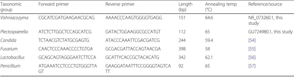
Real-time qPCR measurement targeting microbial indicators
Following the community DNA extraction from stored sugar beet samples obtained under controlled condi-tions, qPCR amplifications using specific primers were conducted in order to quantify distinct taxonomic groups that were selected as disease indicators. Specific
primers targeting Candida, Fusarium, Penicillium,
Lactobacillus, as found in previous literature were im-plemented. Primers forVishniacozymaand
Plectosphaer-ella were designed using the Primer-BLAST tool [53]
and deposited sequences in the NCBI database (Table1). The quantification was performed with a Corbett Research TM thermocycler (Rotor-Gene 6000, Corbett Research, UK) and SYBR Green PCR master mix TM (KAPA Biosystems, USA). The standard curves were ob-tained using a single isolate gene fragment with known copy numbers and further 1:10 dilutions. Three repli-cates of each standard dilution were prepared to calcu-late mean values. The standards were employed to determine the gene copy numbers in the analyzed
samples. Negative controls (using pure dH2O) were
implemented and further subtracted from the analyzed samples to reduce quantification inaccuracies.
Additional file
Additional file 1:Table S1.Summary of performed PERMANOVA test. Pairwise comparison of categories using the unweighted (UUF) and weighted (WUF) UniFrac distance metrics for both, the 16S and ITS, datasets. Table S2: Sampling locations and sample conditions of the implemented sugar beets. Healthy and decaying beets were sampled from beet clamps in Austria (AT) and Germany (DE). At the locations Kleinweichs and Osterhofen, two neighboring beet clamps were sampled (1 and 2). Table S3: Overview of sequencing data. Number of reads,
assigned sequence variants (SVs) using the DADA2 algorithm and Shannon Index of each group is given. Figure S1: Principal component analysis of bacterial and fungal communities from different beet clamps. PCoA using the unweighted UniFrac (UUF) distance metric. Samples are color-coded based on their geographic origin or health status. Figure S2: Principal component analysis of bacterial and fungal communities from different beet clamps. PCoA using the weighted UniFrac (WUF) distance metric. Samples are color-coded based on their geographic origin or health status. Figure S3: Sample visualization, schematic representation of fungal growth in the beet clamps, and geographic locations of the sampling sites. Fungal nests start within the clamp and spread to the surrounding beets (A, B). Healthy, uninfected beets, as well as decaying sugar beets within the same beet clamp were sampled from six different beet clamps in Austria and Germany (C). (DOCX 1605 kb)
Abbreviations
DNA:Deoxyribonucleic acid; ITS: Internal transcribed spacer; PCR: Polymerase chain reaction; qPCR: Real-time (quantitative) polymerase chain reaction
Acknowledgements
The authors gratefully acknowledge Anna Heinzel (Graz) and Barbara Fetz (Graz) for the help with laboratory work, Sebastian Siebauer (Plattling) for his help during sampling, as well as Stefan Hofmann (Offstein) for the help with sugar analysis.
Funding
This work has been supported by the Federal Ministry of Science, Research and Economy (BMWFW), the Federal Ministry of Traffic, Innovation and Technology (bmvit), the Styrian Business Promotion Agency SFG, the Standortagentur Tirol, the Government of Lower Austria, and ZIT - Technology Agency of the City of Vienna through the COMET-Funding Program managed by the Austrian Research Promotion Agency FFG (grant number 282482).
Availability of data and materials
Raw sequencing data for each sample used in this study was deposited at the European Nucleotide Archive (ENA) in the FASTA format and is available under the Bioproject accession number PRJEB28964.
Authors’contributions
CZ, GB, and TC conceived and designed the study. KH, JM, and HE provided specific knowledge for improved study design and oversaw the long-term storage experiments. PK performed the laboratory work as well as evaluation of the microbiome data. PK, TC, CZ, and GB wrote the manuscript. All authors read and approved the final version of the manuscript.
Ethics approval and consent to participate
Not applicable.
Table 1Sequences, annealing temperatures, fragment length, and sources of the implemented qPCR primers. The primers for VishniacozymaandPlectospaerellawere designed with deposited sequences (accession numbers provided) in the NCBI database and the Primer-BLAST tool [53]
Taxonomic group
Forward primer Reverse primer Length
(bp)
Annealing temp (°C)
Reference/source
Vishniacozyma CGCATCGATGAAGAACGCAG AAAACCCAAGTGGGGTGAGG 151 64.6 NR_073260.1, this
study
Plectospaerella ATCTCTTGGCTCCAGCATCG GATACTGGAAGGCGCCATGT 112 65 GU724980.1, this study
Candida TCTAACGTCTATGCGAGTG ATACCCAAATTCGACGATCG 244 59.4 [54]
Fusarium CAACTCCCAAACCCCTGTGA GCGACGATTACCAGTAACGA 398 58 [55]
Lactobacillus GCAGCAGTAGGGAATCTTCCA GCATTYCACCGCTACACATG 342 62.1 [56]
Penicillium ATGAAATCCTCCCTGTGGGTTA
GT
GAAGGATAATTTCCGGGGTAGTCA TT
Consent for publication
Not applicable.
Competing interests
The authors declare that they have no competing interests.
Publisher’s Note
Springer Nature remains neutral with regard to jurisdictional claims in published maps and institutional affiliations.
Author details
1Austrian Centre of Industrial Biotechnology, Petersgasse 14, 8010 Graz, Austria.2Institute of Environmental Biotechnology, Graz University of Technology, Petersgasse 12, 8010 Graz, Austria.3Südzucker AG, Maximilianstraße 10, 68165 Mannheim, Germany.4Agrana Research & Innovation Center, Josef-Reither-Straße 21–23, 3430 Tulln, Austria. 5Roombiotic GmbH, c/o: SciencePark, Stremayrgasse 16/IV, 8010 Graz, Austria.
Received: 16 April 2019 Accepted: 31 July 2019
References
1. Berendsen RL, Pieterse CMJ, Bakker PAHM. The rhizosphere microbiome and plant health. Trends Plant Sci. 2012;17:478–86.
2. Berg G, Grube M, Schloter M, Smalla K. Unraveling the plant microbiome: looking back and future perspectives. Front Microbiol. 2014;5https://doi. org/10.3389/fmicb.2014.00148.
3. Vandenkoornhuyse P, Quaiser A, Duhamel M, Le Van A, Dufresne A. The importance of the microbiome of the plant holobiont. New Phytol. 2015; 206:1196–206.
4. Droby S, Wisniewski M, Teixidó N, Spadaro D, Jijakli MH. The science, development, and commercialization of postharvest biocontrol products. Postharvest Biol Technol. 2016;122:22–9.
5. Berg G, Köberl M, Rybakova D, Müller H, Grosch R, Smalla K. Plant microbial diversity is suggested as the key to future biocontrol and health trends. FEMS Microbiol Ecol. 2017;93https://doi.org/10.1093/femsec/fix050. 6. Castoria R, De Curtis F, Lima G, Caputo L, Pacifico S, De Cicco V.
Aureobasidium pullulans(LS-30) an antagonist of postharvest pathogens of fruits: study on its modes of action. Postharvest Biol Technol. 2001;22:7–17. 7. Tzortzakis NG, Economakis CD. Antifungal activity of lemongrass
(Cympopogon citratusL.) essential oil against key postharvest pathogens. Innov Food Sci Emerg Technol. 2007;8:253–8.
8. Trebbi D, McGrath JM. Fluorometric sucrose evaluation for sugar beet. J Agric Food Chem. 2004;52:6862–7.
9. Osburn RM, Schroth MN, Hancock JG, Hendson M. Dynamics of sugar beet seed colonization byPythium ultimumandPseudomonasspecies: effects on seed rot and damping-off. Phytopathology. 1989;79:709–16.
10. Kiewnick S, Jacobsen BJ, Braun-Kiewnick A, Eckhoff JLA, Bergman JW. Integrated control ofRhizoctoniacrown and root rot of sugar beet with fungicides and antagonistic bacteria. Plant Dis. 2001;85:718–22.
11. Weiland J, Koch G. Sugarbeet leaf spot disease (Cercospora beticolaSacc.)†. Mol Plant Pathol. 2004;5:157–66.
12. Zachow C, Fatehi J, Cardinale M, Tilcher R, Berg G. Strain-specific colonization pattern ofRhizoctoniaantagonists in the root system of sugar beet. FEMS Microbiol Ecol. 2010;74:124–35.
13. Jaggard KW, Clark CJA, May MJ, McCullagh S, Draycott AP. Changes in the weight and quality of sugarbeet (Beta vulgaris) roots in storage clamps on farms. J Agric Sci. 1997;129:287–301.
14. Liebe S, Wibberg D, Winkler A, Pühler A, Schlüter A, Varrelmann M. Taxonomic analysis of the microbial community in stored sugar beets using high-throughput sequencing of different marker genes. FEMS Microbiol Ecol. 2016;92https://doi.org/10.1093/femsec/fiw004.
15. Klotz KL, Finger FL. Impact of temperature, length of storage and postharvest disease on sucrose catabolism in sugarbeet. Postharvest Biol Technol. 2004;34:1–9.
16. Hoffmann C. Lagerfähigkeit geköpfter und entblätterter Rüben. Sugar Ind. 2012;137:458–67.
17. Kenter C, Hoffmann CM. Changes in the processing quality of sugar beet (Beta vulgarisL.) during long-term storage under controlled conditions. Int J Food Sci Technol. 2009;44:910–7.
18. Zachow C, Tilcher R, Berg G. Sugar beet-associated bacterial and fungal communities show a high indigenous antagonistic potential against plant pathogens. Microb Ecol. 2008;55:119–29.
19. Berg G. Plant–microbe interactions promoting plant growth and health: perspectives for controlled use of microorganisms in agriculture. Appl Microbiol Biotechnol. 2009;84:11–8.
20. Janisiewicz WJ, Korsten L. Biological control of postharvest diseases of fruits. Annu Rev Phytopathol. 2002;40:411–41.
21. Kusstatscher P, Cernava T, Harms K, Maier J, Eigner H, Berg G, et al. Disease incidence in sugar beet fields is correlated with microbial diversity and distinct biological markers. Phytobiomes J. 2019;https://doi.org/10.1094/ PBIOMES-01-19-0008-R.
22. Schillinger U, Geisen R, Holzapfel WH. Potential of antagonistic microorganisms and bacteriocins for the biological preservation of foods. Trends Food Sci Technol. 1996;7:158–64.
23. Zhang X-W, Jia L-J, Zhang Y, Jiang G, Li X, Zhang D, et al.In plantastage -specific fungal gene profiling elucidates the molecular strategies of
Fusarium graminearumgrowing inside wheat coleoptiles. Plant Cell. 2012;
https://doi.org/10.1105/tpc.112.105957.
24. Müller T, Ruppel S. Progress in cultivation-independent phyllosphere microbiology. FEMS Microbiol Ecol. 2014;87:2–17.
25. Wu P-C, Su H-JJ, Ho H-M. A comparison of sampling media for environmental viable fungi collected in a hospital environment. Environ Res. 2000;82:253–7. 26. Yu L, Nicolaisen M, Larsen J, Ravnskov S. Molecular characterization of root
-associated fungal communities in relation to health status ofPisum sativum
using barcoded pyrosequencing. Plant Soil. 2012;357:395–405.
27. Lemanczyk G, Sadowski CK. Fungal communities and health status of roots of winter wheat cultivated after oats and oats mixed with other crops. BioControl. 2002;47:349–61.
28. Yurgel SN, Abbey L, Loomer N, Gillis-Madden R, Mammoliti M. Microbial communities associated with storage onion. Phytobiomes J. 2018;2:35–41. 29. van Elsas JD, Chiurazzi M, Mallon CA, ElhottovāD, Krištůfek V, Salles JF.
Microbial diversity determines the invasion of soil by a bacterial pathogen. Proc Natl Acad Sci. 2012;109:1159–64.
30. Schirmer M, Ijaz UZ, D’Amore R, Hall N, Sloan WT, Quince C. Insight into biases and sequencing errors for amplicon sequencing with the Illumina MiSeq platform. Nucleic Acids Res. 2015;43:e37.
31. Ying-Wu S, Kai L, Chun L. Effects of endophytic fungus on sugar content and key enzymes activity in nitrogen and sugar metabolism of sugar beet (Beta vulgarisL.). Acta Agron Sin. 2009;35:946–51.
32. Atkins SD, Clark IM, Sosnowska D, Hirsch PR, Kerry BR. Detection and quantification ofPlectosphaerella cucumerina, a potential biological control agent of potato cyst nematodes, by using conventional PCR, real-time PCR, selective media, and baiting. Appl Env Microbiol. 2003;69:4788–93. 33. Bailey K, Derby J-A, Bourdôt G, Skipp B, Cripps M, Hurrell G, et al.
Plectosphaerella cucumerinaas a bioherbicide forCirsium arvense: proof of concept. BioControl. 2017;62:693–704.
34. Bulgarelli D, Rott M, Schlaeppi K, van Themaat EVL, Ahmadinejad N, Assenza F, et al. Revealing structure and assembly cues forArabidopsis root-inhabiting bacterial microbiota. Nature. 2012;488:91–5.
35. Kolton M, Frenkel O, Elad Y, Cytryn E. Potential role ofFlavobacterialgliding -motility and type IX secretion system complex in root colonization and plant defense. Mol Plant-Microbe Interact. 2014;27:1005–13.
36. Krishnamoorthy R, Kwon S-W, Kumutha K, Senthilkumar M, Ahmed S, Sa T, et al. Diversity of culturable methylotrophic bacteria in different genotypes of groundnut and their potential for plant growth promotion. 3 Biotech. 2018;8:275. 37. Wei W, Zhou Y, Chen F, Yan X, Lai Y, Wei C, et al. Isolation, diversity, and
antimicrobial and immunomodulatory activities of endophyticActinobacteria
from tea cultivars Zijuan and Yunkang-10 (Camellia sinensis var. assamica). Front Microbiol. 2018;9https://doi.org/10.3389/fmicb.2018.01304. 38. Bugbee WM.Penicillium claviformeandPenicillium variabile: pathogens of
stored sugarbeets. Phytopathology. 1975;65:926-7.
39. Snowdon AL. A colour atlas of post-harvest diseases and disorders of fruits and vegetables. Volume 1: General introduction and fruits. London: Wolfe Scientific Ltd., 1990.
40. Calabia BP, Tokiwa Y. Production of d-lactic acid from sugarcane molasses, sugarcane juice and sugar beet juice byLactobacillus delbrueckii. Biotechnol Lett. 2007;29:1329–32.
42. Liebe S, Varrelmann M. Effect of environment and sugar beet genotype on root rot development and pathogen profile during storage.
Phytopathology. 2015;106:65–75.
43. Nirenberg H. Untersuchungen über die morphologische und biologische Differenzierung in derFusarium-Sektion Liseola. Mitt Biol Bundesanst Land-U Forstwirtsch Eerlin-Dahlern. 1976;169:1–117.
44. Kõljalg U, Nilsson RH, Abarenkov K, Tedersoo L, Taylor AFS, Bahram M, et al. Towards a unified paradigm for sequence-based identification of fungi. Mol Ecol. 2013;22:5271–7.
45. ICUMSA. Glucose and fructose in beet juices and processing products by an enzymatic method—accepted. Colney: Bartens; 2007.
46. ICUMSA. Polarisation of sugar beet by the macerator or cold aqueous digestion and aluminium sulphate. Colney: Bartens; 1994.
47. Walters W, Hyde ER, Berg-Lyons D, Ackermann G, Humphrey G, Parada A, et al. Improved bacterial 16S rRNA gene (V4 and V4–5) and fungal internal transcribed spacer marker gene primers for microbial community surveys. mSystems. 2015;1https://doi.org/10.1128/mSystems.00009-15.
48. Lundberg DS, Yourstone S, Mieczkowski P, Jones CD, Dangl JL. Practical innovations for high-throughput amplicon sequencing. Nat Methods. 2013; 10:999.
49. Caporaso JG, Kuczynski J, Stombaugh J, Bittinger K, Bushman FD, Costello EK, et al. QIIME allows analysis of high-throughput community sequencing data. Nat Methods. 2010;7:335–6.
50. Callahan BJ, McMurdie PJ, Rosen MJ, Han AW, Johnson AJA, Holmes SP. DADA2: high resolution sample inference from Illumina amplicon data. Nat Methods. 2016;13:581–3.
51. Quast C, Pruesse E, Yilmaz P, Gerken J, Schweer T, Yarza P, et al. The SILVA ribosomal RNA gene database project: improved data processing and web-based tools. Nucleic Acids Res. 2013;41:D590–6.
52. Nguyen NH, Song Z, Bates ST, Branco S, Tedersoo L, Menke J, et al. FUNGuild: an open annotation tool for parsing fungal community datasets by ecological guild. Fungal Ecol. 2016;20:241–8.
53. Ye J, Coulouris G, Zaretskaya I, Cutcutache I, Rozen S, Madden TL. Primer-BLAST: a tool to design target-specific primers for polymerase chain reaction. BMC Bioinformatics. 2012;13:134.
54. Ogata K, Matsuda K, Tsuji H, Nomoto K. Sensitive and rapid RT-qPCR quantification of pathogenicCandidaspecies in human blood. J Microbiol Methods. 2015;117:128–35.
55. Abd-Elsalam KA, Aly IN, Abdel-Satar MA, Khalil MS, Verreet JA. PCR identification ofFusariumgenus based on nuclear ribosomal-DNA sequence data. Afr J Biotechnol. 2003;2:82–5.
56. Walter J, Hertel C, Tannock GW, Lis CM, Munro K, Hammes WP. Detection of
Lactobacillus, Pediococcus, Leuconostoc, andWeissellaspecies in human feces by using group-specific PCR primers and denaturing gradient gel electrophoresis. Appl Env Microbiol. 2001;67:2578–85.