M
AŁGORZATAT
ROCHA, A
DAMS
ZELĄGThe Role of Calcium Channel−Blocking Drugs
in Preserving Rat Liver for Transplantation
Wpływ leków blokujących kanały wapniowe
na wątrobę przechowywaną do transplantacji
Department of Pharmacology, Silesian Piasts University of Medicine in Wrocław, Poland Adv Clin Exp Med 2007, 16, 5, 609–618
ISSN 1230−025X
ORIGINAL PAPERS
© Copyright by Silesian Piasts University of Medicine in Wrocław
Abstract
Background. Liver transplantation has become a widely accepted therapy for patients with end−stage liver dis− eases. The efficacy of this method depends on many different factors. Calcium plays a crucial role in physiologi− cal processes in liver cells; however, in high concentrations its ions can be pathogenic and lead to cell death. Because calcium ions enter the cell mostly through calcium channels, it has been suggested that calcium channel inhibitors (CCIs) could protect hepatocytes from the action of toxic substances.
Objectives.Using an extracorporeal liver perfusion model, the protective actions of nitrendipine and nifedipine dur− ing ischemia and reperfusion on the structure and function of rat liver preserved for transplantation were evaluated.
Material and Methods.The study was carried out on livers isolated from adult male Wistar rats. The rat livers were preserved for 24 hours in HTK solution (4ºC) with or without nifedipine or nitrendipine. After preservation, the livers were flushed with Ringer’s solution and perfused for two hours with a perfusion fluid. Glucose concen-tration and ALT, AST, and LDH activities were measured during perfusion. After perfusion all the produced bile was collected and the livers were histologically examined.
Results.A statistically significant influence of nitrendipine on the activities of the liver enzymes was found. Livers which were both perfused and rinsed with addition of this drug showed the lowest levels of enzyme activity and histological damage among all the examined groups. No statistically significant differences between the groups in glucose concentration and total amount of bile were observed.
Conclusions.Nitrendipine demonstrated a protective influence on the function and structure of rat liver preserved for transplantation and maintained its action throughout perfusion, especially when added not only to the perfusion fluid, but also to the Ringer’s solution used for rinsing (Adv Clin Exp Med 2007, 16, 5, 609–618).
Key words: calcium channel blockers, liver, preservation−reperfusion injury, extracorporeal perfusion.
Streszczenie
Wprowadzenie. Obecnie przeszczepianie wątroby jest popularną i akceptowaną metodą leczenia pacjentów w ostatnim stadium niewydolności wątroby. Skuteczność tej metody zależy od wielu czynników. Jednym z nich jest prawidłowe stężenie jonów wapnia w tym narządzie. Jeśli nasilenie uszkodzeń w wątrobie podczas jej prze− chowywania do przeszczepienia zależy od wzrostu stężenia wapnia w komórce, to leki, które hamują jego napływ do komórki, powinny działać cytoprotekcyjnie.
Cel pracy. Ocena cytoprotekcyjnego działania nitrendypiny i nifedypiny na strukturę i czynność wątroby szczu− rzej przechowywanej do przeszczepienia.
Liver transplantation is the method of choice in the treatment of end−stage liver failure. Unfortunately, the list of patients waiting for a transplant is very long and hundreds die each year when delivery of an appropriate transplant fails [1]. Therefore, improvements in the transplantation technique and the methods of liver storage prior to transplantation are very significant in clinical prac− tice. Liver damage before transplantation occurs in two stages. Initial injury is caused by ischemia and later the damage is aggravated by reperfusion of the organ. The pathogenesis of these lesions is very complex. Among the factors responsible for cell damage and death are calcium ions, whose concen− tration markedly rises under these circumstances [2]. If the extent of damage in a liver preserved for transplantation is largely dependent on the eleva− tion of the calcium ion concentration in the cells, the view seems justified that drugs inhibiting their influx into the cells should be cytoprotective. Calcium channel blockers (CCBs) act together with immunosuppressive medications [3–5] and fulfill an immunomodulatory function [5]. Thus, from this point of view, there are no contraindica− tions for their potential use during transplantation.
For many years attempts have been made to evaluate the protective action of CCBs on liver preserved for transplantation, but the results of these studies are conflicting [5–7]. The aim of this study was to assess the cytoprotective actions of nitrendipine and nifedipine during ischemia and reperfusion on the structure and function of rat liver preserved for transplantation.
Material and Methods
Animals
The study was carried out on livers isolated from adult male Wistar rats (312.72 ± 20.32 g) obtained from the Animal Laboratory of the Chair and Department of Pathological Anatomy, Silesian Piasts University of Medicine in Wrocław, Poland. The animals were housed individually in chambers with a 12−h/12−h light/dark cycle and a tempera− ture maintained at 21–23°C. Before the experiment
the animals had free access to standard food and water. All steps were performed under sterile con− ditions. For this experiment, five groups were cre− ated: 0K (n = 11) with livers perfused without preservation, 24K (n = 11) with livers preserved without any drug, 24A (n = 13) with livers pre− served with nitrendypine, 24B (n = 10) with livers preserved with nifedipine, and 24C (n = 10) with livers preserved with nitrendypine and also with nitrendypine added to the rinsing solution. The experiments were preformed with the consent of the First Local Ethics Commission on Animal Experiments in Wrocław (no. 70/03).
Substances
Histidine−tryptophane−ketoglutarate (HTK) solution (Dr. Franz Köhler Chemie GmbH, Germany), nitrendipine and nifedipine (Sigma− −Aldrich, Germany), trypan blue (Fluka Chemie, Switzerland), heparin (ampoule of 25 000 IU) and thiopental (ampoule of 0.5 g) (Bioechemie, Austria), and modified Krebs−Henseleit bicarbon− ate buffer (KHB) (118 mM NaCl, 25 mM NaHCO3, 4.8 mM KCl, 1.5 mM CaCl2, 1.2 mM
MgSO4×7H2O, 1.2 mM KH2PO4, and 4.9955 mM
glucose, pH 7.4) and phosphate buffer (prepared at own laboratory) were used in the study.
Liver Isolation and Storage
The animals were anesthetized by intraperi− toneal injection of thiopental at a dose of 70 mg/kg of body weight. A middle incision was made to open the abdominal cavity, the inferior caval vein was lig− ated above the right renal vein opening, and a can− nula was introduced into the portal vein through which Ringer’s solution supplemented with heparin was perfused. Then the chest was opened and a sec− ond cannula was inserted into the inferior caval vein, pointing towards the liver. In the control group (0K) the livers were excised and transferred whole to the chamber for extracorporeal liver perfusion (ELP). In the remaining groups the livers were prepared for cooling. Before isolation from the rat body, the liver was flushed with HTK solution at 4°C for 3 min (5 ml/min). Then the liver was isolated and transferred
Wyniki. W badaniu wykazano statystycznie istotny wpływ nitrendypiny na aktywność enzymów wątrobowych. W grupie, w której dodano nitrendypinę zarówno do płynu HTK, jak i roztworu Ringera aktywność enzymów by− ła najmniejsza, a zmiany histopatalogiczne najsłabiej wyrażone. Nie wykazano żadnych istotnych różnic w stęże− niu glukozy i całkowitej ilości żółci między grupami.
Wnioski. Nitrendypina wykazuje ochronne działanie na czynność i strukturę wątroby szczurzej przechowywanej do przeszczepienia. Działanie to utrzymuje się przez cały okres perfuzji, szczególnie wtedy, gdy została podana za− równo do płynu HTK, jak i roztworu Ringera (Adv Clin Exp Med 2007, 16, 5, 609–618).
to HTK solution (100 ml) maintained at 4°C (group 24K). The HTK solution in groups 24A and 24C was supplemented with nitrendipine (20 µM) and in group 24B it contained nifedipine (20 µM). The livers were placed in a refrigerator at a constant temperature of 4°C. The drug concentration was chosen based on the authors’ previous studies [8]. After 24 hours of storage, the livers were taken out of the refrigerator and flushed with Ringer’s solution at 20°C at a rate of 10 ml/min for 3 min. In group 24C the Ringer’s solution was supplemented with nitrendipine (20 µM).
Extracorporeal Perfusion
The livers were then transferred to the perfu− sion chamber and placed in an anatomical posi− tion. The time from removing the livers from the refrigerator to the beginning of perfusion was 6 min. Perfusion fluid, heated to 37°C and oxy-genated with a mixture of 95% O2and 5% CO2, flowed into the liver through the cannula inserted into the portal vein and left the organ through the cannula introduced into the inferior caval vein. The perfusion fluid (KHB) was propelled by a peristaltic pump at a flow rate of 20 ml/min for the first 10 min of perfusion and at 30 ml/min thereafter. The pressure was monitored throughout the entire perfusion period and kept within the range of 4–5 mm Hg. The livers were perfused using the Universal Perfusion System UNIPER UP-100 (Hugo Sachs Elektronik, Harvard Apparatus GmbH) in an open-circuit mode.
Baseline glucose concentration was deter− mined before ELP began. After 10 min of perfu− sion, the initial glucose concentration and the activities of the hepatocellular enzymes alanine aminotransferase (ALT), aspartate aminotrans− ferase (AST), and lactate dehydrogenase (LDH) were assayed. Samples of perfusion fluid were col− lected every 30 min of ELP. Glucose concentration was determined using a Marcel S330 PRO spec− trophotometer (Analco Medical Trade) and the activities of ALT, AST, and LDH were assayed with an enzymatic method (BioMerieux). At the end of perfusion, all the bile produced during the experiment was collected.
Histological Examination
After 120 minutes of ELP, 300 µM of trypan blue was added to the perfusion fluid and the liv− ers were perfused for 7 min. Then the livers were fixed by perfusion with 2% paraformaldehyde and 2% glutaraldehyde in 0.1 M phosphate buffer, pH 7.6. Slices of different regions of the liver were taken for examination. Some were stained with
eosin/hematoxylin and examined to assess struc− tural changes [9, 10]. Others were used for count− ing the proportion of trypan blue−stained nuclei in the total number of nuclei of non−parenchmal cells in certain regions of the liver [11–13].
Statistics
Activities of enzymes, glucose concentration, and the amount of bile were calculated per gram of rat liver. All values are reported as the mean and standard deviation. Statistical analysis of the effect of factors (drug, time) on enzymatic activities and glucose level was performed using multifactor analysis of variance (MANOVA) with repeats. The amount of bile produced during the experiment and the total amount of glucose released to the per− fusion fluid were compared using one−way analy− sis of variance. Specific comparisons were made with Tukey’s HSD test. STATISTICA 6.0 software was used. Hypotheses were considered positively verified if p≤0.05.
Results
Leakage of Enzymes
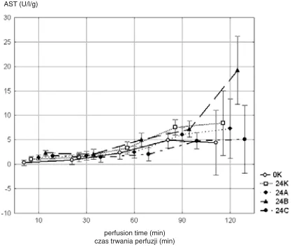
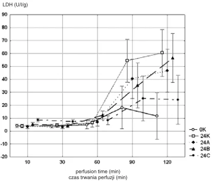
The study showed statistically significant influ− ences of nitrendipine, time of liver storage at 4°C, and duration of perfusion on the activities of AST (Fig. 1), ALT (Fig. 2), and LDH (Fig. 3). Critical changes were seen in the second hour of perfusion, when the activities of the enzymes increased the most, and the dynamics of this increase differed among the groups. At that time the lowest average AST, ALT, and LDH activities of all experimental groups were observed in group 24C, similar to the respective average values measured in group 0K. In all groups, average AST activities were higher than average ALT activities during ELP.
There was a statistically significant difference between the groups in the change in AST and LDH activity during perfusion (p< 0.001 for both comparisons) (Figs. 1 and 3). The increases in AST and LDH activities between the 10thand 120th
minute of perfusion in group 24C were signifi− cantly smaller than in group 24K (AST: p< 0.01, LDH: p < 0.001). Additionally, the increase in LDH activity between the 10thand 120thminute of
perfusion in group 24C was significantly smaller than in group 24B (p< 0.05).
Glucose
Glucose levels were visibly affected by ELP duration (p< 0.001) (Fig. 4). The pattern of time− dependent changes in glucose level was similar in all groups and the changes in glucose concentra− tion in relation to its initial level also did not sta− tistically significantly differ among the groups.
Bile
The amount of bile produced during the whole perfusion period was comparable in all groups and no statistically significant differences among the groups were noted.
Number of Damaged
Non−Parenchymal Cells
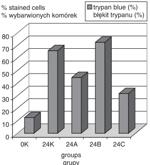
The percentage of damaged non−parenchymal cells in relation to the total number of cells in group 24C was the lowest of the groups of livers which were stored before perfusion and amounted to 31.5%. In group 24A the percentage of damaged cells was higher (44.5%), while the proportion of stained cells in relation to the total non−parenchy− mal cell count in group 24B group was 72.5% (Figs. 5 and 7).
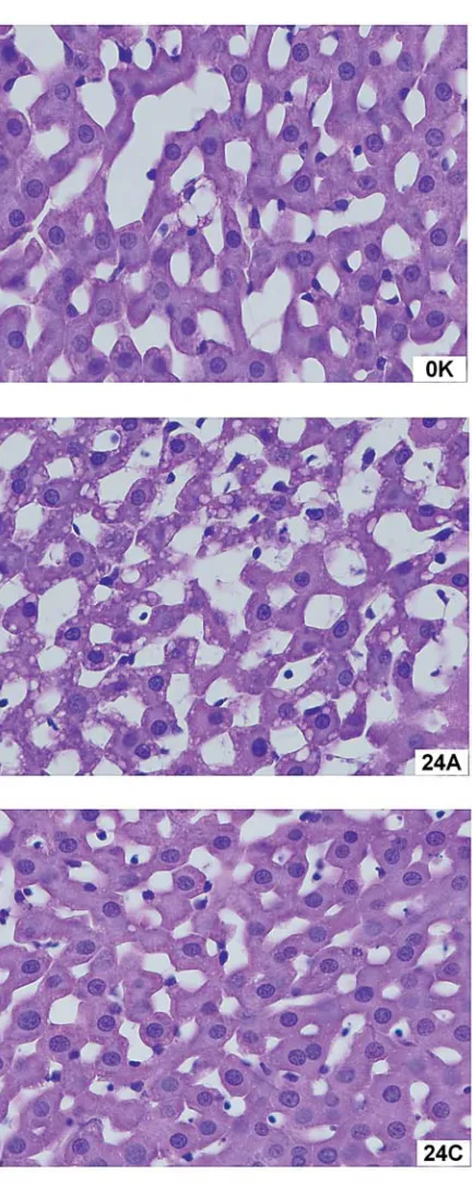
Histopathological Studies
The morphological picture of group 24C livers was similar to that of group 0K (Fig. 6). In both
AST (U/l/g)
perfusion time (min) czas trwania perfuzji (min)
Fig. 1. Differences in average activity of AST (U/l) as a function of the time of perfusion in all the experi− mental groups of livers: non−preserved (0K, n = 11), preserved without any drug (24K, n = 11), preserved with nitrendipine at a dose of 20 µM (24A, n = 13), preserved with nifedipine at a dose of 20 µM (24B, n = 10), and a group in which nitrendipine at a dose of 20 µM was added not only to HTK solution used for preservation, but also to the Ringer’s solution used for rinsing (24C, n = 10). Statistically significant dif− ferences: a change in AST activity during perfusion between the groups (p< 0.001), AST activity at 120 minutes of perfusion (24B : 24C, p< 0.001, and 24B : 24A, p< 0.05), increase in AST activity between the 10thand 120thminute of perfusion
(24K : 24C, p< 0.01)
Ryc. 1. Średnia aktywność AST (U/l/g) jako funkcja czasu trwania perfuzji we wszystkich badanych gru− pach: wątrobach perfundowanych bezpośrednio po izolacji z organizmu szczura (0K, n = 11), przechowy− wanych przez 24 godz. w 4ºC, w płynie HTK (24K, n = 11), przechowywanych w płynie HTK z dodaną nitrendypiną w stężeniu 20 µM (24A, n = 13), prze− chowywanych w płynie HTK z dodaną nifedypiną w stężeniu 20 µM (24B, n = 10) oraz w grupie, w której nitrendypina w stężeniu 20µM została dodana zarówno do płynu HTK, jak i roztworu Ringera służą− cego do przepłukania wątroby po okresie przechowy− wania (24C, n = 10). Różnice istotne statystycznie: zmiana aktywności AST między grupami podczas per− fuzji (p < 0,001), aktywność AST w 120 min perfuzji (24B : 24C, p < 0,001 i 24B : 24A, p < 0,05), wzrost aktywności AST pomiędzy 10 a 120 min perfuzji (24K : 24C, p < 0,01)
ALT (U/l/g)
perfusion time (min) czas trwania perfuzji (min)
Fig. 2. Differences in average activity of ALT (U/l/g) as a function of the time of perfusion in all experimen− tal groups of livers (as described in Fig. 1).
Statistically significant difference: ALT activity at 120 minutes of perfusion (24B : 24C, p< 0.05)
these groups the cytoplasm of the hepatocytes did not contain fat−rich vacuoles and deformations in the trabecular system’s architecture were hardly noticeable. The number of normal endothelial cells linearly adjoining the sinus walls was also the largest. Parts of the endothelium lost their linear structure, but only a few were detached and released to the sinus lumens. The morphological changes in group 24A were more apparent: some vacuoles were present, the trabecular structure was disarranged, the sinus lumens were widened, and the number of normal endothelial cells was lower. Some of the cells protruded into the sinus lumens. The largest morphological changes, indicating damage to the rat liver structure after 120 min of ELP, were observed in groups 24K and 24B (Fig. 6). The cells contained numerous vacuoles, the tra− becular system was disrupted, the structure of
sinuses was disordered, and the lumens were widened. No normal endothelial cells were observed. Only a few of these cells remained attached to the sinus walls and the majority of them was loosely scattered in the lumens.
Discussion
The results demonstrated a protective influ− ence of nitrendipine on the function of the rat liver preserved for transplantation. The analysis of enzyme activities, their increments, the morpho− logical picture of the liver, and the number of undamaged non−parenchymal cells clearly indicat− ed further that the action of nitrendipine was stronger when added not only to the preservation solution (HTK), but also to the Ringer’s solution used for flushing and heating the liver.
It is known that regardless of supplements, liver decays with time.Liver function was similar in all groups during the first 60 minutes. The dif− ferences in enzyme activities between the drug−
LDH (U/l/g)
perfusion time (min) czas trwania perfuzji (min)
Fig. 3. Differences in average activity of LDH (U/l/g) as a function of the time of perfusion in all experimen− tal groups of livers (as described in Fig. 1).
Statistically significant differences: a change in LDH activity during perfusion between the groups (p< 0.001), an increase in LDH activity between the 10thand 120thminute of perfusion (24K : 24C,
p< 0.001, and 24B : 24C, p< 0.05)
Ryc. 3. Średnia aktywność LDH (U/l/g) jako funkcja czasu trwania perfuzji we wszystkich badanych gru− pach: wątrobach perfundowanych bezpośrednio po izolacji z organizmu szczura (0K, n = 11), przechowy− wanych przez 24 godz. w 4ºC, w płynie HTK (24K, n = 11), przechowywanych w płynie HTK z dodaną nitrendypiną w stężeniu 20 µM (24A, n = 13), prze− chowywanych w płynie HTK z dodaną nifedypiną w stężeniu 20 µM (24B, n = 10) oraz w grupie, w której nitrendypina w stężeniu 20 µM została doda− na zarówno do płynu HTK, jak i roztworu Ringera służącego do przepłukania wątroby po okresie prze− chowywania (24C, n = 10). Różnice statystycznie istotne: zmiana aktywności LDH między grupami podczas prefuzji (p < 0,001), wzrost aktywności LDH między 10. a 120. min perfuzji – 24K : 24C (p < 0,001) i 24B : 24C (p < 0,05)
glucose (mM/l/g)
perfusion time (min) czas trwania perfuzji (min)
Fig. 4. Differences in average glucose concentration (mM/g) as a function of the time of perfusion in all experimental groups of livers (as described in Fig. 1). Glucose levels were visibly affected by ELP duration (p< 0.001)
less and drug−treated groups started to be apparent in the second hour of the experiment. Such obser− vations could only be made in experiments based on the ELP model.
The results of many studies on the cytoprotec− tive action of CCBs are very discrepant. Nicardipine [14, 15], nimodipine [16], verapamil [6, 17], and diltiazem [18] were reported to exhib− it such potential. However, in vitrostudy on iso− lated rat hepatocytes subjected to ischemia, vera− pamil, diltiazem, and nifedipine did not inhibit an increase in LDH activity [19]. There are only a few papers that document a hepatoprotective action of nitrendipine [20, 21].
Both AST and ALT belong to a group of indi− cator hepatocellular enzymes whose activities rise during hepatocyte damage. Both enzymes can be present in the cytoplasm, but AST was also found
in mitochondria. In the present experiment, the average AST activities were higher than ALT lev− els at all time intervals, which indicates that ischemia during liver storage, like alcoholic liver injury, is associated mostly with disruption of mitochondria [22].
In the majority of studies, the drugs belonging to this group were added only to the preservation solution, so only their effect on “cold” ischemia was investigated. However, they can also have protective influence during heating of the organ. It was shown that amlodipine was more efficient if it was added in both periods of ischemia. This dif− ference was seen mainly in hepatocytes [23]. In the present study, the protective action of nitrendipine was observed not only in hepatocytes, but also in non−parenchymal cells.
Some earlier studies demonstrated protective effects of drugs of this group on bile production [17, 20]. This is a complex, energy−dependent process. Early bile efflux is an indicator of liver damage during ischemia and reperfusion [24] and its production is observed to increase after supple− mentation of the UW and flushing solutions with amlodipine [23]. The present experiment did not show statistically significant differences in total bile production between the groups.
In line with other experiments [25], the pre− sent study did not demonstrate any significant effect of the CCBs on the glucose concentration in perfusion fluid throughout the two−hour ELP. However, the glucose concentration was influ− enced by the duration of perfusion. The pattern of perfusion−time−dependent changes in glucose con− centrations was similar in all the groups.
The level of disorder in liver structure and function after transplantation is strongly related to the level of endothelial cell damage during storage and reperfusion. It has been suggested that CCBs have a protective effect on endothelial cells, prob− ably by lowering the cytoplasmic calcium concen− tration [10, 23]. Both nitrendipine [20] and dilti− azem [18] diminished the percentage of trypan blue−stained non−parenchymal cells. The present experiments revealed that nitrendipine supplemen− tation during storage and heating of the liver had protective effect on non−parenchymal cells. The proportion of trypan blue−stained cells in this group was the lowest of all the groups of stored livers. This finding was also confirmed by the results of histopathological examination.
A protective effect of nifedipine was not observed. In this group the activities of hepatocel− lular enzymes rose the most, the number of dam− aged non−parenchymal cells was highest, and the morphological picture also indicated equally strong disruption of the liver structure as in the
0 10 20 30 40 50 60 70 80
0K 24K 24A 24B 24C groups
grupy
trypan blue (%) błękit trypanu (%) % stained cells
% wybarwionych komórek
Fig. 5. Trypan blue−stained cells in proportion to all non−parenchymal cells after 120 min of perfusion in all experimental groups of livers (as described in Fig. 1). After fixation, the percentage of nonviable non−parenchymal cells was determined in histological section. In group 0K the percentage of nonviable non −parenchymal cells was 12.54%, in 24K 65.87%, in 24A 44.49%, in 24B 72.54%, and in 24C 31.68%
Fig. 6. Light microscopy of eosin/hematoxylin−stained liver sections in all experimental groups of livers (as described in Fig. 1). Eosin/hematoxylin−stained preparations were examined to assess structural changes in the liver and the degree of cell damage
group stored in the drug−free solution. Earlier investigations documented a weaker action of this drug than of nitrendipine [8] and nicardipine [15].
The difference between nifedipine’s and nitrendipine’s action in the present experiment may result from their physical properties The elapsed
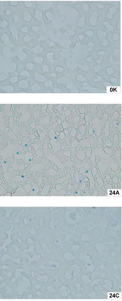
Fig. 7. Light microscopy of eosin/hematoxylin−unstained liver sections in all experimental groups of livers (as described in Fig. 1). Eosin/hematoxylin−unstained sections were used for counting the number of trypan blue−stained nuclei in relation to the total number of nuclei of non−parenchymal cells in certain regions of the liver
time from drug administration to the HTK solution to the end of perfusion was 26 hours. Although nifedipine and nitrendipine have very similar chem− ical structures, nitrendipine is 5–10 times more resistant to photodegradation than nifedipine, which can be significant in this experimental model [26].
The target of CCBs in the liver is not known. Many experiments demonstrated that these drugs changed the calcium concentration in cytoplasm and organelles. It has been suggested that this is a result of their action on calcium channels [27, 28]. However, there are studies in which treatment with drugs belonging to this group was not accompanied by any changes in cytoplasmic calcium ion level [2, 29].
It is worth emphasizing that the cytoprotective action of these drugs already manifests itself at much lower concentrations than those which block receptor−dependent calcium channels. Verapamil, diltiazem, and nifedipine block these channels at concentrations of 100–400 µM, whereas their pro− tective action can be observed already at concen− trations ranging between 15–45 µM [30]. At equally low concentrations (20 and 50 µM), both these drugs have a protective effect on hepatocytes exposed to the harmful action of ethanol [2]. Low concentrations of nitrendipine (10 µM) suppress calcium influx into the Kupffer cells induced by the calcium channel agonist BAY K 8644 [31]. Nifedipine, nitrendipine, and verapamil added to perfusion fluid at 20 and 50 µM improve the meta− bolic capacity of the liver as evaluated by the mea− surement of antipyrine concentration during extra−
corporeal perfusion of this organ [8]. Results of this experiment served as a basis for the choice of the 20−µM concentration of both nitrendipine and nifedipine in the present study, since at this con− centration the drugs showed stronger cytoprotec− tive action on liver cells than at 50 µM.
Since calcium ions are second messengers engaged in many processes, the action of these drugs on liver cells is connected with these ions, if not by directly blocking the channels, then possi− bly by their influence on calcium−related changes in injured liver cells. It has also been suggested that CCBs can stabilize the plasma membrane [32], protect the structure and function of mito− chondria [33] and lysosomes from damage [2], and increase hepatic blood flow [20]. These drugs probably have antioxidant properties [34]. They can also inhibit lipid peroxidation [35] and hypox− ia−induced apoptosis [36].
Despite numerous studies, the role of CCB in liver prepared for transplantation is still unsettled. CRS solution containing nicardipine (among other components) used for flushing the liver after stor− age is considered to be highly efficacious [37]. However, the results of the present study and reports from previous studies indicate different efficacies of the various drugs of this group which depend on many still largely unknown factors. Studies on the improvement of storage conditions should contribute to making this organ available to a wider group of patients for whom liver trans− plantation is the only effective treatment method.
References
[1] Gilbert JR, Pascual M, Schoenfeld DA, Rubin RH, Delmonico FL, Cosimi AB: Evolving trends in liver trans− plantation. Transplantation 1999, 67, 246–253.
[2] Cobreros A, Sainz L, Lasheras B, Cenarruzabeitia E: Hepatotoxicity of ethanol: protective effect of calcium channel blockers in isolated hepatocytes. Liver 1997, 17, 76–82.
[3] De Broin E, Urata K, Giroux L, Lepage R, Huet PM: Effect of calcium antagonists on rat liver during extend− ed cold preservation−reperfusion. Transplantation 1997, 63, 1547–1554.
[4] Jones TE, Morris RG: Pharmacokinetic interaction between tacrolimus and diltiazem. Clin Pharmacokinet 2002, 41, 381–388.
[5] Mies S, Massarollo PC, Figueira ER, Leitao RM, Raia S: Lower incidence of liver graft rejection in patients on diltiazem plus cyclosporine therapy. Transplant Proc 1998, 30, 1437–1438.
[6] Cheng S, Ragsdale JR, Sasaki AW, Lee RG, Deveney CW, Pinson CW: Verapamil improves rat hepatic preser− vation with UW solution. J Surg Res 1991, 50, 560–564.
[7] Lemasters JJ, DiGiuseppi J, Nieminen AL, Herman B: Blebbing, free Ca2+ and mitochondrial membrane potential preceding cell death in hepatocytes. Nature 1987, 325, 78–81.
[8] Szeląg A, Magdalan J, Rutkowska M, Dziewiszek W, Trocha M, Rzepka M, Pieśniewska M, Fereniec L:
Influence of nifedipine, nitrendipine and verapamil at low concentration on antipyrine metabolism examined by extracorporeal rat liver perfusion. Pol J Pharm 2003, 55, 203–208.
[9] Takeda Y, Arii S, Kaido T, Niwano M, Moriga T, Mori A, Hanaki K, Gorrin−Rivas MJ, Ishii T, Sato M, Imamura M: Morphologic alteration of hepatocytes and sinusoidal endothelial cells in rat fatty liver during cold preservation and the protective effect of hepatocyte growth factor. Transplantation 1999, 67, 820–828.
[10] Takei Y, Marzi I, Kauffman FC, Currin RT, Lemasters JJ, Thurman RG: Increase in survival time of liver transplants by protease inhibitors and a calcium channel blocker, nisoldipine. Transplantation 1990, 50, 14–20.
[12] Gao W, Bentley RC, Madden JF, Clavien PA: Apoptosis of sinusoidal endothelial cells is a critical mechanism of preservation injury in rat liver transplantation. Hepatology 1998, 27, 1652–1660.
[13] Schemmer P, Connor HD, Arteel GE, Raleigh JA, Bunzendahl H, Mason RP, Thurman RG: Reperfusion injury in livers due to gentle in situ organ manipulation during harvest involves hypoxia and free radicals. J Pharmacol Exp Ther 1999, 290, 235–240.
[14] Fujita Y, Kimura K, Takaori M: Influence of nicardipine on post−hypoxic injury in the isolated perfused rat liver. Resuscitation 1991, 22, 253–260.
[15] Umeshita K, Monden M, Ukei T, Gotoh M, Nakano Y, Endoh W, Okamura J, Mori T: Different cytoprotec− tive effects of calcium blockers in hypothermic liver preservation. Transplant Proc 1989, 21, 1290–1291.
[16] Iimuro Y, Ikejima K, Rose ML, Bradford BU, Thurman RG: Nimodipine a dihydropyridine−type calcium channel blocker prevents alcoholic hepatitis caused by chronic intragastric ethanol exposure in the rat. Hepatology 1996, 24, 391–397.
[17] Karwinski W, Garcia R, Helton WS: Protective effects of the calcium channel blocker verapamil on hepatic function following warm ischemia. J Surg Res 1999, 64, 150–155.
[18] Liang DC, Thurman RG: Protective effects of the calcium antagonists diltiazem and TA3090 against hepatic injury due to hypoxia. Biochem Pharmacol 1992, 44, 2207–2211.
[19] Gasbarrini A, Borle AB, Van Thiel DH: Ca2+ antagonists do not protect isolated perfused rat hepatocytes from anoxic injury. Biochim Biophys Acta 1993, 1177, 1–7.
[20] Matsuda S: Protective effects of calcium antagonist (nitrendipine) on calcium ionophore A 23187−induced liver cell injury. Bull Tokyo Med Dent Univ 1991, 38, 35–44.
[21] Thurman RG, Apel ED, Lemasters JJ: Protective effect of nitrendipine against hypoxic injury in perfused liv− ers from ethanol−treated rats. J Cardiovasc Pharmacol 1988, 12, Suppl. 4, S113–S116.
[22] Habior A: Liver diseases (Polish). In: Interna. Eds.: Januszkiewicz W, Kokot F, PZWL, Warsaw 2002, 558–591.
[23] Piratvisuth T, Dunne JB, Williams R, Tredger JM: Amlodipine improves hepatic hemodynamic and metabolic function in the isolated perfused rat liver after sequential cold and warm ischemia. Transplantation 1995, 60, 23–28.
[24] Bowers BA, Branum GD, Rotolo FS, Watters CR, Meyers WC: Bile flow – an index of ischaemic injury. J Surg Res 1987, 42, 565–569.
[25] Churchill TA, Green CJ, Fuller BJ: The importance of calcium−related effects on energetics at hypothermia: effects of membrane−channel antagonists on energy metabolism of rat liver. Cryobiology 1995, 32, 477–486.
[26] Bayomi MA, Abanumay KA, al−Angary AA: Effect of inclusion complexation with cyclodextrins on photosta− bility of nifedipine in solid state. Int J Pharm 2002, 243, 107–117.
[27] Haddad P, Cabrillac JC, Piche D, Musallam L, Huet PM: Changes in intracellular calcium induced by acute hypothermia in parenchymal, endothelial, and Kupffer cells of the rat liver. Cryobiology 1999, 39, 69–79.
[28] Lidofsky SD, Xie MH, Sostman A, Scharschmidt BF, Fitz JG: Vasopressin increases cytosolic sodium con− centration in hepatocytes and activates calcium influx through cation−selective channels. J Biol Chem 1993, 268, 14632–14636.
[29] Konrad T, Bloechle C, Haller G, Broelsch CE, Usadel KH, Kusterer: Verapamil and flunarizine protect the isolated perfused rat liver against warm ischemia and reperfusion injury. Res Exp Med 1995, 195, 61–68.
[30] Wu J, Danielsson A, Lindstrom P, Karlsson K, Sehlin J: Protective effects of calcium channel blockers on acute bromobenzene toxicity to isolated rat hepatocytes. Inhibition of phenylephrine−induced calcium oscillations. Scand J Gastroenterol 1995, 30, 590–600.
[31] Hijioka T, Rosenberg RL, Lemasters JJ, Thurman RG: Kupffer cells contain voltage−dependent calcium chan− nels. Mol Pharmacol 1992, 41, 435–440.
[32] Kurita K, Tanabe G, Aikou T, Shimazu H: Inhibition of the increase of intrahepatic Ca2+ by diltiazem in rats with liver ischemia. J Hepatol 1994, 21, 567–571.
[33] Michael AD, Whiting RL: Cellular action of nicardipine. Am J Cardiol 1989, 64, 3H–7H.
[34] Stein HJ, Oosthuizen MM, Hinder RA: Effect of verapamil on hepatic ischaemic−reperfusion injury in normal and glutathione−depleted rats. Gastroenterology 1989, 96, A662–A663.
[35] Mak IT, Weglicki WB: Comparative antioxidant activities of propranolol nifedipine verapamil and diltiazem against sarcolemmal membrane lipid peroxidation. Circ Res 1990, 66, 1449–1452.
[36] Crenesse D, Tornieri K, Laurens M, Heurteaux C, Cursio R, Gugenheim J, Schmid−Alliana A: Diltiazem reduces apoptosis in rat hepatocytes subjected to warm hypoxia−reoxygenation. Pharmacology 2002, 65, 87–95.
[37] Abdennebi HB, Steghens JP, Margonari J, Ramella−Virieux S, Barbieux A, Boillot O: Evaluation of parenchymal and nonparenchymal cell injury after different conditions of storage and reperfusion. Transpl Int 1998, 11, 365–372.
Address for correspondence:
Małgorzata Trocha
Department of Pharmacology
Silesian Piasts University of Medicine Mikulicza−Radeckiego 2
50−345 Wrocław Poland
Tel.: +48 71 784 14 42 E−mail: [email protected]
Conflict of interest: None declared