Sebastian Niedźwiecki
A–F, Janusz Piekarski
A–F, Arkadiusz Jeziorski
A–FThe Clinical and Histopathological Factors
in Patients Operated on for Gastric GIST Tumors
with Unclear Diagnosis
Department of Surgical Oncology, Medical University of Łódź, Poland
A – research concept and design; B – collection and/or assembly of data; C – data analysis and interpretation;
D – writing the article; E – critical revision of the article; F – final approval of article; G – other
Abstract
Background. The preoperative radiological diagnosis of GIST is complicated by its varied macroscopic morphol-ogy. Moreover, the precision of preoperative histopathological diagnostics is reduced by the submucosal localiza-tion of the lesion.
Objectives. The goal of the study was to perform a retrospective analysis of the clinical and histopathological fac-tors seen in patients operated on for a stomach GIST tumor with unclear diagnosis.
Material and Methods. Two groups of GIST patients treated in our department were compared with regard to their histopathological and clinical data. The first group (9 patients, group 1) comprised patients with a histopatho-logical diagnosis for stomach GIST confirmed before the surgical procedure, while the second group (10 patients, group 2) comprised patients with no solid histopathological diagnosis before surgery. The following clinical and histopathological variables were analyzed in the study: age, gender, presence or absence of metastases, anatomi-cal location of metastases, symptoms, tumor size, surgianatomi-cal mortality, tumor recurrence, treatment with imatinib, patient survival in months, histological subtype, mitotic index, cellular atypia, necrosis, tumor ulceration and Ki-67. The results were analyzed statistically.
Results. The mean survival time differed significantly between the two study groups: group 1 being 12 months and group 2 being 8 months. The lower survival time in group 2 was connected with the higher stage of the disease at the moment of diagnosis.
Conclusions. Our findings suggest that GIST tumors with an unclear diagnosis are recognized at a late stage of the disease. The more advanced stage of the tumor probably results from faster tumor growth caused by higher prolif-eration activity. These GIST tumors are characterized by a lower survival rate due to the later stage of the disease at the time of diagnosis (Adv Clin Exp Med 2014, 23, 4, 567–573).
Key words: gastric GIST tumor, difficult diagnostic GIST, difficulties in diagnosis. Adv Clin Exp Med 2014, 23, 4, 567–573
ISSN 1899–5276
ORIGINAL PAPERS
© Copyright by Wroclaw Medical University
Gastrointestinal stromal tumors (GIST) occur in only roughly 2% of gastric neoplasms [1]. How-ever, GISTs have recently become the most com-mon mesenchymal malignancies diagnosed in the stomach [2]. The stomach is also the most com-mon location of gastrointestinal GIST (60% of cas-es) [3]. The annual incidence of GIST is estimated between 1 and 2 cases per 100000 population [4].
The malignant transformation of interstitial Cajal cells has been proven to be crucial for GIST development. As the molecular background of the neoplastic changeover of Cajal cells is believed to
a key method in GIST management [8]. Radiother-apy is not in common use as a treatment for GISTs and is suggested only in selected cases [9].
Size and mitotic index have been identified as risk factors in GIST malignancy [10]. Other fea-tures, such as location, proliferation index and mutation type, are suggested to influence the pres-ence or abspres-ence of metastases, and recurrpres-ence of tumors [10]. Many risk scores (e.g. NIH criteria, AFIP criteria) have been formulated regarding the above-mentioned factors, enabling the prediction of tumor recurrence, metastases and survival [11].
GISTs present a wide range of clinical mani-festations from totally asymptomatic to symptoms typical of the malignancy (e.g. weight loss, abdomi-nal tumor) [3]. Some authors estimate that 15–30% of GIST tumors are asymptomatic and discovered incidentally [4, 12, 13]. The most commonly used imaging methods for GIST diagnosis are ultra-sonography and computer tomography [14], while PET/CT is used more for monitoring the course of treatment than for diagnosis [15]. The preoper-ative radiological diagnosis of GIST is complicat-ed by its diverse macroscopic morphologies [16]. Some authors speculate that it is hard to deter-mine the origin organ of a GIST tumor larger than 3 centimeters [13]. Preoperative histopathological diagnostics appear to be imprecise when the lesion is located in the submucosal area, in which case the specimen collected during biopsy is often nondiag-nostic [17]. Therefore, only 15% of individuals are suspected of being GIST patients prior to the op-eration [18].
The literature about GIST tumors with ob-scure diagnoses is poor, and no analysis of the sur-vival rate of patients with the condition could be found.
Patients and Methods
The goal of the study was to perform a ret-rospective analysis of clinical and histopatholog-ical factors in patients operated on due to stomach GIST tumors which presented difficult diagnoses. The present retrospective study analyzes a num-ber of anathomopathological and clinical factors seen in patients operated on for GIST tumors in the stomach, over a period of 10 years. Two groups of GIST patients treated in our department were compared, with regard to histopathological and clinical data. The first group (group 1) consisted of patients with a histopathological diagnosis for stomach GIST confirmed before the surgical pro-cedure, while the second group comprised those operated on without a solid histopathological di-agnosis before surgery.
Patients
From 2001 to 2010, 19 patients (10 women, 9 men) received a laparotomy due to stomach GIST or suspected stomach GIST tumor. In each patient, an endoscopy was carried out preoperatively with tissue sample collection or core needle biopsy.
A group of 9 patients (group 1) was composed of individuals with preoperative proof of stomach GIST, diagnosed by tissue sample collection dur-ing endoscopy. The GIST tumors diagnosed pre-operatively were located in the stomach.
The remaining individuals (10 patients) were enrolled to group 2. These subjects were not found to be GIST patients during histopathological diag-nosis based on tissue sample collection or core nee-dle biopsy during endoscopy. The patients whose preoperative histopathological diagnosis suggest-ed other neoplasms were excludsuggest-ed from the anal-ysis. Other exclusion criteria were the primary lo-calization of the tumor being elsewhere than the stomach as indicated by preoperative diagnostics (endoscopy and/or computer tomography and/or ultrasonography) or a previous history of abdomi-nal malignancy.
Clinical Parameters
The following clinical variables were analyzed in the study: age, gender, presence or absence of metastases, anatomical location of metastases, symptoms, tumor size, surgical mortality, tumor recurrence, treatment with imatinib, patient sur-vival in months.
Histopathological Parameters
The tumor samples were conventionally insert-ed in 10% neutral formalin, embinsert-eddinsert-ed in paraffin and then deparaffinized. Finally, the histopatho-logical sections were stained with hematoxylin– eosin for further evaluation.
The following histopathological variables were evaluated: histological subtype (fusocellular, epi-thelioid, or mixed), mitotic index, cellular atypia (low, moderate, and high), necrosis, tumor ulcer-ation and Ki-67 (proliferative index).
Statistical Analysis
and the log-rank test. P values < 0.05 were consid-ered statistically significant. All statistical analyz-es were done using Statistica 10 PL (StatSoft, Tul-sa, USA).
Results
Characteristics of the Patients
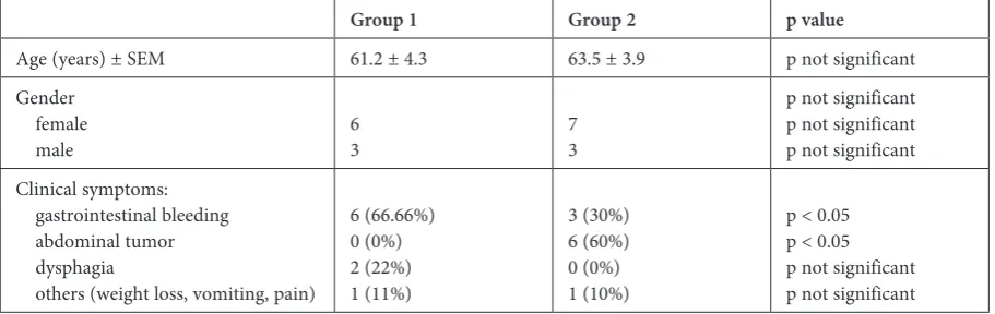
The clinical characteristics of the patients are presented in Table 1. The mean age of the patients was 62.3 (from 45 to 82 years). All patients of group 1 were clearly symptomatic, mostly with up-per gastrointestinal bleeding and dysphagia. Two of these patients presented gastrointestinal bleed-ing and abdominal pain simultaneously at the time of diagnosis. Although all patients in group 2 were also symptomatic, an abdominal tumor was the first and only symptom of GIST in sixty percent of them. Gastrointestinal bleeding was significant-ly more common in patients of group 1, while ab-dominal tumors were significantly more common in group 2. No other statistically significant differ-ences were seen between the groups regarding oth-er clinical symptoms.
Histopathological
and Immunohistochemical
Characteristics
The histopathological and immunohistochem-ical features are shown in Table 2. At the time of diagnosis, the metastases were noted in 70% of group 2 compared to 44% of group 1 (statistically significant, p < 0.05). Tumor size was significantly greater in group 2 (2.3 cm vs. 6.2 cm; p < 0.05).
Ki-67 expression was significantly higher in group 2 (70% vs. 33%, p < 0.05). Elevated Ki-67 expression was associated with high mitotic in-dex and high histological grade (p < 0.05). High
Ki-67 index was associated with low survival. Tu-mor necrosis and intense cellular atypia tended to be more common in group 2, although this ten-dency was not found to be significant.
The metastases were located in the liver and peritoneum in both groups.
Survival
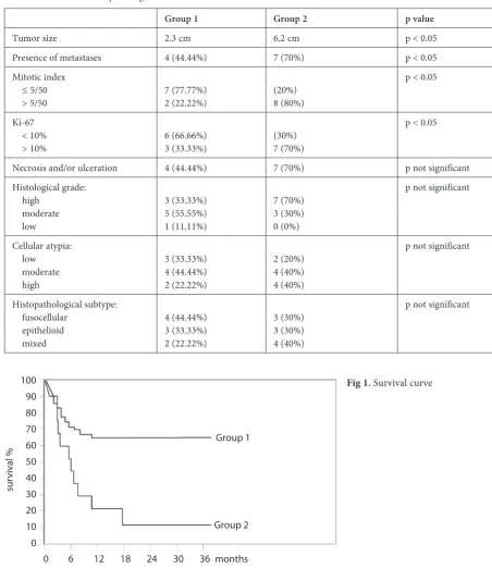
No patients from either study group died in the perioperative period. The maximal follow-up time of some analyzed patients was 38 months. The two-year survival rate varied significantly between both groups and was 66.66% and 10%, respectively (95% CI = 19.2–32.3 and 95% CI = 6.3–24.2) (Fig. 1).
The mean survival time differed significant-ly between the two study groups. The mean sur-vival in group 1 was 12 months, compared to 8 months in group 2 (95% CI = 4.2–13.3 and 95% CI = 2.3–9.2).
All patients in group 1, and only 3 individu-als in group 2, received adjuvant therapy with ima-tinib. The survival difference among group 2 be-tween patients treated with imatinib and patients without adjuvant therapy was not statistically significant.
Complete resection (R0) was feasible in 5 pa-tients of group 1 and, among those, GIST recur-rence occurred in 3 cases. Complete resection (R0) was achieved by partial resection in 3 patients of group 1, and total gastrectomy in 2 of group 1.
Radical resection (R0) was performed in only 3 individuals of group 2. In all patients of group 2, the GIST recurrence rate was 100%, despite total gastrectomy being performed.
Discussion
GISTs may present a wide spectrum of biolog-ical activity from totally indolent to highly
malig-Table 1. Patient characteristics
Group 1 Group 2 p value
Age (years) ± SEM 61.2 ± 4.3 63.5 ± 3.9 p not significant
Gender female
male 63 73
p not significant p not significant p not significant Clinical symptoms:
gastrointestinal bleeding abdominal tumor dysphagia
others (weight loss, vomiting, pain)
6 (66.66%) 0 (0%) 2 (22%) 1 (11%)
3 (30%) 6 (60%) 0 (0%) 1 (10%)
nant. Molecular targeted therapy has changed the treatment and prognosis for GIST patients, though surgical resection remains a key therapy. Howev-er, total R0 resection has a significant influence on treatment outcome (survival) and is attained in ap-proximately 40% of all cases of GIST [8]. The type of surgical procedure depends on tumor location and size. However, the main goal of the surgery is the achievement of negative margins. Thus, local resection of GIST lesions is recognized as a gold standard for surgical treatment [19]. Although all
patients from our study groups were treated surgi-cally, R0 resection was completed in only approxi-mately 55% of group 1 and in 20% of group 2. The reason for the incomplete resection in a high num-ber of patients was the advanced stage of the disease at the moment of diagnosis. Two factors which in-fluence survival are GIST tumor size and the pres-ence of a negative microscopic surgical margin. In some situations, it is necessary to perform a more extensive procedure to achieve microscopic nega-tive margins [20].
Fig 1. Survival curve
Group 1
Group 2
0 6 12 18 24 30 36 months 100
90 80 70 60 50 40 30 20 10 0
sur
vival
%
Table 2. Clinical and histopathological features
Group 1 Group 2 p value
Tumor size 2.3 cm 6.2 cm p < 0.05
Presence of metastases 4 (44.44%) 7 (70%) p < 0.05
Mitotic index ≤ 5/50
> 5/50 7 (77.77%)2 (22.22%) (20%)8 (80%)
p < 0.05
Ki-67 < 10%
> 10% 6 (66.66%)3 (33.33%) (30%)7 (70%)
p < 0.05
Necrosis and/or ulceration 4 (44.44%) 7 (70%) p not significant
Histological grade: high
moderate low
3 (33.33%) 5 (55.55%) 1 (11.11%)
7 (70%) 3 (30%) 0 (0%)
p not significant
Cellular atypia: low moderate high
3 (33.33%) 4 (44.44%) 2 (22.22%)
2 (20%) 4 (40%) 4 (40%)
p not significant
Histopathological subtype: fusocellular
epithelioid mixed
4 (44.44%) 3 (33.33%) 2 (22.22%)
3 (30%) 3 (30%) 4 (40%)
Recent publications have shown complete re-section in 40–60% of operated patients [10, 21, 23]. However, the mean tumor size given in these stud-ies was 1.5–4.5 cm, compared to 2.3–6.5 cm in the present study. Moreover, the mitotic index was significantly lower in the above-mentioned studies compared to the present study.
It is estimated that approximately 15–30% of GIST patients are asymptomatic with incidental findings of GIST [4, 12, 13]. In our study, all pa-tients were symptomatic, mostly with gastrointes-tinal bleeding, and no GIST case was discovered incidentally. Most frequent clinical manifesta-tions described in the literature are gastrointesti-nal bleeding, abdomigastrointesti-nal pain, weight loss, abdom-inal tumor, dysphagia, nausea and vomiting [12]. Its symptoms and incidence in our patients did not vary significantly from the literature data.
Statistical studies indicate that most patients with gastric GIST are 60–70 years old [24, 25]. In our study, the mean age of patients was within the above-mentioned range and no patients were younger than 45 years old.
A variety of problems can potentially be in-volved in the preoperative diagnosis of gastroin-testinal stromal tumors, some of which result from the low incidence of GIST and limited experience of clinicians and radiologists. However, the main reason for a lack of a final preoperative histopatho-logical diagnosis is the diversity of macroscop-ic GIST morphologies [13, 26]. Nowadays, due to its high availability, the first diagnostic of ab-dominal symptoms is ultrasonography. An ultra-sound image of GIST usually shows a hypoecho-genic lesion.[27] However, while the echohypoecho-genic pattern of a small tumor is homogeneous, the pat-tern of a larger lesion is heterogeneous. The vari-ety of ultrasound GIST images comprises a wide range of images including cyst, solid tissue or both in one tumor. The establishment of the exact GIST organ of origin is usually difficult in cases where the tumor size is greater than 3 cm [13, 27]. In ad-dition, the possibility of detecting GIST lesions of the gastrointestinal tract by ultrasound scans is sig-nificantly reduced because they develop inside the wall. However, ultrasonography is a valuable meth-od for locating liver and peritoneum metastases. In our series, ultrasound diagnostics were only car-ried out in patients with abdominal tumors or pain at the beginning of the diagnostic process, and was useful only to confirm the suspicion of malignancy based on the presence of metastases.
Computer tomography is also limited in the diagnosis of gastric GIST due to a lack of any char-acteristic features of GISTs [26]. The computer to-mography image of gastric GIST is very similar to other tumors of the gastrointestinal wall (e.g.
sarcomas, carcinoid tumors etc.) [26]. Moreover, in the case of large lesions, the point of origin is difficult to ascertain due to invasions of surround-ing organs [26].
In the present study, no imaging method fa-cilitated a correct diagnosis of GIST. Neither CT scans nor ultrasound scans showed the point of or-igin of large tumors and the results of these im-aging methods were also uncharacteristic of small lesions.
It seems that the best diagnostic method for gastric GIST is endoscopy. However, the lesion may be impossible to detect endoscopically due to its submucosal location [13]. A typical endo-scopic image of gastric GIST registers a submu-cosal tumor or wall elevation as possessing a nor-mal mucosal surface. An ulceration on the surface of a lesion is observed during endoscopy in some cases of gastric GIST. Ulceration of the GIST sur-face usually contains a normal mucous cell [11]. Therefore, a tissue sample collected from the ulcer-ation cannot usually provide a definite histopatho-logical diagnosis.
Due to the submucosal location of the GIST le-sion, a preoperative histopathological diagnosis of gastric GIST based on endoscopy with tissue sam-ple collection is unclear in 15–50% of GIST cas-es [11, 13]. Fine needle aspiration biopsy guided with ultrasonography is characterized by low ac-curacy [13].
Fine needle aspiration biopsyis a method in-tended for palpable tumors whose origin and char-acter is difficult to establish; these tumors usually present a solid-cystic architecture. The main dis-advantage of GIST biopsy is the possibility of the sample being collected from the non-diagnostic area of the cystic part of the tumor. In all group 1 patients, the GIST diagnosis was given based on endoscopy with tissue sample collection.
The Ki-67 protein is a protein involved in cell proliferation. Neto et al. demonstrate a strong sta-tistical correlation between Ki-67, histological grade, mitotic index and presence of necrosis [1]. The authors also suggest a statistically significant relationship between Ki-67 expression and poor survival rate. The present study shows comparable results. A significant difference was seen in Ki-67 expression between both study groups. A signifi-cantly lower survival rate was seen in patients with difficulties ingastric GIST diagnosis (group 2), as well as a correlation between Ki-67 expression, mi-totic index and histological grade.
Only the survival rate of group 2 patients differed dramatically from published data.
Mitotic count and size are the most recognized prognostic factors for GIST tumors [30]. The mi-totic count and size were significantly greater in group 2 than in group 1, as well as in data from re-cent publications. Besides other factors such as cel-lular atypia, a high histological grade and the pres-ence of metastases determined high proliferation activity and low survival rate in group 2 patients.
This study is limited by being retrospec-tive and the small number of patients in the sub-groups. However, our findings suggest that GIST tumors with a difficult diagnosis are diagnosed in a late stage of the disease. The more advanced stage of the tumor probably results from faster tumor growth due to higher proliferation activity.
GIST tumors with a difficult diagnosis are characterized by a lower survival rate due to the later stage of the disease at the time of diagnosis.
Acknowledgements. The authors thank Ed Lowczowski, language consultant.
References
Neto RA, Logullo AF, Stávale JN, Lourenço LG:
[1] Ki-67 expression score correlates to survival rate in gastrointes-tinal stromal tumors (GIST). Acta Cirúrgica Brasileira 2012, 27, 315–321.
Halpern J, Kim YJ, Sultana R, Villani G:
[2] Effectiveness of radiation therapy in GIST: A case report. J Gastrointest Oncol 2012, 3, 143–146.
Zhou L, Liu C, Bai JG, Wei JC, Qu K, Tian F, Tai MH, Wang RT, Meng FD:
[3] A rare giant gastrointestinal
stro-mal tumor (GIST) of the stomach traversing the upper abdomen: a case report and literature review. World J Surg Oncol 2012, 10, 66–70.
Valls-Ferrusola E, García-Garzón JR, Ponce-López A, Soler-Peter M, Fuertes-Cabero S, Moragas-Solanes M, [4]
Riera-Gil E, Carrió-Gasset I, Lomeña-Caballero F: Patterns of extension of gastrointestinal stromal tumors (GIST) treated with imatinib (Gleevec)® by 18F-FDG PET/CT REV ESP ENFERM DIG 2012, 104, 360–366.
Hirota S, Isozaki K, Moriyama Y, Hashimoto K, Nishida T, Ishiguro S, Kawano K, Hanada M, Kurata A, [5]
Takeda M, Muhammad Tunio G, Matsuzawa Y, Kanakura Y, Shinomura Y, Kitamura Y: Gain-of-function mutations of c-kit in human gastrointestinal stromal tumors. Science 1998, 279, 577–580.
Singer S, Rubin BP, Lux ML, Chen CJ, Demetri GD, Fletcher CD, Fletcher JA:
[6] Prognostic value of KIT mutation
type, mitotic activity, and histologic subtype in gastrointestinal stromal tumors. J Clin Oncol 2002, 20, 3898–3905.
Dasanu CA:
[7] Length of adjuvant imatinib therapy in GIST: Weighing benefits, side effects and costs. J Oncol Pharm Pract 2012, 18, 379–380.
Valadăo M, Linhares E:
[8] The role of the surgeon in the management of GIST. Rev Col Bras Cir 2009, 36, 261–265.
Ciresa M, D’Angelillo RM, Ramella S, Cellini F, Gaudino D, Stimato G, Fiore M, Greco C, Nudo R, Trodella L: [9]
Molecularly targeted therapy and radiotherapy in the management of localized gastrointestinal stromal tumor (GIST) of the rectum: a case report. Tumori 2009, 95, 236–233.
Sanchez Hidalgo JM, Rufian Peña S, Ciria Bru R, Naranjo Torres A, Muñoz Casares C, Ruiz Rabelo J, Briceño [10]
Delgado J: Gastrointestinal Stromal Tumors (GIST): A Prospective Evaluation of Risk Factors and Prognostic Scores. J Gastrointest Canc 2010, 41, 27–37.
Miettinen M, Lasota J:
[11] Gastrointestinal stromal tumors. Review on morphology, molecular pathology, prognosis, and differential diagnosis. Arch Pathol Lab Med 2006, 130, 1466–1478.
Stamatakos M, Douzinas E, Stefanaki C, Safioleas P, Polyzou E, Levidou G, Safioleas M:
[12] Gastrointestinal
strom-al tumor. World J Surg Oncol 2009, 7, 61–69.
Wroński M, Cebulski W, Pawłowski W, Krasnodębski IW:
[13] Diagnostic difficulties in patients with gastrointesti-nal stromal tumour. Przegl Gastroenterol 2006, 1, 115–120.
Burkill GJ, Badran M, Al-Muderis O, Meirion Thomas J, Judson IR, Fisher C, Moskovic EC:
[14] Malignant
gas-trointestinal stromal tumor: distribution, imaging features, and pattern of metastatic spread. Radiology 2003, 226, 527–532.
Otomi Y, Otsuka H, Morita N, Terazawa K, Furutani K, Harada M, Nishitani H:
[15] Relationship between FDG
uptake and the pathological risk category in gastrointestinal stromal tumors. J Med Invest 2010, 57, 270–274.
Wang CM, Fu H, Zhao GF, Wang J, Shi YQ:
[16] CT Scan is not Everything in the Evaluation of a Patient with Gastrointestinal Tumors (GIST) Under Imatinib Therapy. Pathol Oncol Res 2012, 18, 1095–1097.
Kim CJ, Day S, Yeh KA:
[17] Gastrointestinal stromal tumours: analysis of clinical and pathologic factors. Am Surg 2001, 67, 135–137.
Ludwig DJ, Traverso LW:
[18] Gut stromal tumours and their clinical behavior. Am J Surg 1997, 173, 390–394.
DeMatteo RP, Lewis JJ, Leung D, Mudan SS, Woodruff JM, Brennan MF:
[19] Two hundred gastrointestinal stromal
tumors: recurrence patterns and prognostic factors for survival. Ann Surg 2000, 231, 51–58.
Demetri GD, von Mehren M, Antonescu CR, DeMatteo RP, Ganjoo KN, Maki RG, Pisters PW, Raut CP, [20]
Riedel RF, Schuetze S, Sundar HM, Trent JC, Wayne JD: NCCN Task Force report: update on the management of patients with gastrointestinal stromal tumors. J Natl Compr Canc Netw 2010, 8, Suppl 2, 1–41.
El-Hanafy E, El-Hemaly M, Hamdy E, El-Raouf AA, El-Hak NG, Atif E:
[21] Surgical management of gastric
Dematteo RP, Gold JS, Saran L, Gönen M, Liau KH, Maki RG, Singer S, Besmer P, Brennan MF, Antonescu CR: [22]
Tumor mitotic rate, size, and location independently predict recurrence after resection of primary gastrointestinal stromal tumor (GIST). Cancer 2008, 1, 112, 608–615.
Henckens T, Van de Putte D, Van Renterghem K, Ceelen W, Pattyn P, Van Nieuwenhove Y:
[23] Laparoendoscopic
single-site gastrectomy for a gastric GIST using double-bended instruments. J Laparoendosc Adv Surg Tech A 2010, 20, 469–471.
Novitsky YW, Kercher KW, Sing RF, Heniford BT:
[24] Long-term Outcomes of Laparoscopic Resection of Gastric Gastrointestinal Stromal Tumors. Ann Surg 2006, 243, 738–745.
Bümming P, Ahlman H, Andersson J, Meis-Kindblom JM, Kindblom LG, Nilsson B:
[25] Population-based study of
the diagnosis and treatment of gastrointestinal stromal tumours. Br J Surg 2006, 93, 836–843.
Werewka-Maczuga A, Osiński T, Chrzan R, Buczek M, Urbanik A:
[26] Characteristics of computed tomography
imaging of gastrointestinal stromal tumor (GIST) and related diagnostic problems. Pol J Radiol 2011, 76, 38–48.
Kawamoto K, Yamada Y, Utsunomiya T, Okamura H, Mizuguchi M, Motooka M, Hirata N, Watanabe H, [27]
Sakai K, Kitagawa S, Kinukawa N, Masuda K: Gastrointestinal submucosal tumours: evaluation with endoscopic US. Radiology 1997, 205, 733–740.
DeMatteo RP, Lewis JJ, Leung D, Mudan SS, Woodruff JM, Brennan MF:
[28] Two Hundred Gastrointestinal Stromal
Tumors Recurrence Patterns and Prognostic Factors for Survival Annals of Surgery 2000, 231, 51–58.
Sánchez Hidalgo JM, Muñoz Casares FC, Rufian Peña S, Naranjo Torres A, Ciria Bru R, Briceño Delgado J, [29]
López Cillero P: Gastrointestinal stromal tumors (GIST): factors predictive of survival after R0-cytoreduction. Rev Esp Enferm Dig 2007, 99, 703–708.
Aparicio T, Boige V, Sabourin JC, Crenn P, Ducreux M, Le Cesne A, Bonvalot S:
[30] Prognostic factors after surgery
of primary resectable gastrointestinal stromal tumours. Eur J Surg Oncol 2004, 30, 1098–1103.
Address for correspondence:
Sebastian Niedźwiecki
Department of Surgical Oncology Medical University of Łódź Paderewskiego 4
93-509 Łódź Poland
Tel.: 042 689 54 41
E-mail: [email protected]
Conflict of interest: None declared