R E S E A R C H
Open Access
Bright field microscopic cells counting method
for BEVS using nonlinear convergence index
sliding band filter
Dong Sui
1, Kuanquan Wang
1*, Heemin Park
2and Jinseok Chae
3** Correspondence:[email protected]; [email protected]
1Biocomputing Research Center,
School of Computer Science and Technology, Harbin Institute of Technology, Harbin, China 3Department of Computer Science
and Engineering, Incheon National University, Incheon, Korea Full list of author information is available at the end of the article
Abstract
Background:The Baculovirus Expression Vector System (BEVS) is a very popular expression vector system in gene engineering. An effective host cell line cultivation protocol can facilitate the baculovirus preparation and following experiments. However, the counting of the number of host cells in the protocol is usually performed by manual observation with microscopy, which is time consuming and labor intensive work, and prone to errors for one person or between different individuals. This study aims at giving a bright field insect cells counting protocol to help improve the efficient of BEVS.
Method:To develop a reliable and accurate counting method for the host cells in the bright field, such as Sf9 insect cells, a novel method based on a nonlinear Transformed Sliding Band Filter (TSBF) was proposed. And 3 collaborators counted cells at the same time to produce the ground truth for evaluation. The performance of TSBF method was evaluated with the image datasets of Sf9 insect cells according to the different periods of cell cultivation on the cell density, error rate and growth curve.
Results:The average error rate of our TSBF method is 2.21% on average, ranging from 0.89% to 3.97%, which exhibited an excellent performance with its high accuracy in lower error rate compared with traditional methods and manual counting. And the growth curve was much the manual method well.
Conclusion:Results suggest the proposed TSBF method can detect insect cells with low error rate, and it is suitable for the counting task in BEVS to take the place of manual counting by humans. Growth curve results can reflect the cells’growth manner, which was generated by our proposed TSBF method in this paper can reflected the similar manner with it’s from the manual method. All of these proven that the proposed insect cell counting method can clearly improve the efficiency of BEVS.
Keywords:Cell counting, Transformed sliding band filter, BEVS, Microscopy image processing
Background
Biology background
The baculoviruses are double-stranded DNA viruses containing a large genome up to 180,000 bp, and are arthropod specific viruses with two different phenotypes during different infection stages [1,2] including budded viruses (BVs) and occlusion-derived viruses (ODVs). Depending on the distinct structural and biological characteristics, the
© 2014 Sui et al.; licensee BioMed Central Ltd. This is an Open Access article distributed under the terms of the Creative Commons Attribution License (http://creativecommons.org/licenses/by/4.0), which permits unrestricted use, distribution, and reproduction in any medium, provided the original work is properly credited. The Creative Commons Public Domain Dedication waiver (http:// creativecommons.org/publicdomain/zero/1.0/) applies to the data made available in this article, unless otherwise stated.
Suiet al. BioMedical Engineering OnLine2014,13:147
baculoviridae family can be divided into four genera: α-baculovirus, β-baculovirus, γ -baculovirus, and δ-baculovirus [2,3]. Traditionally, these kinds of arthropod specific viruses are employed to control insect pests in soybean fields, cotton bollworm and helicoverpa armigera. Very recently, with the development of microbiology and vir-ology, the special properties of high level expression for very late genes make them suitable vectors for the delivery of foreign genes. So far, Autographa California Multiple Nucleopolyhedrovirus (AcMNPV) and lesser extent Bombyxmori Nucleopolyhedrovirus (BMNPV) are broadly applied as alien gene vectors to produce recombinant proteins in many hosts [2,3].
The baculovirus expression vector system (BEVS) has been widely used to express heterologous genes in various cell lines since the mid 1980’s [4]. With recent advances in cell culture and molecular manipulations, many special media, transfection reagents and expression vectors have been developed for applications. As an excellent expression system, BEVS contains efficient promoters that can provide ideal production of the re-combinant proteins. Its host insect cell lines, serum-supplemented or serum-free growth media and infection strategies also allow optimal virus production and gene ex-pression. In addition, the scalable process for the culture of insect cell lines makes the downstream processing more convenient in labs. Together, these advantages enable its large-scale applications in gene expression and protein production. Furthermore, the broad range of susceptible cell lines and the nature in cells entrancing without toxic and replication make baculovirus an excellent tool for studying the expression and function of genes, which allows the BEVS to be successfully applied to gene therapy, pharmaceutical and vaccine productions.
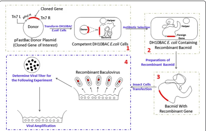
Figure 1 explains the classical protocol of recombinant-baculoviruses production and gene expression using the Bac-to-Bac Expression System. Firstly, a recombinant donor plasmid is constructed, and then the plasmid was transformed into DH10BAC E.coli
competent cells (step 1 in Figure 1) to produce recombinant Bacmid through homolo-gous recombination (step 2 in Figure 1). After preparing of recombinant Bacmid (step 3 in Figure 1), the host insect cells, are transfected by the extracted Bacmid. Finally, the re-combinant baculovirus containing a cloned gene is prepared from the product of insect cell disruptions (step 4 in Figure 1).
As Figure 1 shows, the hosts, such as Sf9 insect cells, are critical for producing the recombinant baculovirus and the insect cells’density (1 × 10⁶-2 × 10⁶cells/ml) are very important for the follow-up experiments. An effective culture protocol of insect cells can facilitate the virus preparation. In spite of its key rolls, the counting of insect cells usually takes lots of time and is labor intensive by traditional methods in lab because it is usually manipulated by humans under microscopy. Moreover, traditional methods are even prone to cause errors without being repeated by different people. It should be noted that there are still no efficient computer-aided methods to solve these problems in regard to the BEVS protocol. In this paper, we propose a bright field insect cell counting method based on the Nonlinear Convergence Index Sliding Band Filter to im-prove the protocol efficiency.
Related works
Cell counting is an indispensable and essential problem because it directly affects the efficiency of many cell-based gene expression systems like BVES. Traditionally, this task is usually performed by microscopic-based counting. For example, the Neubauer, Burker and Fuchs-Rosenthal chambers are well known methods for counting cells in different cell concentration of interest [5]. However, all of these methods have to be manually manipulated and therefore are prone to cause errors for the same person or different persons. Furthermore, most of them require frequent repetitions for valida-tions [6]. In the 1940s, Wallace Coulter introduced a suspended particles counting method in a fluid to provide an automatic cell counting tool without lab worker de-pendencies, which is a milestone in solving cell counting automatically [5]. Following this milestone, automated human blood counting tools based on microscopic image analyses with high performance became commercially available. However, there are still many defects to be improved [7-9]. All of these defects should be addressed in order to develop automatic cell counting and analysis tools to facilitate the cell based experi-ments. In this paper, we focus on an image processing based insect cell counting method for BVES. To the best of our knowledge, there are no existing methods that have the same purpose with our proposed method. Beyond that, the proposed method can also be applied to other kinds of cells under microscopy with a bright field.
As a basic problem of computer-aided biology experiments, microscopic cell image analysis in a dark field has been explored by many researchers. Image processing based cell counting methods have been widely used for cell-based biological experiments dur-ing the past decades, especially for red blood cells and white blood cells [10]. Detectdur-ing and counting cells in microscopy images for biological systems is a key factor that af-fects system efficiency. Traditionally, classical computer-aided methods solve this prob-lem by applying image segmentation, which is a fundamental but difficult probprob-lem in computer vision. These methods mainly focus on cell images with high contrast be-tween cells and their background; this can be described as the dark field cell identifica-tion problem. Several image filters and segmentaidentifica-tion methods were employed for cell
Suiet al. BioMedical Engineering OnLine2014,13:147 Page 3 of 17
identification and counting in microscopy images, such as, watershed transform-based methods [11], morphological operators-based methods [12], gray level threshold based methods [13], contour or region-based methods [14], minimum-error-threshold histogram-based methods [14], and Artificial neural network (ANN) was also used for investigating the same problem [14]. On the other hand, various commercial or free software have been developed for solving cell counting in different image datasets, such as ImageJ and Cellprofiler [7]. However, most of these methods and software mainly fo-cused on processing some types of cells or cell lines and cannot be applied to other cell types. Another problem regarding these methods is that they are either semi-automated or require re-adjusting parameters on the same images to obtain accurate results. Additionally, the aforementioned methods and commercial software cannot ob-tain accurate results without human interventions, and some methods must be manip-ulated by experienced biology technical staffs that are trained in digital image processing. It can draw a conclusion that more robust and reliable tools are needed for the biotechnologist in larger cell image data sets. Furthermore, another challenge we have faced is the bright field cell counting which has had rare attentions so far.
In this paper, we propose an effective cell counting method for the insect-baculovirus expression system using Transformed Sliding Band Filter (TSBF), which is a new image-based microscopy cell counting method under a bright field. The TSBF method is based on a nonlinear filter, Sliding Band Filter (SBF), which was proposed for lung nodule detection and dark field fluorescein stained cell nuclei identification [15-17], and was also employed for density packed cell counting in another work of our group. This filter is based on gradient vector convergence which is capable of detecting low contrast cell nuclei and cytoplasm information lost in the background noise [15]. It can also reduce the uncertainty caused by noise. Furthermore, the parameters of the filter are directly related to cell shape and size, which leads to easy and intuitive setup by the biotechnologist who knows little about image processing techniques. In our method, we evaluated the gradient vector convergence in the bright field, and transformed the convergence index in the SBF filter to fit the location we want to detect. The results show that the TSBF method can be applied to insect cells in different infecting stages, and can improve the efficiency in the BEVS protocol.
Methods
Workflow of our proposed pipeline
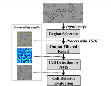
Firstly, after collecting the image dataset, images of region A1 through A5 in Figure 2 were selected manually as shown in Figure 2. Our approach to insect cell counting in bright field microscopy images is based on local TSBF image filtering. The distributions of gradient vectors for cells in bright and dark fields were compared, and then the SBF filter was transformed according to the gradient vectors in the bright field to make it match the cell gradient vector distribution manner well. Like most work on cell identi-fication and counting, our approach conducts cell image enhancement as an initial step. Given the enhanced images by TSBF, a non-maximal suppression filter was applied to search local maxima. Subsequently, cell centers were tentatively associated with the lo-cations of maxima responses.
section, the characteristics for cell images in both dark and bright fields are introduced. Finally, an evaluation method is introduced for comparing with manual counting and some computer-aided approaches.
Host cell preparation
As the host of baculovirus, there are four types of insect cell lines commonly used for BVES applications as listed in Table 1. These insect cell lines support various levels of ex-pression and differential glycosylation with the same recombinant protein [18]. We choose the most widely used cell type, Sf9, a clonal isolated from theSpodoptera frugiperdacell line IPLB-Sf21-AE, as the host cells, which was support by Harbin Veterinary Research Institute, CAAS.
After recovering from liquid nitrogen, Sf9 insect cells were diluted by SF900 II complete medium (Cat. No. 10902-088, Invitrogen Inc., USA) with 10% fetal bovine serum (Cat. No. A15-043, PAA Inc., Austria) and 1% double-antibiotic (Penicillin and Streptomycin, Takara Inc., Japanese). Cell suspension with a density of 5 × 10⁵cells/mL was prepared at 27°C for the counting task in different concentrations. After the
Figure 2Cell detection diagram, using example images from the Sf9 insect cells dataset illustrating our approach.
Table 1 Commonly used insect cell lines in BVES
Insect species Cell line
Spodoptera frugiperda Sf9
Spodoptera frugiperda Sf-21
Trichoplusia ni Tn-368
Trichoplusia ni High-Five™BTI-TN-5B1-4
Suiet al. BioMedical Engineering OnLine2014,13:147 Page 5 of 17
density reaches 2 ~ 3 × 10⁶cells/mL, the population of the insect cells grow exponen-tially and various kinds of experiments were conducted during this period. When its density reaches 5 ~ 6 × 10⁶cells/mL, it comes into the stationary phase with no growth of cells and descends.
Manual counting data collection
Insect cell densities in a suspension culture medium are usually calculated by a Hemacytometer, because it includes enough regions to evaluate both low and high con-centration of insect cells [5], see Figure 2 for details.
Traditionally, the insect cells are diluted at a certain ratio using Sf900 II medium ac-cording to the period of cell cultivation. Then, they are injected into the chamber where the volume (the depth and area) is standardized. The cell numberSquireInsect_cellfrom the
field was obtained by the visual inspection in 1 mm2regions in the chamber. Finally, the density of insect cellsVolINCis calculated by Eq. 1.
V olIN C ¼SquireInsectcellCDepthCellcct ð1Þ
Where Cdepthis the depth of the chamber and Cellcctis the concentration of the
in-sect cells.
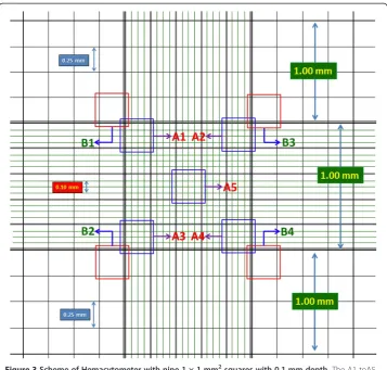
Typically, visual evaluation for insect cells is calculated from 4 or 5 regions. An ex-ample of the 4 regions and 5 regions are B1, B2, B3, B4and A1, A2, A3, A4, A5, respect-ively, as shown in Figure 3. In this paper, we mainly focus on region A1through A5.
Image data set acquisition
Sf9insect cell images were collected using optical model of Nikon Te2000 confocal mi-croscopy with 20 × 10 (objective × eyepiece) for finding counting region and 40 × 10 (objective × eyepiece) for performing the cell counting task in BEVS. For each 24 hours until cells grow into the stationary phase, 3 samples were collected separately and marked as sample 1, sample 2 and sample 3 according to the collected sequence. Microscopic im-ages of region A1 through A5 were collected separately from each sample.
Differences of cell images in dark and bright field
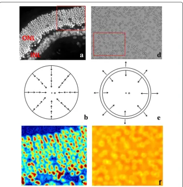
Traditionally, the cells in the dark field appear to have a higher intensity in the center than the edge, and this can be described as a rounded convex region [15]. This region is defined as: the region’s equi-contours of intensity are concentric and all gradient vec-tors in the area toward the center. Figure 4(a) presents the round convex region related to image intensity considered in this paper. Figure 4(b) shows a typical round convex region image in a dark background Red arrows indicated the convergence directions of gradient vectors.
The retinal ONL cells were stained by nuclei dye, TO-PRO (excitation wavelength 642 nm, emission wavelength 661 nm). Image data sets was published in [19]. The ret-inal cell DNA concentrated at the center of each cell leading to the intensity of cell nu-clei appears much higher than the edge in the single plane cell image. Thus, the individual cell in the dark field can be simulated as simple, nearly circular shapes, which refers to a rounded convex region. Figure 5(b) presents the gradient vector dis-tribution of each cell. In addition, after being enhanced by the SBF filter, as depicted in Figure 5(c), the center was enhanced.
Figure 3Scheme of Hemacytometer with nine 1 × 1 mm2squares with 0.1 mm depth.The A1 toA5 region is divided into 1/25 mm2regions, and the B1 to B4 region is divided into1/16 mm2regions.
Figure 4Scheme of rounded convex region. (a)Map of Round convex region with image intensity;
(b)Distribution of Gradient Vectors in the Convex Region.
Suiet al. BioMedical Engineering OnLine2014,13:147 Page 7 of 17
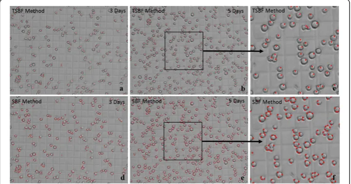
Unlike the dark field, insect cells in the bright field usually shows the whole living cell body and the cytoplasm is not visible compared with the cell membrane. Figure 5(d) shows the insect cells in the bright field compared with the retinal cells in the dark field (Figure 5(a)). The boundaries of an insect cell in the bright field shows a clearly bound-aries of cytoplasm and the edge exhibited discontinuity between inside and outside of the cell. The gradient vector around the cell membrane area shows convergence toward the cytoplasm center and divergence away from the cell membrane toward the back-ground, and this was modeled as a transformed rounded convex region enhanced by our proposed method discussed in part B. Figure 5(e) indicated the gradient vector dis-tribution inside and outside of the insect cells. Unfortunately, as shown in Figure 5(f ), the SBF filter does not work in the bright field, and it can only detect the region of the cell membrane instead. Figure 6(a) to (f ) exhibited the detection results of comparison between the TSBF and SBF method in the 3rdand 5thday. TSBF can detect the center of each cell as one, but the SBF can only detect the cell membrane region and it detect one cell as two, three or even more cells.
Figure 5Comparison of gradient vector distribution and SBF filter enhancement results between the dark field and bright field. (a)Retinal cells image in the dark field.(b)Distribution of gradient vector for each retinal cell in(a).(c)Result of SBF filter enhancement on(a).(d)Sf9 insect cells in the bright field.
Cell detector design using TSBF
Cell detection is an important task in cell counting and recognition, which is usually achieved by intensity threshold or image segmentation [20]. However, these methods assume that cells are isolated from each other in a dark field [21,22]. In this paper, in-sect cells in images appeared as ring structures with different intensities. The most challenging problem in cell detection is that images in this case are obtained by bright field microscopy. In addition, most of the aforementioned approaches require ideal en-vironments that do not have irregular illumination, channel cross talk and noise caused by the microscopy [20].
In this paper, a new method for insect cells counting is proposed, which uses image filtering to enhance certain features on gray level images [23]. Considering most of the linear filters have small support regions, i.e., m × m pixels wherem∈{2, 3, 5...}, in this paper, a Transformed Sliding Band Filter with larger support regions is employed to en-hance insect cells in a bright field. TSBF was originated from a nonlinear filter, the Sliding Band Filter (SBF), a member of the Convergence Index (CI) family.
The CI family was design for enhancing the rounded convex region in digital images, and was based on the maximization of CI at each pixel of spatial coordinates (x,y). The Convergence Index is defined by the following formulations:
Convergence Index xð ;yÞ ¼ 1 Pn
X
mR;nR
ð Þ
cosαðmR;nRÞ ð2Þ
where (x,y) is the coordinate of the interesting pixel,Pnis the number of pixels in the support regionR,α(mR,nR) is the angle between the gradient vector computed at pixel
(mR,nR) and the line connected (x,y) and (mR,nR).
There are several members in the CI family, such as CF (Coin Filter), IF (Iris Filter), ARF (Adaptive Ring Filter) and the SBF [24,25]. The differences in these members are the definition of the support region R. The SBF filter has a band with a fixed width
Figure 6Sf9 insect cell detection comparison between TSBF and SBF method. (a)TSBF detecting result on the 3rdday.(b)TSBF detecting result on the 5thday.(c)Magnification of part of(b).(d)SBF detecting result on the 3rdday.(e)SBF detecting result on the 5thday.(f)Magnification of part of(e).
Suiet al. BioMedical Engineering OnLine2014,13:147 Page 9 of 17
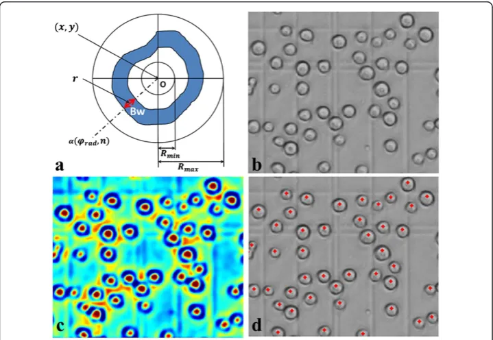
support region, the average convergence index in band width can be maximized by changing the position of the band in each radius direction. Figure 7(a) depicts the scheme of the support region in SBF. The definitions of the SBF filter is as follow:
SBF xð ;yÞ ¼ 1 Pn
XPn
rad¼1
max
Rmin<r<Rmax 1 Bwþ1
X
rþðBw=2Þ
r−ðBw=2Þ
Conv radð ;nÞ 0
@
1
A ð3Þ
With
Conv radð ;nÞ ¼ cosðφrad−αðφrad;nÞÞ
φrad¼
2πðrad−1Þ N
αðφrad;nÞ ¼ arctan
GradnC
GradnR
whereGradnCandGradnRare the column and row gradients at image position n,
respect-ively,Pnis the number of support region lines irradiated from the center pixel (x,y),Bwis
the sliding band width,ris the position of the band center in the support region line ran-ging fromRmintoRmax, and cos(φrad−α(φrad,n)) is the angle between the gradient vector
at (φrad,n) and the direction ofφrad.
The SBF was employed to develop a cell segmentation method in Quelhas’work [15], but it detects only convergence or divergence in the dark field. However, the cell mem-brane in the bright field is distinct from the background with the location of both con-vergence and dicon-vergence, see Figure 7(c) and (d), and cytoplasm is not visible. This leads to theConv(rad,n) in SBF filter does not fit the bright field cell area. By ignoring the affection of divergence and convergence in the bright field, we transform the
convergence index in SBF to facilitate it to fit the cell membrane area well. In this paper, the Transformed Sliding Band Filter (TSBF) is given by:
TSBF xð ;yÞ ¼ 1 Pn
XPn
rad¼1
max
Rmin<r<Rmax 1 Bwþ1
X
rþðBw=2Þ
r−ðBw=2Þ
ABS CIð Þ þωGv radð ;nÞ
ð Þ
0 @
1
A ð4Þ
with
ABS CIð Þ ¼kConv radð ;nÞk Gv radð ;nÞ ¼ ffiffiffiffiffiffiffiffiffiffiffiffiffiffiffiffiffiffiffiffiffiffiffiffiffiffiffiffiffiffiffiffiffiffiffiffiffiffiffiffiGradnC2þGradnR2
p
where ABS(CI) is the absolute value of the convergence index at pixel (rad,n), this eliminates the characteristic of divergence and convergence at the edge of cells, Grad
(rad,n) is the gradient value at pixel (rad,n),ωis a weight parameter designed for the image variation on uneven illumination and out of focus (we set ω= 1 in this paper as the default value). After applying the TSBF filter to insect cells, filter output results in-dicate that the cell centers are associated with the locations of filter local maximal value.
The classical SBF filter was employed by the image processing community to enhance the edge and center in the rounded convex region. In this paper, we use transformed SBF, named TSBF to detect cells in the bright field. The given insect cell image, as shown in Figure 7(b), was processed by our designed TSBF filter, and the results show that the center of cell was enhanced by this method, as shown in Figure 7(c). Further-more, the enhanced center was assumed as the center of each cell.
2
1 1
1 1
1
1
( , ) { , 2
1,...}
[0,
] [0,
]
2 (max , max )
( , );
3
( , ) [ ,
] [ ,
]
4
( , )
(max , max )
5 (max , max )
( ,
k l
n n
W
n
H
n
k
l
k l
k l
k k
n
l l
n
I k l
I
k
l
k
l
k l
∈
+
∩
− ×
−
←
∈
+ ×
+
>
←
forall
do
o
d
l
l
a
r
o
f
n
e
h
T
f
i
1 1 1 1 1);
6
( , ) [max
, max
] [max
, max
] [ ,
] [ ,
]
7
( , )
(max , max )
8 goto failed;
9 MaxlocAt( max , max );
10
k l
k
n
k
n
l
n
l
n
k k
n
l l
n
I k l
I
k
l
k
l
∈
−
+ ×
−
+ −
+ ×
+
>
forall
do
n
e
h
T
f
i
failed;
Algorithm 1: 2D (n+1) × (n+1)-Block NMS
Suiet al. BioMedical Engineering OnLine2014,13:147 Page 11 of 17
A local Non-Maximum Suppression (NMS) was applied to detect the local maximal value which was the assumed cell center, details of the NMS were listed in Algorithm 1, where W and H represent the width and height of a given image, and position (k, l) and (k1, l1) are pixels in the (n + 1) × (n + 1) region [26,27]. In this part, the NMS can be for-mulated as the local maximum search, where one local maximum, excluding itself is greater than all its neighbors. 2D square images of (n + 1) × (n + 1) centered on the pixel were under consideration in this paper.
Cell detector evaluation
The proposed TSBF method for detecting cells in a bright field was tested on the insect cell image datasets collected using the protocol of BEVS. To evaluate the accuracy of the proposed method, an error rate estimation process was employed. We used manual counting results as the GT (Ground Truth), and then the performance of the proposed cell detector was evaluated by the error criterion according to equation five and equa-tion six.
ERT ¼jDA−GTj
GT ð5Þ
TERT¼ERT
NT ð6Þ
with
GT ¼Ns1þNs2þNs3 3 DA¼Ds1þDs2þDs3
3 Nsi¼
SampleisuiþSampleidaiþSampleizhang
3 ;i∈f1;2;3g
where Error Rate of TSBF (ERT) is the relative error rate at a certain cell density calcu-lated using the TSBF filter, Total Error Rate of TSBF (TERT) is the total error rate of all cell density calculated using TSBF, NT is the number of cell samples in different densities, GTis the Ground Truth,Sample_iis cell density of a sample from a certain time counted by our lab collaborators. Nsiis the average number ofSample_i. For
ex-ample, after recovering from the liquid nutrition, cell suspension with a density of 5 × 10⁵cells/mL was made by an experienced lab collaborator as the starting point, 3 sam-ples were collected each 24th hour until the 216th hour (the 9th day) and then counted by 3 lab collaborators and the proposed method.
Results and discussion
The proposed method was implemented on a desktop computer with an Intel 1.86 GHz CPU and 2GB RAM memory. The proposed approach is currently fully imple-mented with MATLAB R2012a. Although the TSBF filter is a computationally intensive operation, our method currently takes approximately 22 mins to perform the cell detec-tion of a 512 × 512 pixel image and it will be improved to perform in less time by using GPUs in the near future.
Cell counting is a complicated and tedious task in many cell-based experiments and usually performed manually by microscopic observations. This leads to many problems such as labor intensity, time consumption, and proneness to errors intro-and inter-persons. Thus, effective cell detectors are needed for biotechnicians to master this task during cell based experiments. Being an excellent cell detector in bright fields, it must satisfy some criteria such as providing the accuracy and reliability of manual counting results. In this paper, the introduced cell counting method in the bright field for insect cells can facilitate the BEVS’s manipulation and improve its efficiency. We test our method on confocal images of insect cells collected from different periods of cultiva-tion. For each sample of insect cells, masks were employed to cover the A1 through A5 regions of the chambers separately in Figure 2, and then we manually calculated each sample by 3 lab collaborators to create the ground truth for investigating the variations among manual counting, the TSBF method and the SBF method.
Counting insect cells is usually performed manually by chambers under microscopy. In addition, the results have to be repeated several times in the same view and then an average number is taken as the relative accurate number. Compared with the manual counting, the proposed method can obtain results with an intuitive parameter setting and the variance on cell density per mL ranging from 7,000 cells to 200,000 cells. We choose 3 samples each day to count the results by our method and manual counting during the cultivation period. The samples were collected from the cultivation medium by sequence and named sample 1, sample 2 and sample 3. Figure 8 compared the aver-age density of cells between manual, TSBF, and SBF methods during the cultivation period. After applying the TSBF method to the cell counting task, there is still a vari-ance in counting results up to 200,000 cells per milliliter. Although there is an enormous variation between these methods, it can be eliminated during the huge cultivation of in-sect cells after evaluation by an experienced biotechnologist.
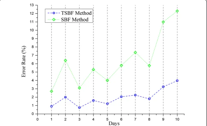
The error rate is an important criterion to evaluate the accuracy of a cell detector. The average error rate of our TSBF method is 2.21% on average, ranging from 0.89% to 3.97% as shown in Figure 9. As an estimated value, the ideal cell density in the log phase for experiments is 5.5 × 10⁶. As an experimental experience in cell cultural, this density is permitted during 5.3 × 10⁶~ 5.7 × 10⁶, and the difference is higher than 2%, so this error rate can be eliminated in cell counting. In our research, the insect cells were cultivated using the suspension method and they usually crowded together when they were out of the shaker. Some of the cells still exhibited crowding after being injected into the chamber, and these cells were usually identified as one cell by manual counting but were detected as 2, 3 or more cells by the TSBF method. This may lead to increase the total error rate as insect cells grow. To verify this hypothesis, we diluted the cell suspension to make sure cells were separated from each other at the 4th and 8th day, and the results exhibited a decrease in error rate. But after cells grew into the stationary phase, some cells were corrupted for lacking nutrition, and the TSBF method
Suiet al. BioMedical Engineering OnLine2014,13:147 Page 13 of 17
detected the bigger cell corruption as a single cell, which leads to an increase in the error rate. Fortunately, insect cells in this phase were out of use for experiments. So, this defect can be omitted.
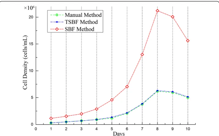
The growth curve can reflect the insect cells growth manner. Drawing an accurate growth curve in the pre-culture stage can enable bio-technologists to decide when to seed baculovirus into insect cells and when to perform follow-up experiments. We compared the TSBF method and manual counting method on the preparation of the growth curve. Figure 10 shows the growth curve where the solid green line and dotted blue line correspond to the manual and TSBF methods, respectively. The two curves
Figure 9Error rate evaluation of the TSBF method and SBF method.
match well before and during the exponential growth phase with little separation. But, they do not match so well after the stationary phase. The reason for this is that the TSBF method detects the cell corruptions as a single cell and therefore leads to a de-tected cell density larger than the real density. Fortunately, the insect cells in this period were in the decline phase and the cells were out of use which has no effect on follow-up experiments, so the negative effect can be eliminated.
Cell counting is an important process in cell based experiments. Although various methods and software have been developed to aid biotechnologists in performing this task, most of the methods are limited in their application scopes and cannot be applied to other kinds of cells or cell lines. The TSBF method can be applied to most cells like insect cells in bright field microscopy to perform the cell counting task. Comparison results between the TSBF and traditional image enhancement methods demonstrated an excellent performance of our proposed method in insect cell image enhancement. Figure 10 shows the comparisons between Laplacian of Gaussian (LOG), image thresh-old and the TSBF method. Results show that the TSBF method proposed in this paper can detect the insect cells with a higher filter resonance value in the center of the cell. Comparing with the other two traditionally methods, only the LOG method can detect the single cell in the image, but the background noise heavily affects the filter response and the detecting result was not as good as the TSBF method. The image threshold method merely detects the cell membrane area in the bright field images. More details were shown in Figure 11(a) to (d). In addition, the TSBF method can also eliminate the background noise in the image, as it is depicted in Figure 11(c).
Additionally, as a common scene for biological experimental workers, most of cells cultivated in vitro usually exhibited the same morphology in bright field, and thus, all of these cell lines’counting task which needs Hemacytometer can be solved by our pro-posed method.
Figure 10Growth curve comparison results between the Ground truth, TSBF method and SBF method.
Suiet al. BioMedical Engineering OnLine2014,13:147 Page 15 of 17
Conclusion
In contrast to traditional methods, in this paper we presented a semi-automated cell counting protocol for BEVS in bright field microscopy image datasets. Comparing with the SBF filter, a transformed non-linear filter (TSBF) was employed to detect the insect cells in the protocol according to the cells gradient vector distribution manner. Since all four of the cell lines exhibited the same gradient vector distribution in the bright field, we tested on the Sf9 insect cell rather than all the 4 cell lines. The proposed method is adaptive and efficient.
To test the accuracy of the proposed cell counting method, we evaluated the error rate. In experiments, the TSBF method showed an excellent performance with an aver-age error rate of 2.21%, ranging from 0.89% to 3.97%, compared with human manual counting by 3 lab collaborators. Also, the error rate of 2.21% can be eliminated in cell counting during cell cultivation. In addition, growth curve evaluation indicates that the TSBF method can be applied to the protocol of BEVS for its host cell density evaluation and growth stage identification.
The ultimate goal of this study is to help biotechnologists count the host cells of baculovirus in the BEVS protocol. As demonstrated in our experiments, the proposed method is very accurate compared with manual counting. Therefore, the application of our proposed bright field cell counting method can clearly benefit the protocol of BEVS. Finally, as a non-linear filter, the TSBF method is computationally expensive. Thus, our fu-ture work is aiming to improve the method so it runs in less time by using GPUs.
Part of this work was introduced in a conference paper of EMBC 2012 [28].
Competing interests
The authors declare that they have no competing interests.
Authors’contribution
DS carried out the insect cells cultural protocol, KW designed the workflow of the cell counting. HP and JC provided the manuscript revise suggestion and protocol optimization. All authors read and approved the final manuscript.
Acknowledgement
This work was partly supported by the University of Incheon International Cooperative Research Grant in 2012 and National Nature Science Foundation of China (NSFC) Grant No. 61173086.
Author details
1
Biocomputing Research Center, School of Computer Science and Technology, Harbin Institute of Technology, Harbin, China.2Department of Computer Software Engineering, Sangmyung University, Cheonan, Korea.3Department of Computer Science and Engineering, Incheon National University, Incheon, Korea.
Received: 14 June 2014 Accepted: 2 September 2014 Published: 24 October 2014
References
1. Blissard GW, Rohrmann GF:Baculovirus diversity and molecular biology.Annu Rev Entomol1990,35:127–155. 2. Jehle JA, Blissard GW, Bonning BC, Cory JS, Herniou EA, Rohrmann GF, Theilmann DA, Thiem SM, Vlak JM:On the
classification and nomenclature of baculoviruses: a proposal for revision.Arch Virol2006,151:1257–1266. 3. Jehle JA, Lange M, Wang H, Hu Z, Wang Y, Hauschild R:Molecular identification and phylogenetic analysis of
baculoviruses from Lepidoptera.Virology2006,346:180–193.
4. Marek M, van Oers MM, Devaraj FF, Vlak JM, Merten OW:Engineering of baculovirus vectors for the manufacture of virion-free biopharmaceuticals.Biotechnol Bioeng2011,108:1056–1067.
5. Pollard JW, Walker JM:Basic cell culture protocols.2nd edition. Totowa, N.J.: Humana Press; 1997.
6. Bracke ME, Boterberg T, Bruyneel EA, Mareel MM:Collagen invasion assay.Methods Mol Med2001,58:81–89. 7. Lamprecht MR, Sabatini DM, Carpenter AE:Cell Profiler: free, versatile software for automated biological image
analysis.Biotechniques2007,42:71–75.
8. Preston K:High-resolution image analysis.J Histochem Cytochem1986,34:67–74.
9. Preston K:Image processing in medical microscopy.InProceedings of the Annual Symposium on Computer Application in Medical Care.American Medical Informatics Association; 1986:213.
10. Seliger C, Schaerer B, Kohn M, Pendl H, Weigend S, Kaspers B, Hartle S:A rapid high-precision flow cytometry based technique for total white blood cell counting in chickens.Vet Immunol Immunopathol2012,145:86–99. 11. Malpica N, de Solorzano CO, Vaquero JJ, Santos A, Vallcorba I, Garcia-Sagredo JM, del Pozo F:Applying watershed
algorithms to the segmentation of clustered nuclei.Cytometry1997,28:289–297.
12. Nedzved A, Ablameyko S, Pitas I:Morphological segmentation of histology cell images.InPattern Recognition, 2000 Proceedings 15th International Conference on. IEEE; 2000:500–503.
13. Demandolx DDJ:Multiparameter image cytometry: from confocal micrographs to major subcellular structures in fluorescence microscope images of hela cells.Bioimaging1997,5:159.
14. Usaj M, Torkar D, Kanduser M, Miklavcic D:Cell counting tool parameters optimization approach for electroporation efficiency determination of attached cells in phase contrast images.J Microsc2011, 241:303–314.
15. Quelhas P, Marcuzzo M, Mendonca AM, Campilho A:Cell nuclei and cytoplasm joint segmentation using the sliding band filter.IEEE Trans Med Imaging2010,29:1463–1473.
16. Pereira CS, Fernandes H, Mendonça AM, Campilho A:Detection of lung nodule candidates in chest radiographs.InPattern Recognition and Image Analysis.Springer; 2007:170–177.
17. Pereira CS, Mendonça AM, Campilho A:Evaluation of contrast enhancement filters for lung nodule detection. InImage Analysis and Recognition.Springer; 2007:878–888.
18. Hink W, Thomsen D, Davidson D, Meyer A, Castellino F:Expression of three recombinant proteins using baculovirus vectors in 23 insect cell lines.Biotechnol Prog1991,7:9–14.
19. Byun J, Verardo MR, Sumengen B, Lewis GP, Manjunath BS, Fisher SK:Automated tool for the detection of cell nuclei in digital microscopic images: application to retinal images.Mol Vis2006,12:949–960.
20. Harder N, Mora-Bermúdez F, Godinez WJ, Ellenberg J, Eils R, Rohr K:Automated analysis of the mitotic phases of human cells in 3D fluorescence microscopy image sequences.InMedical Image Computing and Computer-Assisted Intervention–MICCAI 2006.Springer; 2006:840–848.
21. Chen X, Zhou X, Wong ST:Automated segmentation, classification, and tracking of cancer cell nuclei in time-lapse microscopy.IEEE Trans Biomed Eng2006,53:762–766.
22. Schmitt O, Hasse M:Morphological multiscale decomposition of connected regions with emphasis on cell clusters.Comput Vis Image Understand2009,113:188–201.
23. Gonzalez RC, Woods RE:Digital image processing.3rd edition. Upper Saddle River, N.J: Pearson Prentice Hall; 2008. 24. Kobatake H, Hashimoto S:Convergence index filter for vector fields.IEEE Trans Image Process1999,8:1029–1038. 25. Wei J, Hagihara Y, Kobatake H:Detection of rounded opacities on chest radiographs using convergence index
filter.InImage Analysis and Processing, 1999 Proceedings International Conference on. IEEE.; 1999:757–761. 26. Neubeck AVGL:Efficient non-maximum suppression.Int Conf Pattern Recogn2006,3:850–855. 27. Nixon MS, Aguado AS:Feature extraction and image processing.2nd edition. Oxford: Academic Press; 2008. 28. Wang KQ, Sui D, Wang W, Yuan Y, Zuo W:A cell counting method for BEVS based on nonlinear Transformed
Sliding Band Filter.Conf Proc IEEE Eng Med Biol Soc2012, 118–21.
doi:10.1186/1475-925X-13-147
Cite this article as:Suiet al.:Bright field microscopic cells counting method for BEVS using nonlinear convergence index sliding band filter.BioMedical Engineering OnLine201413:147.
Suiet al. BioMedical Engineering OnLine2014,13:147 Page 17 of 17