Patron: Her Majesty The Queen Rothamsted Research Harpenden, Herts, AL5 2JQ
Telephone: +44 (0)1582 763133 Web: http://www.rothamsted.ac.uk/
Rothamsted Research is a Company Limited by Guarantee Registered Office: as above. Registered in England No. 2393175. Registered Charity No. 802038. VAT No. 197 4201 51. Founded in 1843 by John Bennet Lawes.
Rothamsted Repository Download
A - Papers appearing in refereed journals
Kleczkowski, J. and Kleczkowski, A. 1952. Effect of Specific
Polysaccharides from the Host Bacteria and of Ribonuclease on the
Multiplication of Rhizobium Phages. Biochemical Journal. 7 (3-4), pp.
340-349.
The publisher's version can be accessed at:
•
https://dx.doi.org/10.1099/00221287-7-3-4-340
The output can be accessed at:
https://repository.rothamsted.ac.uk/item/97065/effect-of-
specific-polysaccharides-from-the-host-bacteria-and-of-ribonuclease-on-the-multiplication-of-rhizobium-phages
.
© 1 November 1952, Portland Press Ltd.
340
KLECZKOWSKI, J. & KLECZKOWSKI, A. (1952). J . gen. Microbial. 7 , 340-349.
Effect of Specific Polysaccharides from the
Host Bacteria
and of Ribonuclease on the Multiplication of
Rhizobium
Phages
BY J. KLECZKOWSKI AND A. KLECZKOWSKI
Rotharnsted Experimental Station, Harpenden, Hertfordshire
SUMMARY : Two serologically unrelated strains of nodule bacteria produced two different polysaccharides, only one of which precipitated with antiserum to its parent bacterium. Both polysaccharides interfered with the multiplication of two bacteriophages in liquid cultures of the two bacterial strains, each of which was susceptible to only one of the two bacteriophages. One polysaccharide was slightly more effective than the other in interfering with multiplication of both bacterio- phages; one phage was much more susceptible than the other to the interfering action of both polysaccharides. Crystallized pancreatic ribonuclease interfered with multiplication of bacteriophages much more strongly than did the polysaccharides. Xeither the polysaccharides nor ribonuclease destroyed the phage particles. The chance that a bacteriophage particle will multiply in a liquid culture of Rhizobium bacteria decreases with increasing age of the culture (Kleczkowski
& Kleczkowski, 1951). This can have various causes. Bacteria may become increasingly resistant to infection as their cultures age, or some metabolic products accumulating in the medium might interfere with phage activity. The literature contains many references suggesting that polysaccharides, or materials containing polysaccharides, produced by individual bacterial species specifically interfere with the activity of bacteriophages that attack those
species (Levine & Frisch, 1933; Burnet, 1934; Gough & Burnet, 1934; White,
1936; Miller & Goebel, 1949). Some authors have also related polysaccharides capable of interfering specifically with bacteriophages to those that confer
serological specificity to their host bacteria (see Burnet, Keogh & Lush, 1937).
On the other hand, Ashenburg, Sandholzer, Scherp & Berry (1940) found no
evidence for specific inhibition of activity of bacteriophage by a polysaccharide produced by its host bacteria, for polysaccharides of widely different origins inhibited similarly. Some polysaccharides have been found to interfere with the multiplication of certain animal viruses in the allantoic sac of the chick
embryo (Green & Wooley, 1947; Ginsberg, Goebel & Horsfall, 1948) and with
infectivity of plant viruses (Takahashi, 1942, 1946; Bawden & Freeman,
1952). There is no evidence of any specificity in the action of polysaccharides against individual viruses, but different plants differ in the degree to which
their susceptibility is influenced (Bawden & Freeman, 1952).
The work described in the present paper was done to see whether strains
of Rhizobium bacteria that differ serologically and in their susceptibility t o
Multiplication
of
Rhixobiurn phages
341
For comparison, tests were made to see whether pancreatic ribonuclease, which is known to be a powerful inhibitor of activity of plant viruses (Loring,
1942; Kassanis & Kleczkowski, 1948), has any effect on activity of Rhizobium
bacteriophages. It reduces the numbers of lesions formed by plant viruses on
leaves of susceptible plants, but has no effect on numbers of plaques formed
by one of the phages (Kassanis & Kleczkowski, 1948).
MATERIAL AND METHODS
Two strains of nodule bacteria, 317 derived from pea (obtained from the
Agricultural Research Station, Wisconsin) and C15 from clover and two
homologous bacteriophages were used. Each bacterial strain was susceptible only to its homologous phage and was agglutinated only by its homologous antiserum. The phages were originally obtained from soil and isolated from single plaques. The media, the poured plate method of obtaining plaques, and
other methods of cultivation were the same as used previously (Kleczkowski &
Kleczkowski, 1951).
The polysaccharides were isolated from 3-week-old, liquid cultures of the
bacteria. The cultures were centrifuged, 10 ml. 20
yo
NaCl were added to eachlitre of the clear supernatant fluid followed by 2 1. 96
yo
(v/v) ethanol. Theprecipitate was filtered off arid suspended in 100 ml. of a 20
yo
solution ofsodium acetate neutralized with glacial acetic acid. Material that did not
dissolve within 24 hr. was removed by centrifugation. Further procedure
was the same as that described by Heidelberger, Kendall & Scherp (1936) for
isolating pneumococcal polysaccharides, except that, before removing protein by Scvag's procedure, the material was precipitated with ethanol, dissolved in 0.06 M-phosphate buffer at pH 7.0 and incubated for 24 hr. a t 37" with the
addition of vol. of filtered saliva. (This removed a material which gave
the same colour as glycogen on treatment with iodine.) The polysaccharide
was then precipitated with ethanol, dissolved in acidified 20% solution of
sodium acetate, and Sevag's treatment was applied. (This removed a material that gave a positive biuret test). The polysaccharide was precipitated from
the resulting solution by ethanol, washed in 96
yo
ethanol, in absolute ethanol,in ether, dried and ground to a white powder. The yield was about 100 mg.
from 1 1. bacterial culture.
Antisera were produced by injecting rabbits intraveneously with bacterial
suspensions prepared by washing with physiological saline 3- to 5-day old
cultures from agar slopes. Agglutinin tests with bacterial suspensions were
made as previously described (Kleczkowski & Thornton, 1944). Precipitin
tests with the bacterial polysaccharides were made similarly to the agglutinin tests except that the antiserum was used a t a constant dilution and the poly- saccharide concentration varied.
Crystallized beef pancreatic ribonuclease was prepared by the method
described by Kunitz (1940).
For paper chromatography the polysaccharides were hy drolysed from
342
J .
Klecxlcowski
and
A .
Klecxkowski
by adding an excess of BaCO,, and centrifuged after a few hr. incubation at
room temperature.
One-dimensional chromatograms were prepared as described by Partridge
(1948), using two different solvents ; water-saturated phenol (with ammonium- saturated atmosphere) and a water-saturated n-butanol-ethanol mixture with
the volume ratio butanol : ethanol : water=4*5 : 0.5 : 5.0.
Benzidine reagent (Horrocks, 1949) was used as a developer. For the
quantitative estimation of sugars from the different spots after development, water extracts were made from areas corresponding with the spots but cut from part of the filter-paper that had not been sprayed with the developer. The reducing power of the extract was then determined by the Somogyi
(1945) titrimetric procedure.
RESULTS
Some properties of the polysaccharides
Both polysaccharides were obtained as white powders, which formed clear
colourless solutions in water and were highly viscous a t 1
yo.
Solutions ofpolysaccharide 317 were obviously more visc'ous than those of Cl,. They
contained no nitrogen, did not stain with iodine and did not reduce Fehling's
solution unless previously hydrolysed. After hydrolysis with 5
%
H,SO, for1 hr. a t 120' and subsequent neutralization with NaOH, their reducing power
was about S5% that of glucose. The intensity of colour developed when
solutions of polysaccharides were heated with ten times their volume of
0.2% orcinol in 6 6 % H,SO,, did not differ appreciably from that formed under the same conditions by an equally concentrated solution of glucose.
The polysaccharides had different compositions. The 317 produced only
two spots on the paper chromatograms. Their positions corresponded with
those of glucose and of a uronic acid. The C1, produced three spots; the
positions of two of them were identical with those of the components of the
317, that of the third corresponded with the position of fructose. This spot
was weak, and it was absent if the time of hydrolysis was 2% hr., and the
temperature was raised to 125', under which conditions fructose is destroyed
and gives no spot on the chromatogram. Over 90% of the total reducing
power of each hydrolysed polysaccharide was due to the component identified as glucose.
If hydrolysed for only 20 min., the hydrolysate of each polysaccharide
contained a component that gave an additional sport on chromatograms. This occurred only when phenol was used as a solvent, and then the component
moved very slowly (R, about 0.015). The component was presumably
stationary with the butanol-ethanol mixture so that its position coincided with that of the uronic acid. The component may be an aldobionic acid or an oligosaccharide that was hydrolysed when hydrolysis was prolonged.
ikhltiplication
of Rhixobiurn
phages
aldobionic acid (Hopkins, Peterson & Fred, 1931; Cooper, Daker & Stacey,
1938; Schluchterer & Stacey, 1945).
The two polysaccharides differed serologically. The 317 polysaccharide
precipitated with an antiserum to 317 bacteria (Table 1) but not with an
antiserum to Cl, bacteria. The polysaccharide C1, did not precipitate with
either antiserum. Each antiserum agglutinated suspensions of its homologous
bacterium up to a titre of l[SOOO, but neither of the bacterial strains was
agglutinated by antiserum to the other.
Although polysaccharides produced by different strains of clover Rhixobiurn
may difTer serologically, some of them may be closely related to polysac- charides produced by bacteria that do not belong to the Rhizobium group.
The polysaccharide isolated by Schluchterer & Stacey (1945) from ' Bartel ,4 '
strain of clover Rhixobiurn was found by Dr
1cI.
Heidelberger to give a positiveprecipitin test at a high dilution with pneumococcus I11 antiserum and also
with mixed antisera to other types of pneumococcus.
The possibility that one or both of the polysaccharides 317 and C1, was
a mixture cannot be excluded. However, the high dilution end point a t
which the polysaccharide 317 precipitated with the antiserum to 317 bacteria
makes it unlikely that the polysaccharide was a mixture of which the sero- logically active component was only a minor part.
Esect of the polysaccharides and of riboizuclease orb the
bacteriophages
Three types of experiment were made to see whether the polysaccharides and ribonuclease interfere with bacteriophage activity. Experiments of the first type were designed to show whether contact with these materials per-
manently inactivates phage particles. Bacteriophage 317 was incubated with
the polysaccharide 317 and with ribonuclease, then diluted to obtain a con-
veniently countable number of plaques, mixed with host bacteria and plated.
The details and results are given in Tables 2 and 3 which show that the
incubation did not inactivate any phage particles.
Experiments of the second type were designed to test whether the presence of the polysaccharide or ribonuclease in the bacterial culture, to which a suitably diluted phage preparation was added and which was then plated
directly, could render some phage particles inactive. Table 4 gives the details
of one such experiment arid shows that the polysaccharides had no such effect, for the number of plaques formed was not affected.
Experiments of the third type were designed to show whether the presence of the polysaccharide or of ribonuclcase in a liquid culture of host bacteria
interferes with phage multiplication in the culture. Table 5 shows that the
polysaccharide 317, added to a concentration of 0 . 2 5 % to a culture of
317 bacteria, which was then inoculated with 317 bacteriophage, did interfere
with niultiplication of the phage, which, within the first 3 hr., increased only
about 50 times compared to about 100 times in the control. The polysaccharide
Cl, interfered with multiplication of 317 phage only slightly, though signi-
Table 1. Precipitin yeaction qf the polysaccharide 317 with an azztiserum to 317 Time of Reciprocal of dilution of a 0.05 solution of the polysaccharide reading r-- (hr.) 1 2 4 8 16 32 64 128 -I
++
+
+
k+
+++
+++
++
++
+
+
+++
+++
+++
+++
++
++
- --
1 2 4
-
-
-
bacteria __
\ 256 512 1024 1 ml. of the antiserum diluted 1/10 was added to 1 ml. of variously diluted solution of the polysaccharide, and incubated at 50".
+
+
+
,+
+
,+
and 2 signs show the presence and the extent of precipitation ; - sign shows the absence of precipitation. Table 2. Incubation of bacteriophage 317 with the polysaccharide 31 7 beforemixi
fig with host bacteria and plating Composition of the mixtures ~ 3 0-5yo
solution Phage culture Mixture polysaccharide H2O diluted 1/50 Mean no. 1 1.0 - 1.0 41 2-
1-0 1.0 38 no. (ml.) (ml.) (ml.) plaques/plate The mixtures were incubated for 1 hr. at room temperature, then diluted in 24 hr. bacterial cultures and plated. Four platings were made with each mixture. Table 3. Incubation qf bacteriophage 317 with r.ibonuclease before mixing with host t-acteria and plating Mean no. plaques/plate Composition of the mixtures A f \ formed by mixture 0.4yo
Phage culture incubated for dil. Mixture ribonuclease 1 1 0-25 0.75 0.1 103 95 2 - 1 -00 0.1 106 1 04 H*O A 24 hr. no. (ml.1 (ml.) (ml.) 0 hr. The mixtures were incubated for 24 hr. at 25". Samples were taken at the beginning and at the end of incubation, diluted l/lOO in 24 hr. bacterial cultures and plated. Each plating was made in four replications.Multiplication
of
Rhixobiurn
phages
345
although considerably less. Rabbit serum albumin used at the same con- centration had no effect.
Ribonuclease when present a t a concentration of 0.01
o/b,
i.e. a t &th of thecoiicentration of the polysaccharides, inhibited phage multiplication much
more strongly than did the polysaceharicies, and at a coiicentration of 0.1
yo
it stopped its multiplication completely during the first 3 hr.
After 24 lir. incubation the ratios of the concentrations reached by bacterio-
phage in the presence of the interfering materials to that reached in their
absence were the same as, or greater than, after 3 hr. incubation. I t is
obvious. therefore, that the inhibiting effect of the materials tested lasted less
than 24 hr.
Preparations of crystallized ribonuclease obtained from beef pancreas by
Kunitz's (1940) method contain a proteolytic enzyme (Kleczkowski. 1948)
and possibly some other enzymes and proteins. Proteolytic activity of the
preparations can be destroyed or greatly decreased by heating for 5 min. to
90-100" at pH 7.5, preserving about 30
yo
of the original riboriuclease activity(Kleczkowski, 1948). As almost all known enzymes are also destroyed by
heating in these conditions, heated preparations of crystallized ribonuclease can be assumed to be free from active enzymes other than ribonuclease.
After heating in 0.06 M-phosphate buffer a t pH 7.5 for 5 min. in a water-bath
at 100", the ribonuclease preparation still interfered with phage multiplication
t o an extent proportional to its remaining ribonuclease activity.
The results given in Table 6 show that the extent of inhibition depends on
the host bacterium. The growth of phage Cl, in C15 bacterial culture was much
more strongly inhibited by polysaccharides 317 and Cl, than was the growth
of phage 317 in a culture of 317 bacteria. Again the polysaccharide 317
inhibited somewhat more strongly than did the polysaccharide Cl,.
To see whether inhibition of phage multiplication by the tested materials
could be a direct result of inhibition of growth of host bacteria, 24 hr. bac-
terial cultures were incubated a t 25" with the materials a t concentrations a t
which they inhibited phage growth. Samples were taken a t intervals for
haeniocytometer counts. The presence of 0.25
yo
of the polysaccliarides or of0.01
yo
ribonuclease had no effect on multiplication of the bacteria, but thepresence of 0.1
yo
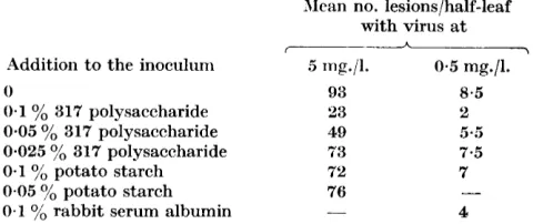
ribonuclease decreased it considerably.Experiments were also made to test whether the polysaccharide 317 inter-
feres with infectivity of tobacco mosaic virus. Details and results of one of
such experiments are given in Table 7, which shows that the presence of the
polysaccharide in the inoculum reduced numbers of lesions formed on leaves
of Il'icotiana glutinosa more than did the presence of potato starch or rabbit
serum albumin. The reduction was very slight by comparison with that
caused by the polysaccharide obtained from yeast by Takahashi (1942), or by
such substances as beef pancreatic ribonuclease or a protein isolated from
Phytolacca escuknta (Kassaiiis & Kleczkowski, 1948).
Table 4. Eflect of adding polysaccharide 317 or ribonuclease to bacterial cultures with which bacteriophage 317 was plated Composition of the mixtures 1
yo
solution 0-4yo
solution 24 hr. bacterial Phage culture Mixture polysaccharide ribonuclease H2O culture diluted Mean no. A I > no. (ml.) W*) (ml.) W) (ml.) plaqueslplate - 0.75 0.1 38 0.75 0.1 31 0.25 0.75 0.1 33 1 0.25 - 2-
0.25 - 3 - - The mixtures were mixed with 10 ml. molten agar medium, which had been cooled to 42", and poured into Petri dishes. Four plates were made from each mixture. Table 5. Effect of the polysaccharides and of riboikuclease on the multiplication of 317 bacteriophage Concentration of bacteriophage in terms of no. of plaqueslml. of the mixtures I Composition of the mixtures 3 ml. 24 hr. culture of 317 bacteria+
1 ml. of: 1yo
317 polysaccharide 1yo
C15 polysaccharide 1yo
potato starch 1yo
rabbit serum albumin 0.4yo
ribonuclease 0.04yo
ribonuclease 0404yo
ribonuclease H2OImmediately 21
x 102 18 x 102 20 x 102 19 x 102 17 x lo2 17 x lo2 19 x 102 19 x 102 After 3 hr. 10 x 104 13 x 104 17 x 104 24 x 104 23 x 104 25 x 104 15 x lo2 26 x lo2 After 24 hr. 65 x
lo8
85 xlos
106 x lo8 120 x 108 4-5 x 10⁸ 46 x lo8 125 xlo8
129 x lo8 0.4 ml. of a culture of phage 317 diluted lo-, was added to each mixture. The mixtures were then incubated at 25" and samples were taken at intervals for phage estimation. Table 6. Eflect of polysaccharides (PSH) on multiplication of Cl, bacteriophage Composition of mixtures I A \ Concentration of bacteriophage in terms PSH PSH H2O diluted-
7 24 hr. culture 1yo
317 1 % c1, Phage C1, of no. plaques/ml. of the mixtures A Mixture C1, bacteria no. (ml-) (ml.) (ml*) (ml.) (ml.) Tmmediately After 24 hr.1 2 3
0-75 0.75 0-75
0.1 0.1 0.1
Multiplication
of Rhixobium
phuges
347
Table '7. Efiect of polysucchnride 317 on a,ctivity of tobacco mosaic virus
Mean no. lesions/half-leaf with virus a t
I A \
Addition to the inoculum 5 mg./l. 0.5 mg./l.
0 93
0.1
yo
317 polysaccharide 330.05
yo
317 polysaccharide 490.025 yo 317 polysaccharide 73
0.1
Yo
potato starch 7'20.1 yb rabbit serum albumin
0-05
yo
potato starch 76-
8.5
2
5 a . 5 7.5 7
4
-
Each inoculum was applied to twelve half-leaves distributed among six Nicotiana
glutinosa plants according to the principle of the Latin square. Each column of numbers
was obtained in a separate experiment.
DISCUSSION
The fact that specific bacterial polysaccharides can interfere with phage
multiplication shows clearly that their accumulation in a liquid bacterial
culture may decrease the chance of a phage particle to multiply. Howel-er, the ability to interfere with phage multiplication is shared by various other materials such as potato starch and beef pancreatic ribonuclease, the last one being by far the most effective. This suggests that, apart from specific poly- saccharides, various other products of bacterial metabolism that accumulate
in the medium are also likely to decrease the chance of a phage particle to
multiply.
There is obviously no specific affinity between the bacterial polysaccharides and phage particles, because interference by the polysaccharides with phage multiplication is unspecific, i.e. not restricted to phages whose hosts produced the polysaccharide. ,!!lso it is not connected with the ability of the polysac- charides to precipitate specifically with antisera to host bacteria. The un- specific nature of the interference is further emphasized by the fact that the polysaccharides can also interfere with infectivity of tobacco mosaic virus.
The mechanism of interference by the polysaccharides or ribonuclease remains unknown, but some possibilities can be excluded. First, phage par- ticles are not destroyed by these interfering materials. Secondly, the materials do not interfere with phage multiplication by aflecting the rate of bacterial
multiplication. It is true that ribonuclease, when present a t a concentration
of 0.1
yo
did interfere with bacterial multiplication, but it did not do so at0.01
Yo,
although it interfered strongly with phage multiplication a t this concentration. It is possible, thereforc, that ribonuclease interferes with phage multiplication by affecting bacterial metabolism. The latter is obviouslyaffected by 0.1
yo
ribonuclease. It may therefore also be affected by 0.01yo
ribonuclease though not sufficiently to influence bacterial multiplication and
it may be similarly affected by the polysaccharides. It is also possible, how-
ever, that 0.01
yo
ribonuclease affects only the bacterial surface, making itless penetrable for bacteriophage, or preventing its adsorption; and the poly-
348
J . Klecxkowski
and
A .
Klecxkowski
saccharides may interfere with phage activity by forming a protective coating on the bacterial surface.
A feature of the inhibitory effect of the polysaccharides and of ribonuclease
on phage multiplication is that with young bacterial cultures it is only
temporary; with 24 hr. cultures of 317 bacteria inoculated with 317 phage it
lasts less than 24 hr. Various explanations can be suggested. For example, if
the inhibitory mechanism is based on a protective coating of the bacterial surface, in a young fast-growing culture the surface may outgrow its protective coating and thus increase its vulnerability; if it is based on interference with bacterial metabolism, the metabolism may recover its original course either
by adaptation or through some process directed against the inhibitor.
Although the interference by the polysaccharides is unspecific, different host-phage systems are affected to different extents. This is shown by the
fact that the phage C1, was much more affected by each of the two polysac-
charides, 317 and Cl,, than was the phage 317.
It may seem that there is a contradiction between a polysaccharide or
ribonuclease interfering with phage multiplication in a liquid bacterial culture and having no effect on the numbers of plaques formed on a solid medium. The
contradiction, however, is apparent only. To be plated, a phage-bacteria
mixture is diluted 1/10 in molten agar medium, and this may suffice to make
the inhibitor ineffective. However, should it remain effective, plaque numbers are still unlikely to be affected, because the inhibitor does not destroy phage particles or have more than a temporary effect on phage multiplication in a young bacterial culture.
Bawden & Freeman (1952) have discussed the interference by a polysac-
charide and by ribonuclease with the activity of some plant viruses, and have
concluded that it is most likely to occur because these substances affect the
metabolism of the host cells. Thus there may be no basic difference between the effect of polysaccharides and of ribonuclease on the activity of plant
viruses, on the one hand, and on that of bacteriophage, on the other. If so,
the fact that these materials decrease the numbers of local lesions formed by
plant viruses and have no effect on the numbers of plaques formed by a
bacteriophage would mean that the lesions are not analogous to the plaques. There would be a closer analogy between the formation of lesions and lysis of liquid bacterial cultures. This analogy is also apparent when a comparison is made between the dilution curves obtained with numbers of lesions caused by plant viruses and the dilution curves obtained with proportions of liquid
bacterial cultures lysed by a bacteriophage (Kleczkowski & Kleczkowski,
1951).
REFERENCES
ASHENBURG, N. J., SANDHOLZER, L. A., SCHERP, H. W. & BERRY, G. P. (1940). The influence of bacterial and non-bacterial polysaccharides upon bacterio- phagy. J. Bact. 39, 71.
BAWDEN, F. C. & FREEMAN, G. G. (1952). The nature and beliaviour of inhibitors produced by Trichothecium roseurn Link. J . gen. Microbiol. 7 , 154.
iWultiplication of
Rhixobium
phages
349
BURNET, b'. &I., KEOGH, E. V. & LUSH, D. (1937). Immunological reactions of filterable viruses. Aust. J . exp. Biol. vrbed. Sci. 15, 227.
COOPER, E. A., DAKER, W. D. & STACEY, M. (1938). Enzyme formation and poly- saccharide synthesis by bacteria. 111. Polysaccharides produced by ' nitrogen fixing' organisms. Biochevn. J . 32, 1752.
GINSBERG, PI. S., GOEBEL, W. F. & HORSFALL, E'. L. (1948). The inhibitory effect of polysaccharide on mumps multiplication. J . exp. M e d . 87, 385.
GOUGH, G. A. C. & BURNET, F. R1. (1934). The chemical nature of the phage- inactivating agent in bacterial extracts. J . Path. Bact. 38, 301.
GKEEX, R. M. & WOOLEY, D. W. (1947). Inhibition by certain polysaccharides of
haemagglutination and multiplication of influenza virus. J . exp. M e d . 86, 55.
HEIDELBERGER, M., KENDALL, F. E. & SCHERP, H. W. (1936). Specific polysac- charides of types I, I1 and I11 pneumococcus. J . exp. Med. 64, 559.
HOPKINS, E. W., PETERSON, W. H. & FRED, E. B. (1931). Glucoronic acid, a con- stituent of the gum of root nodule bacteria. J . Amer. chem. SOC. 53, 306.
HORROCKS, R. H. (1849). Paper partition chromatography of reducing sugars with benzidine as a spraying reagent. Suture, Lorid. 164, 444.
KASSANIS, B. & KLECZKOWSKI, A. (1948). The isolation and some properties of
a virus-inhibiting protein from Phytolncca esculenta. J . gen. Microbiol. 2, 143.
KLECZKOWSKI, A. ( 1948). Proteolytic activity of preparations of crystallized ribonuclease. Biochem. J . 42, 523.
KLECZKOWSKI, A. & KLECZKOWSKI, J. (1931). The ability of single phage particles to
form plaques and to multiply in liquid cultures. J . geri. 31icrobiol. 5, 346.
KLECZKOWSKI, A. & THORNTON, H. G. (1044). A serological study of root nodule bacteria from pea and clover inoculation groups. J . Bctct. 48, 661.
KVNITZ, M. (1940). Crystalline riboliuclcase. J . gen. Physiol. 24, 15.
LEviwE, P. & FRISCII, A. W. (1933). Specific inhibition of bacteriophage by bac- terial extracts. Proc. SOC. exp. Biol., X.17. 30, 993.
LORING, H. S. (1942). The reversible inactivation of tobacco mosaic virus by crystalline ribonuclease. J . gen. Physiol. 25, 497.
MILLER, E. J1. & GOEBEL, W. F. (1949). Studies on bacteriophage. I. The relation-
ship between the somatic antigens of Shigella sonnei and their susceptibility
to bacterial viruses. J . ezp. M e d . 90, 255.
PARTRIDGE, S. M. (1O48). Filter paper partition chromatography of sugars. Biochem. J . 42, 238.
SCNLUCHTER~CR, E. & STACEY, M. (1945). The capsular polysaccharide of Rhizobiurn radicicolum. J . chem. SOC. 776.
SOMOGYI, R;I. (1945). A new reagent for the determination of sugars. J . biol. Chem.
160, 61.
TAKAHASIII, W. W. (1942). A virus inactivator from yeast. Science, 95, 586. TAKAHASHI, W. W. (1946). Properties of a virus inactivator from yeast. Science, WHITE, 104, P. B. 377. (1936). Differential fixation of cholera phages by extracts of V . cholerae.
J . Path. Bact. 43, 591.