DOI: 10.1534/genetics.109.109686
A
Caenorhabditis elegans
RNA-Directed RNA Polymerase in Sperm
Development and Endogenous RNA Interference
Jonathan I. Gent,* Mara Schvarzstein,
†Anne M. Villeneuve,*
,†Sam Guoping Gu,
‡Verena Jantsch,
§Andrew Z. Fire*
,‡,1and Antoine Baudrimont
§*Department of Genetics,†Department of Developmental Biology, and‡Department of Pathology, Stanford University School of Medicine, Stanford, California 94305 and§Department of Chromosome Biology,
Max F. Perutz Laboratories, University of Vienna, A-1030 Vienna, Austria Manuscript received September 11, 2009
Accepted for publication October 1, 2009
ABSTRACT
Short interfering RNAs (siRNAs) are a class of regulatory effectors that enforce gene silencing through formation of RNA duplexes. Although progress has been made in identifying the capabilities of siRNAs in silencing foreign RNA and transposable elements, siRNA functions in endogenous gene regulation have remained mysterious. In certain organisms, siRNA biosynthesis involves novel enzymes that act as RNA-directed RNA polymerases (RdRPs). Here we analyze the function of aCaenorhabditis elegansRdRP,RRF-3, during spermatogenesis. We found that loss ofRRF-3function resulted in pleiotropic defects in sperm development and that sperm defects led to embryonic lethality. Notably, sperm nuclei in mutants of either rrf-3 or another component of the siRNA pathway, eri-1, were frequently surrounded by ectopic microtubule structures, with spindle abnormalities in a subset of the resulting embryos. Through high-throughput small RNA sequencing, we identified a population of cellular mRNAs from spermatogenic cells that appear to serve as templates for antisense siRNA synthesis. This set of genes includes the majority of genes known to have enriched expression during spermatogenesis, as well as many genes not previously known to be expressed during spermatogenesis. In a subset of these genes, we found that RRF-3 was required for effective siRNA accumulation. These and other data suggest a working model in which a major role of theRRF-3/ERI pathway is to generate siRNAs that set patterns of gene expression through feedback repression of a set of critical targets during spermatogenesis.
R
EPRESSION of gene expression by small RNAs of 20–30 nt in length is important for many aspects of multicellular eukaryotic development. A variety of classes of small RNA with distinct structural features, modes of biogenesis, and biological functions have been identified (reviewed in Hutvagnerand Simard2008). We are particularly interested in a class of small RNAs, called endogenous short interfering RNAs (siRNAs), that are similar to intermediates in exogenously triggered RNA interference (RNAi) in their perfect complementarity to mRNA targets. High-throughput sequencing technology has provided a valuable tool for characterization of endogenous siRNA populations from many diverse sources, including mouse embryonic stem cells (Babiarz et al. 2008), Drosophila heads (Ghildiyal et al. 2008), and Arabidopsis pollen (Slotkinet al.2009). These siRNAs have been proposed to function in the regulation of both cellular processes and genome defense through downregulation of geneexpression.Caenorhabditis elegans, like plants and fungi, utilizes RNA-copying enzymes called RNA-directed RNA polymerases (RdRPs) as part of the RNAi machinery (Smardonet al.2000; Sijenet al.2001). While two of the C. elegans RdRPs are nonessential (RRF-1 andRRF-2), mutations in either of the remaining two (EGO-1or RRF-3) lead to fertility defects (Smardonet al.2000; Simmer et al.2002).RRF-3is functionally distinct fromEGO-1in that the RRF-3requirement in fertility is temperature dependent. In addition,RRF-3activity has an inhibitory effect on exogenously triggered RNAi (resulting in an ERI, or enhanced RNAi, mutant phenotype in rrf-3
mutants). Mutants lacking eitherRRF-3or another ERI factor, ERI-1, have been used as experimental tools because of their enhanced sensitivity in RNAi-based screens. One proposed mechanism for the enhance-ment in RNAi in rrf-3 and eri mutants has been a competition for cofactors between the exogenously triggered RNAi pathway and an endogenous RNAi pathway. Consistent with this hypothesis, siRNAs corre-sponding to several genes have been shown by Northern analysis to depend uponRRF-3and other ERI factors for their accumulation (Duchaineet al.2006; Leeet al.2006; Yigitet al.2006). Global microarray analyses have also Supporting information is available online athttp://www.genetics.org/
cgi/content/full/genetics.109.109686/DC1.
1Corresponding author:Stanford University, 300 Pasteur Dr., Room L235,
Mail Stop M5324, Stanford, CA 94305-5324. E-mail: [email protected]
been undertaken to identify messenger RNAs whose expression is affected byRRF-3andERI-1(Leeet al.2006; Asikainenet al.2007).
A functional significance of theRRF-3/ERI pathway has been inferred by the inability ofrrf-3,eri-1,eri-3, and
eri-5mutant strains to propagate at a high growth tem-perature (Simmer et al. 2002; Duchaine et al. 2006). Rather than producing temperature-sensitive mutant protein effects,RRF-3and other ERI proteins are thought to act in a temperature-sensitive process, as evidenced by the predicted truncated and presumed nonfunctional protein fragments that would result from the available deletion alleles and by their shared temperature-sensitive phenotypes.rrf-3mutant animals have been observed to exhibit X-chromosome missegregation (Simmer et al. 2002) and an unusual persistence of a chromatin mark on the X chromosome during male spermatogenesis (Maineet al.2005). X-chromosome missegregation and defective spermatogenesis have been referred to in pre-vious studies oferi-1(Kennedyet al.2004) anderi-3and eri-5(Duchaine et al.2006). Furthermore,eri-3mutant sterility can be rescued by insemination with wild-type sperm (Duchaineet al.2006).
Here we investigated the role of RRF-3 during spermatogenesis. We found defects evident at multiple stages, including after fertilization, where defects inrrf-3
mutant sperm can produce subsequent nonviable embryos. By using high-throughput sequencing, we characterized a large population of siRNAs present in spermatogenic cells and found a strong enrichment for antisense siRNAs from genes with known mRNA expres-sion during spermatogenesis. While the majority of siRNA production during spermatogenesis does not requireRRF-3, we found a set of genes for which siRNA production was dependent uponRRF-3. Existing data indicate increased expression for these genes in rrf-3
and/or eri-1 mutants. Taken together, our analyses suggest a working model in which the RRF-3/ERI pathway generates siRNAs that downregulate specific genes during spermatogenesis, with this regulation playing a key role in generating functional sperm.
MATERIALS AND METHODS
C. elegansstrains, growth conditions, and spermatogenic cell isolation
Strains:Except as noted,C. elegansBristolN2(Brenner1974) was used as the wild-type strain for these studies. The other strains used in this study were the following:GE24—pha-1(e2123) I(Schnabel and Schnabel 1990); GR1378—eri-1(mg366) IV (Kennedy et al. 2004); JK816—fem-3(q20) IV (Barton et al. 1987); JK987—tra-2(q276)/mnC1 dpy-10(e128) unc-52(e444) II (Okkemaand Kimble1991);NL2099—rrf-3(pk1426) II(Sijen et al. 2001); PD3303—rrf-3(pk1426) II; pha-1(e2123) III; PD3330—rrf-3(pk1426) II; him-8(e1489) IV; PD3331— him-8(e1489) IV; VC407—rrf-3(ok629)/mIn1[mIs14 dpy-10(e128)] II (http://www.celeganskoconsortium.omrf.org); WM48—rde-4 (ne299) III (Tabaraet al.1999); andWM161—prg-1(tm872) I (Yigitet al.2006),tra-2(q122) II(Schedland Kimble1988).
Growth conditions: As stated in the description of each experiment, animals were grown at 16°, 23°, or 25°. Therrf-3 anderi-1mutant fertility phenotypes were strongest at 25°, but certain experiments were carried out at the milder tempera-ture of 23° due to the insufficient numbers of embryos produced at 25°. Self-fertilizedrrf-3(pk1426)hermaphrodites had an approximate surviving brood size of 2 at 25°, but 30 at 23°.rrf-3(pk1426)males were also significantly more fertile at 23°. Self-fertilized rrf-3(ok629) hermaphrodites also showed temperature-sensitive decreased fertility (they also exhibited a temperature-independent egg-laying-defective phenotype). Animals used for RNA extraction and sequencing were grown and harvested at 25°(see below).
Spermatogenic cell isolation: A population of mixed stage fem-3(q20)hermaphrodites grown at 16° was treated with a 1.2% sodium hypochlorite, 0.5nNaOH solution with periodic vortexing to dissolve everything but eggs. After washing in M9 buffer to remove hypochlorite, eggs were hatched overnight in M9 solution and allowed to starve as L1 larva. These were put ontoOP50-seeded, peptone-enriched plates at 25°to mature to early adult stage (55 hr), when animals were washed off plates with M9 buffer. After repeated washes to remove bacteria, the animals were diced with a razor blade in a 10-cm tissue culture dish to release spermatogenic cells into solution (other cells remained attached to carcasses). The mixture of carcasses and released spermatogonic cells was filtered through a double layer of 10mm Nitex bolting cloth (Wildlife Supply) and washed three times in M9 buffer before flash freezing in liquid N2. Visual inspection of a portion that was transferred to a microscope slide prior to freezing indicated that the sample contained a mix of spermatogenic cell types, including primary spermatocytes, secondary spermatocytes, residual bodies, and spermatids (supporting information, Figure S5). This procedure is optimized for small-scale isolation of mixed-stage spermato-genic cells to be immediately frozen [we note that studies of mature sperm function optimally utilize a Percoll gradient and SM buffer (L’Hernaultand Roberts1995; Machaca et al. 1996) rather than M9 buffer (Klassand Hirsh1981; Miller 2006)].
Male isolation: Set 1:After sodium hypochlorite treatment of mixed stage animals grown at 16°, released eggs were hatched overnight in M9 solution to starve as L1 larva. Fifty-one hours after being put ontoOP50-seeded NGM plates at 25°, the starved L1’s had reached adulthood, and males were separated from hermaphrodites by filtration through 35-mm Nitex bolting cloth (Wildlife Supply). Embryos that passed through the filter were separated from males by putting both males and embryos on an unseeded area of a plate and allowing males to crawl toward food. Adult males were washed off plates in EN buffer (0.1mNaCl, 10 mmEDTA) and filtered a second time immediately prior to freezing in liquid N2, 53 hr after initially being put on seeded plates as L1’s. Approxi-mately 95% of the him-8(e1489) sample and 90% of the rrf-3(pk1426);him-8(e1489) sample was male.
Assaying embryonic viability
Mated tra-2(q122) females were allowed to lay eggs for several hours on NGM plates with thin, clear lawns ofOP50 (#1 day after seeding). After removing the mothers from the plates, the newly laid embryos were immediately counted. The surviving progeny were counted when they had reached L4 or adult stage. For temperature-shift experiments, matings were set up between L4 stagetra-2(q122)females and L4 or adult males raised at the starting temperature, where they were allowed to mate overnight. The gravid females were then separated from the males and shifted to the new temperature, where they were left for sufficient time to flush out previously fertilized embryos (6 hr at 23°and 25°or 12 hr at 16°). After this period they were allowed to lay eggs for several more hours (3 hr at 23° and 25° or 6 hr at 16°). In each experiment, multiple matings were set up, and a minimum of four gravid hermaphrodites were evaluated at each growth temperature. Males were raised at 23°rather than at 25°[because matings with rrf-3(pk1426) males at 25° are inefficient in terms of embryo production]. Twenty-three degrees represents an intermediate temperature at which enough competent sperm are produced to allow for continued egg production many hours after mating.
Cytological techniques
RAD-51, REC-8, HIM-8, and SYP-1 immunostaining:
Hermaphrodite gonads were dissected and fixed as pre-viously described for anti-RAD-51, anti-HIM-8, anti-SYP-1 (Martinez-Perez and Villeneuve 2005), and anti-REC-8 (Pasierbeket al.2001) stainings. Animals were synchronized by bleaching to kill everything but embryos. Embryos were then allowed to hatch overnight on NGM plates seeded with Escherichia coli(OP50) at 16°, after which time embryos were shifted to grow at 25°until the animals were ready for dissection. After fixation, samples were washed three times in 13PBS for 5 min and then blocked with 3% bovine serum albumin (BSA) in 13 PBS for 20 min at room temperature. Antibodies were diluted in 13PBS containing 3% BSA as follows: 1:80 anti- RAD-51(Alpiet al.2003), 1:100 anti-REC-8(Pasierbeket al.2001), 1:500 anti-HIM-8(Phillipset al.2005), and 1:200 anti-SYP-1 (MacQueenet al. 2002). Samples were then incubated over-night at 4°in a humid chamber. After washing three times in 13
PBS plus 0.1% Tween 20, secondary antibodies were applied at the following dilutions: anti-rabbit DyLight 547 nm (1:200), anti-rabbit Alexa Fluor 568 (1:500), and anti-guinea pig Alexa Fluor 488 (1:500). After 2 hr at room temperature, slides were washed three times in 13 PBS plus 0.1% Tween 20 and mounted in Vectashield antifading medium (Vector Laborato-ries, Burlingame, CA) containing 2mg/ml DAPI. Samples were examined with a Zeiss Axio Imager M1microscope. Images were recorded with a Spot camera (Diagnostics Instruments). For multicolor immunostaining, monochrome stacks of images were captured separately for each emission wavelength (Meta-Vue software; Molecular Devices, Sunnyvale, CA). Three-dimensional stacks of images were deconvolved (AutoQuant software; AutoQuant Imaging, Troy, NY) and projected (Heli-con Focus software;http://helicon.com.ua/heliconfocus/).
a-Tubulin immunostaining: We used fixation procedures optimized for preservation of microtubules (Gonczy et al. 1999; Oegemaet al.2001), with modifications as previously described (Martinez-Perez et al. 2008). FITC-conjugated anti-a-tubulin monoclonal antibody DM 1A (Sigma) was used at a concentration of 1mg/ml. Images were acquired as stacks of optical sections at 0.2-mm intervals using a Deltavision deconvolution microscopy system. We quantified the
inci-dence of microtubule wreaths surrounding sperm nuclei in reproductive tracts (gonads and spermathecae) dissected either from self-fertilizing hermaphrodites or from tra-2(q122)females that had been mated with either wild-type or rrf-3(pk1426) mutant males. Quantification was performed using animals that had been raised and maintained at 23°. Microtubule wreaths were frequent in sperm from bothrrf-3 self-fertilizing hermaphrodites (15 worms; 237/287 sperm nuclei with microtubule wreaths) andtra-2(q122)females that had been mated withrrf-3males (1 worm; 17/20 nuclei with microtubule wreaths). Self-fertilized eri-1 hermaphrodite sperm had microtubule wreaths in 59% of their sperm (5 worms; 152/256 nuclei with tubulin wreaths). Microtubule wreaths were not detected in either controlN2self-fertilizing hermaphrodites (2 worms; 0/93 nuclei with tubulin wreaths) or controltra-2(q122)females mated withN2males (4 worms; 0/86 nuclei with tubulin wreaths). Although discrete tubulin wreaths were not detected in control sperm, some hazy/ diffuse tubulin staining was detected in 4/179 control sperm. For experiments analyzing wild-type sperm, successful micro-tubule staining in mitotic germ cells from the same gonad served as an internal positive control.
Preparation of libraries of small RNAs for sequencing
Small RNA was extracted from frozen tissue samples with the mirVana microRNA (miRNA) isolation kit (Ambion). Libraries of small RNAs were prepared using a protocol similar to one previously described for miRNA cloning (Lauet al. 2001), in which small RNA species were flanked by adapter sequences to allow for PCR and sequencing. We modified the original protocol at several key steps to allow for capture of triphosphorylated species and for sequencing with the Illu-mina genome analyzer system. To remove the bias for 59
monophosphorylated species but preserve the bias toward 39
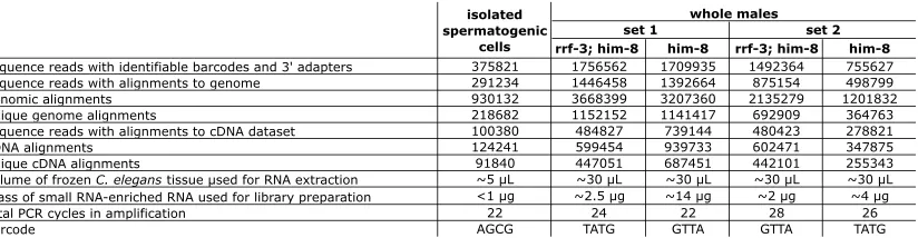
hydroxylated species, we removed both monophosphates and polyphosphates from the 59-end of the small RNA with Antarctic Phosphatase (New England Biolabs) following the addition of the 39adapter with T4 RNA ligase 1 (New England Biolabs). The resulting hydroxylated 59-ends were phosphor-ylated by treatment with T4 polynucleotide kinase (New England Biolabs) and ATP to form a subtrate for T4 RNA ligase 1-mediated addition of the 59adapter. T4 polynucleo-tide kinase treatment after addition of the 39 adapter is important because of its 39 phosphatase and 29,39 cyclic phosphodiesterase activity, which can convert unwanted RNA degradation products into a substrate for the 39adapter ligation reaction. After ligation of both the 39and 59adapters, the adapters were extended during the reverse transcription and PCR steps to include complete primer binding sites for subsequent amplification and sequencing reactions. To pro-tect against cross-contamination and to allow for pooling of samples, we modified the 59adapter sequence to include the Illumina genomic sequencing primer immediately followed by a 4-nt barcode. The pair of barcodes used in the set 1 wild-type and mutant samples was switched in the set 2 wild-wild-type and mutant samples to guard against any potential biases introduced by adapter sequences. The samples were PAGE purified at three stages: after initial RNA extraction and after each ligation step (except with the isolated spermato-genic cell sample, where the first PAGE purification was skipped to conserve material). Additional details for each sample are provided inTable S1. The following oligos were used:
59 adapter (i.e., AF-PP-341 DNA/RNA hybrid oligo with a 59
amino modifier and 4-nt barcode at the 39-end): ACGCTCTTCCGATCTrGrUrUrA;
Dual reverse transcription primer and PCR primer (AF-JIG-37, complementary to IDT linker-1): CAAGCAGAAGACGGCA TACGAattgatggtgcctacag;
PCR primer (AF-JIG-40, containing 59 adapter sequence): AATGATACGGCGACCACCGACACTCTTTCCCTACACGA CGCTCTTCCGATCT.
Sequence analyses
Thirty-six-nucleotide sequencing reads were generated using the Illumina Genome Analyzer system. The 39adapter sequences were trimmed offin silicoby scanning from the 39 -end of the sequence for the first instance of the first 4 nt of the adapter: CTGT. Sequence reads that lacked CTGT in the 39
most 13 nt were excluded from further analysis. After barcode sorting, the 4 nt of barcode were trimmed off the 59-ends to yield 19- to 28-nt reads corresponding to the captured small RNAs. siRNA alignment counts were produced by aligning the reads to a cDNA reference set using a local installation of BLAT (Kent2002), with default parameters, except tileSize was set to 10 and stepSize to 5. The term ‘‘siRNA alignment count’’ (above) refers to the number of matches to an individual cDNA obtained from the set of reads. The cDNA data set, derived from theC. elegans WS190 genome assem-bly, was downloaded from http://www.wormbase.org:80/ biomart/martview. The data set of known transposons sequen-ces was downloaded fromhttp://www.sanger.ac.uk/Projects/ C_elegans/REPEATS/elegans.lib. Read counts and alignment counts are listed inTable S1.
Statistical methods
P-value calculations for identification of genes with RRF-3-dependent siRNAs: Gene-by-gene siRNA alignment counts were tallied and compared for each sample. To measure fold-changes in alignment count between samples for a particular gene, the alignment count was first converted to a proportion by dividing the alignment count of that gene by the sum of the alignments of the complete cDNA reference set. To obtain a measure of the statistical significance of differences between samples, we assumed that there was no difference in population proportions for each gene and then calculated the probability (P-value) of measuring a difference in sample proportions at least as extreme as the one observed. A one-tailed test was used since we had prior expectations that the proportions for certain genes would decrease in therrf-3(pk1426)samples (seeFile S1 for a representative calculation). The standard errors of the means for sample proportion differences were calculated on the basis of predicted normal distributions of the sample proportions, which is justified by the large sample size and the central limit theorem (the proportions here are equivalent to means). The sample proportion differences were standard-ized toZ-scores by dividing by the standard errors of the means for the proportion differences.
P-values and fold-enrichment calculations for overlap between sets of genes with RRF-3-dependent siRNAs and genes with elevated mRNA levels inrrf-3anderi-1mutants:P -values were derived using Fisher’s exact test from the following numbers: 16,887 of the genes represented in the cDNA data set had probes on the microarray used by Asikainen et al. (2007). Of the 21 genes that we identified as spermatogenesis candidateRRF-3targets, 18 were included in this set. Of the
16,887 total genes, 178 had increased mRNA levels in rrf-3(pk1426), while 46 had decreased levels. Of the 18 spermato-genesis candidateRRF-3targets, 3 had increased mRNA levels inrrf-3(pk1426)(15.8-fold enrichment;P-value¼8.83104);
none had decreased levels. Of the 16,887 genes, 706 had increased mRNA levels in eri-1(mg366), while 263 had de-creased levels. Of the 18 spermatogenesis candidateRRF-3 targets, 8 had increased mRNA levels ineri-1(mg366)(10.6-fold enrichment;P-value¼2.93107). These 8 included the 3 with
elevated mRNA levels inrrf-3(pk1426). None of the 18 had decreased levels ineri-1(mg366).When we considered all 44 candidateRRF-3targets rather than just the 18 with siRNAs present in the spermatogenic cell sample, the numbers were still highly significant: fold enrichments of 6.5 and 4.4 withP -values of 1.53102and 4.43104forrrf-3(pk1426)and
eri-1(mg366), respectively.
RESULTS
Compromised spermatogenesis accounts for re-duced brood size and X-chromosome loss from rrf-3(pk1426)hermaphrodites:We first set out to determine whether the rrf-3(pk1426) reduced brood size and X-chromosome missegregation phenotypes were attribut-able to defects in sperm or oocytes.C. elegansexists in two sexes: males, which produce only sperm, and hermaph-rodites, which produce first sperm and then oocytes in the same gonad. Defects in either gamete line can result in sterility of self-fertilizing hermaphrodites. rrf-3(pk1426)self-fertilizing hermaphrodites produced very few fertilized embryos at 25°, with 90% of these non-viable (Figure 1A). Here and in subsequent experi-ments, we defined ‘‘nonviable’’ as failure to give rise to L4 stage larva or adults. We found that a preponderance (or all) of the nonviability was accounted for by embryonic lethality prior to hatching. To determine the source of the reduced embryo count and viability, we set up matings between rrf-3(pk1426) hermaphrodites and wild-typeN2males at 25°. Fertilization by wild-type sperm rescued therrf-3(pk1426)brood size reduction in terms of both numbers of embryos produced and embryonic viability (Figure 1, A and B), indicating that both phenotypes can be attributed to defects during spermatogenesis.
To determine whether X-chromosome missegrega-tion is linked to spermatogenesis in rrf-3(pk1426)
hermaphrodites, we utilized a genetic test that takes advantage of the C. elegans sex determination system. The ratio of X-chromosomes to autosomes inC. elegans
up matings with tra-2(q276) XX-transformed males, which contribute an X from each sperm (like a her-maphrodite). Unless the X chromosome is lost during oogenesis, hermaphrodites mated withtra-2(q276)males give rise to almost entirely XX hermaphrodite progeny (Figure 1C) (Okkema and Kimble 1991). We set up matings between either rrf-3(pk1426) or wild-type her-maphrodites with tra-2(q276) males. To select for out-crossed progeny, the hermaphrodites also carried a temperature-sensitive mutation in pha-1 that causes self-fertilized progeny to die as embryos (Schnabel and Schnabel 1990). The progeny from this cross
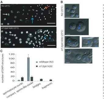
prior to and during completion of spermatogenesis at 25°. We investigated chromosome organization during meiotic prophase using antibodies labeling RAD-51, which functions in recombination and decorates chias-mata (Alpi et al. 2003; Colaiacovo et al. 2003); the synaptonemal complex componentsREC-8 andSYP-1 (Pasierbeket al.2001; MacQueenet al.2002); and HIM-8, which marks a specific domain on the X chromosome (Phillipset al.2005). This analysis did not reveal any overt defects in meiotic prophase (Figure S1andFigure S2). In addition, DAPI-stained nuclei in the process of transitioning between prophase and the first meiotic division appeared normal inrrf-3mutants (Figure 2A). Cytological abnormalities inrrf-3mutant germ cells first started to become evident after the primary spermato-cyte stage, where DAPI staining revealed nuclear abnor-malities such as small or bridged structures (Figure 2A). Further analysis of unfixed late-stage spermatocytes by DIC and Hoechst staining revealed both unusual cell morphology and the presence of multiple nuclear structures within individual mutant spermatocytes (Fig-ure 2B). Other spermatocytes within the same gonads appeared to develop without obvious morphological abnormalities and to produce residual bodies and sperm of the normal size and shape, although in smaller
numbers than wild type. Additional DAPI staining of released gonads revealed that the production of com-pact sperm-like nuclei was both delayed and decreased. At the expected time of spermatogenesis completion (prior to ovulation but after appearance of oocytes), rrf-3mutants had a fivefold reduction in compact, sperm-like nuclei relative toN2(Figure 2C). By the time of the first ovulation, the number inrrf-3mutants of sperm-like nuclei relative toN2increased to half of theN2 levels (data not shown), but these gonads still contained cells with multiple or misshapen nuclei. Together, these phenotypes indicate defects in developmental progres-sion and cell diviprogres-sion processes during spermatogenesis inrrf-3(pk1426).
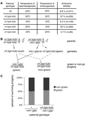
nonviable (37% at 23°). This effect on embryos de-pended on the temperature at which spermatogenesis occurred but was independent of the temperature at which embryogenesis occurred (Figure 3A). The pater-nally derived lethality was not due to the presence of a second, unrelated recessive mutation because trans -heterozygous males bearingrrf-3(pk1426)and another deletion allele, rrf-3(ok629), also produced nonviable embryos. Such males should be heterozygous for any extraneous mutations outsiderrf-3, so the contribution of any such mutations, if any, should be minimized. In setting up the test for therrf-3requirement, we utilized
trans-heterozygote rather than ok629 homozygotes be-cause of a male-tail-defective phenotype in the strain (which we have not definitively assigned took629). In matings betweentra-2(q122)females andok629/pk1426 trans-heterozygote males at 25°, the resulting embryos showed similar levels of nonviability as embryos sired by pk1426 homozygous males: 84% nonviability from
pk1426/pk1426and 72% nonviability fromok629/pk1426, contrasted with 2% nonviability fromN2(Figure S3).
To distinguish a paternal effect from an embryonic requirement for a paternally deliveredrrf-3(1) allele,
we used the scheme in Figure 3B, setting up matings betweenrrf-3(pk1426)homozygous hermaphrodites and
rrf-3(pk1426)/mIn1heterozygous males.mIn1is a chro-mosome inversion of chrochro-mosome II containing the wild-typerrf-3locus and a transgene insertion that drives pharyngeal GFP expression (Edgleyand Riddle2001), enabling us to determine which paternal allele is inherited by the progeny. We found that survival was not affected by physical inheritance of the wild-typerrf-3
allele (onmIn1), indicating thatrrf-3expression is not required after separation of joined secondary sperma-tocytes into spermatids nor in embryogenesis (Figure 2 and Figure 3C). Together with the early temperature-sensitive period (prior to fertilization of the embryo as shown in Figure 3A), this result implicates spermato-genesis as the crucial timing for bothRRF-3expression and activity.
the presence of abnormal microtubule structures in a subset of the mutant embryos at the one-cell stage (i.e., from formation of the maternal meiotic spindle through metaphase of the first mitotic division). Spe-cifically, we detected supernumerary microtubule asters associated with the sperm pronucleus or tripolar spin-dles in 5 of 35rrf-3mutant one-cell embryos (Figure 4A), whereas such structures were never observed in control embryos (0 of 34;P-value: 0.029).
Immunofluorescence analysis also revealed striking microtubule-based abnormalities inrrf-3mutant sperm. At 23°, we observed bright wreaths of microtubules surrounding the nuclei of both hermaphrodite and male-derived sperm (Figure 4B andFigure S4). These microtubule wreaths were clearly visible in 83% (n ¼ 307) ofrrf-3mutant sperm. In contrast, only 2% of wild-type sperm exhibited any detectable tubulin staining (which had a diffuse appearance) other than the sperm centrioles. Microtubule wreath structures were never
observed in wild-type sperm (n ¼179). The fact that tubulin was largely undetectable is expected on the basis of previous observations that microtubules in secondary spermatocytes are effectively segregated to an enucle-ated cytoplast called the residual body (Roberts et al. 1986; Ward1986; Wardet al.1986). At 16°, where we did not see dead embryos, the mutant sperm lacked the microtubule wreath phenotype.
Because meiotic DNA damage resulting from com-promised small RNA machinery in Drosophila results in microtubule polarization defects (Chen et al. 2007; Klattenhoff et al.2007; Pane et al.2007), we tested the possibility that the microtubule wreaths in rrf-3
around sperm nuclei at 23° in double-mutant worms lacking SPO-11 (the enzyme required for inducing meiotic DSBs) (Keeney et al. 1997; Dernburg et al. 1998) andRRF-3. Microtubule wreaths were present in sperm from the double mutant, indicating that these structures do not depend on meiotic DSBs induced by SPO-11(data not shown).
Given the similarities between the fertility and RNAi phenotypes of rrf-3 and eri-1 mutants, we examined tubulin distribution in sperm and embryos from eri-1(mg366)hermaphrodites raised at 23°. We found both microtubule wreaths in sperm (59%, n ¼ 256) and microtubule/spindle defects in embryos (4/20) pro-duced byeri mutants (Figure 5, A and B). We further confirmed a role for ERI-1 in producing functionally normal sperm by showing that mating eri-1(mg366)
males with tra-2(q122) females resulted in nonviable embryos at elevated temperatures (Figure 5C).
Identification of siRNAs present in spermatogenic cells:During the progression through the distinct stages of spermatogenesis from a premeiotic syncitium to a mature sperm, a large number of genes must be choreo-graphed to ensure that the proper genes are expressed at the appropriate level and developmental stage (e.g., Johnstonet al.2008). Since several RNAi-related factors have been implicated in spermatogenesis through mutant studies, we expected that RNA-based regulatory machinery might contribute at several levels to this cho-reography. Consequently, we characterized the small RNAs present in the male germline. We took a sequencing-based approach to this, adapting sperm isolation and optimizing high-throughput sequencing methods to gen-erate an extensive data set of small RNA sequences from isolated spermatogenic cells. Spermatogenic cells were isolated from adultfem-3(q20) XX animals using a tech-nique that yields a large proportion of sperm precursors in addition to mature sperm (adapted from Lamunyonand
Ward1995, which describes a dicing method, and from Miller2006, which uses M9 buffer and does not require a Percoll step to obtain sperm for biochemical analysis). fem-3(q20)animals produce an excess of sperm and no oocytes or embryos at 25°(Bartonet al.1987). Functionality of fem-3(gf) XX sperm has been demonstrated previously by their ability to sire live embryos following artificial in-semination (Lamunyonand Ward1994). The popula-tion of cells isolated and used for sequencing is shown inFigure S5.
number of alignments that it produced from the complete set of small RNA sequences) These data are tabulated inFile S1. Also included in the NCBI short read archive are alignment files in psl format, which can be directly uploaded onto the UCSC Genome Browser (http://www.genome.ucsc.edu) for viewing. Two notable features of the siRNA alignments are a bias toward antisense orientation relative to the annotated mRNA and a diversity of positions within the individual target genes, as illustrated by siRNAs of the gene C05C12.5
(Figure 6). The antisense orientation of the spermato-genesis siRNAs suggests synthesis by RdRP and is consistent with potential modulatory roles of these small RNAs through a ‘‘standard’’ gene-silencing model in which an Argonuate-bound short antisense RNA seg-ment catalytically locates and silences mRNAs with extended regions of homology (reviewed in Hutvagner and Simard2008).
The full small RNA-ome from sperm is large, com-plex, and likely to reflect numerous distinct regulatory processes. We began by analyzing small RNAs with likely roles in the regulation of specific mRNAs by compiling siRNA alignment counts for each annotated mRNA-encoding gene in this data set. While a comprehensive list of spermatogenesis-expressed mRNAs is unavailable forC. elegans, several studies have categorized mRNAs with higher relative expression in spermatogenic cells than in other tissues. We compared the list of genes with siRNAs present in the spermatogenic cell sample with two gene lists, ‘‘spermatogenesis-enriched’’ and ‘‘moun-tain 4.’’ The spermatogenesis-enriched category was derived by comparison of mRNA expression from mutant hermaphrodites that were either masculinized to produce only sperm or feminized to produce only oocytes (Reinke et al. 2004). Mountain 4 was derived from a study that grouped genes according to correlated
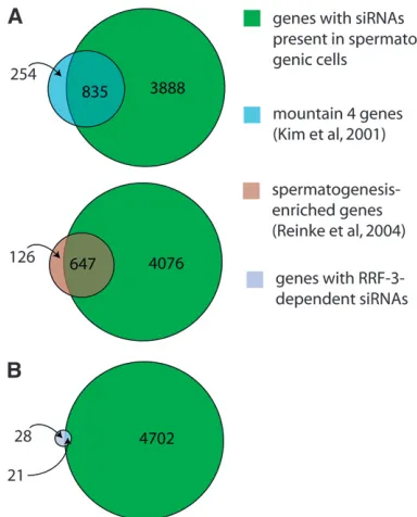
expression patterns over multiple growth conditions, developmental time points, and mutant backgrounds (Kimet al.2001). For a gene to be categorized as having siRNAs in spermatogenic cells, we required that the number of alignments to its mRNA from our isolated spermatogenic cell siRNAs be at least three. A total of 4723 of the 21,133 genes in our cDNA reference set met this criterion. These 4723 genes show a 3.7-and 3.5-fold greater-than-expected overlap with the spermatogenesis-enriched and mountain 4 expression category, respectively (accounting for 84% of the total spermatogenesis-enriched category and 77% of the mountain 4 category, Figure 7A). Conversely, genes from the spermatogenesis-enriched and mountain 4 categories were significantly underrepresented among the group of mRNAs identified in our isolated sper-matogenic cell sample with siRNA alignment counts of fewer than 3 (4.8- and 3.3-fold less-than-expected, respectively). These results indicate that siRNA abun-dance in spermatogenic cells correlates with mRNA expression during spermatogenesis and that siRNA production is a general feature of gene activity during spermatogenesis, involving the majority of genes with enriched expression during spermatogenesis.
Identification of RRF-3-dependent siRNAs:To iden-tify siRNAs that depend upon RRF-3 for their pro-duction, we analyzed siRNA expression profiles from
rrf-3mutant samples. To avoid any possible confound-ing effects of thefem-3mutant background (sinceFEM-3 affects the control of spermatogenesis), we carried these experiments out using populations of karyotypically normal XO males. For bothrrf-3mutant and wild-type control samples, ahim-8(e1489) mutation that compro-mises X-chromosome pairing primarily in oocytes was used to generate populations with large numbers of males (Hodgkinet al.1979).
siRNA profiles from the him-8(e1489) whole males and from the isolated fem-3(q20) hermaphrodite sper-matogenic cells were generally similar in that genes with abundant siRNAs in one sample tended to be abundant in the other, but a number of genes did show genotype/ tissue specificity (Figure S6). To increase the stringency of our analysis, we prepared two sets of males of each genotype at 25°using different population synchroni-zation methods for each set. We aligned the sequences from each sample to the cDNA data set and compared the siRNA alignment count for each gene in therrf-3
mutant and control male sample. For the vast majority of genes, we saw little or no effect ofrrf-3(pk1426)on siRNA levels in these assays:,5% of the genes had reproduc-ible, greater-than-twofold decreases in siRNAs levels. A subset of genes showed dramatic effects, however, and
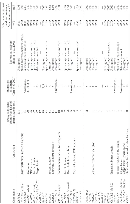
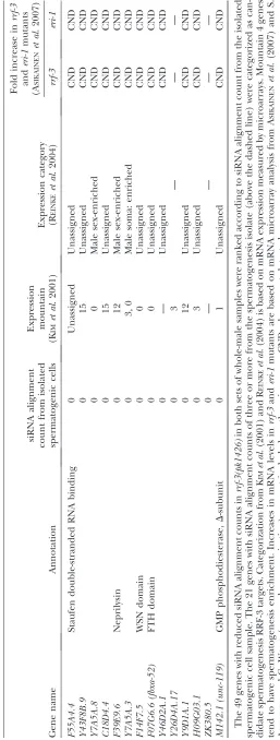
we chose to track 49 genes whose expression is likely dependent uponRRF-3by the criterion of a threefold-reduced alignment count with a P-value of,0.005 in both sets of experiments (Table 1; materials and methods).
This list of 49 genes is intended as exemplary rather than complete: other genes appear to have similar properties without quite reaching our arbitrary statisti-cal threshold criteria, while still more may produce siRNAs that are expressed at levels insufficient for statistical significance in these analyses. In addition, alignment of the small RNA sequences to the genome without regard to predicted coding status revealed additional unannotated loci with small RNAs dependent upon RRF-3, such as the previously identified RRF-3 -dependent locus on the X chromosome called the X cluster (Ambroset al.2003; Duchaineet al.2006). The two genes on the list of 49 with the highest siRNA levels,
C44B11.6andK02E2.6, have previously been shown by siRNA Northern analysis to require RRF-3 for siRNA accumulation, providing an independent confirmation of our experimental approach (Duchaine et al. 2006; Leeet al.2006).
For any of these genes to be regulated by anRRF-3 -driven mechanism during spermatogenesis, one would expect that both their mRNAs and siRNAs would be expressed during spermatogenesis. We found 5.2-fold enrichment for the mountain 4 expression category and 4.6-fold enrichment for the spermatogenesis-enriched category in our list of 49 candidateRRF-3target genes, indicating that a substantial fraction ofRRF-3-regulated genes are expressed during spermatogenesis (Table 1; Figure 7B). We also used an siRNA alignment count from spermatogenic cells as a means of ranking the candidate gene list in terms of the likelihood of spermatogenesis gene regulation. As before, we split the list into two categories using a spermatogenesis siRNA alignment count of 3 as the threshold, resulting in 21 genes with spermatogenesis siRNAs and 28 without (Table 1). These 21 genes with spermatogenesis siRNAs and dependency on RRF-3 represent a sample of candidate RRF-3 regulatory targets during spermatogenesis. An impor-tant result of this analysis was thatRRF-3had little or no effect on gene-specific siRNA counts for the majority of the 4723 genes with spermatogenesis siRNAs (Figure 8): ,4% of the genes had reproducible, greater-than-two-fold decreases in siRNA levels inrrf-3(pk1426)relative to controlhim-8(e1489)males (69 of 1784 genes; to exclude background noise from genes with low siRNA alignment counts, only genes with counts of at least 25 in both sets of control males were included). This result is consistent with the existence of many other RNAi-related factors with substantial germline phenotypes (Coxet al.1998; Smardonet al.2000; Knightand Bass2001; Topset al. 2005; Chenet al.2007).
In addition to mRNA-encoding genes, we examined small RNAs corresponding to a large number of repeated Figure 7.—Overlap between siRNA- and mRNA-based
and/or transposable elements (http://www.sanger. ac.uk/Projects/C_elegans/REPEATS/elegans.lib). From this analysis we found that many transposons, especially Helitron (264 siRNAs aligned to a HELICOP1 sequence) and Tc1/mariner transposons (87 siRNAs aligned to a Tc1 sequence), were associated with high siRNA alignment counts from the spermatogenic cell sample. We did not find any transposons that requiredRRF-3by the criteria of a threefold reduction in siRNA alignment count and aP -value of,0.005 between mutant and control from both sets of samples. Intriguingly, we found three cases in which siRNAs corresponding to a transposon were significantly more abundant in rrf-3 mutants: CELE1, LINE2, and TURMOIL2. Of these, only TURMOIL2 had siRNAs present in the spermatogenic cell sample. The increased siRNAs from these transposons could reflect an activation
of RNAi-like processes as has previously been observed in exogenous RNAi and transgene silencing (Sijenet al.2001; Simmeret al.2002).
siRNAs produced by RRF-3 and ERI-1 function to downregulate mRNA levels during spermatogenesis:In the absence of RRF-3 or ERI-1, at least one putative target, K02E2.6, has been shown to have increased mRNA levels (Duchaine et al.2006). To test whether RRF-3 and ERI-1 generally act to downregulate their spermatogenesis targets, we compared theRRF-3 regu-latory candidates with a global microarray analysis of RRF-3andERI-1effects on mRNA levels in hermaphro-dites undergoing spermatogenesis (Asikainen et al. 2007; S. Asikainenand G.Wong, personal communi-cation). We first note some limitations in the compari-son with the microarray analysis: Three of the 21 genes that we identified as differentially regulated in males were not included in the microarrays used by Asikainen et al. (2007). Furthermore, Asikainen et al. (2007) reported data for whole L4 hermaphrodites, in which sperm or sperm precursors may have been a relatively rare component (so that substantial differences in spermatogenic mRNA abundance could have been masked by pools of unaffected somatic RNA). Similarly, spermatogenic cell-type-specific silencing could be hard to detect in the background of other sperm precursor cells still expressing the transcript at normal levels. Despite these limitations, it is of considerable interest to compare our siRNA data with the mRNA abundances determined by Asikainenet al.(2007; Table 1). Of the 18 genes that were included in the microarray analysis and with RRF-3-dependent spermatogenesis siRNAs, 3 showed increased mRNA levels in both rrf-3 and eri
mutants, and an additional 5 showed increased mRNA levels ineri-1mutants alone (15.8-fold enrichment with a
P-value of 8.8 3 104 for the rrf-3 mutant; 10.6-fold
enrichment with a P-value of 2.9 3 107 for the eri-1
mutant; seematerials and methods).
DISCUSSION
Evidence for a developmental role for RRF-3 during spermatogenesis: Small RNA pathway functions are commonly categorized as defensive (e.g., suppression of transposable elements and viruses) or regulatory (e.g., downregulation of mRNA levels during develop-ment). Either function can be essential, as exemplified by the critical roles of microRNA developmental regu-lators (reviewed in Flynt and Lai 2008) and by the roles of piRNAs in transposon control (reviewed in Klattenhoff and Theurkauf 2008). These develop-mental/defense roles of small RNAs can overlap, as illustrated by a human cellular microRNA that targets a viral RNA (Lecellier et al. 2005) and by the recent description of a C. elegans small RNA family (21U-or piRNAs) that apparently modulates both transpo-Figure8.—Global RRF-3 effects on spermatogenesis siRNA
sons and cellular transcripts during spermatogenesis (Batistaet al.2008; Daset al.2008; Wangand Reinke 2008). Although products of cellular RdRPs apparently differ from piRNAs in both structure and biosynthetic mode, some RdRPs can participate in responses to foreign nucleic acids, including quelling inNeurospora crassa(Cogoniand Macino1999) and viral defense in Arabidopsis (Mourrain et al. 2000). On the basis of developmental phenotypes in mutant strains, other RdRP components have been hypothesized to carry out endogenous regulatory roles (Smardonet al.2000; Shiuand Metzenberg2002).
Several characteristics of RRF-3 (the temperature-dependent character and the requirement for RRF-3 in a defined developmental pathway such as spermatogen-esis) facilitate a detailed analysis of siRNAs associated withRRF-3function and their potential regulatory roles. Using high-throughput sequencing as a tool for charac-terizing RNA populations, we found that antisense siRNAs corresponding to a variety of both transposable elements and cellular mRNAs are present in spermato-genic cells. Among the large and diverse set of cellular mRNAs reflected in the pool of siRNAs, we found a strong enrichment for those with expression during spermatogenesis (in particular, with mRNAs known to have elevated expression during spermatogenesis; Kim et al. 2001; Reinke et al. 2004). By comparing whole males either mutant or wild type for rrf-3, we have identified exemplary cellular mRNAs for which siRNA production during spermatogenesis depends onRRF-3. These cellular mRNAs show strong enrichment for previously identified genes whose mRNA levels are elevated in populations of whole animals in bothrrf-3
anderi-1mutants (Asikainenet al.2007).
RRF-3 and paternal contributions to embryogenesis: In principle, paternal-effect lethality associated with RRF-3 and ERI-1 mutants could result either from a lack of sperm-provided factors essential for normal embryogenesis or from pathological effects of inappro-priate sperm content. We favor the latter possibility for several reasons of parsimony. First, RNAi pathways are known to function broadly in downregulation of gene expression, such that loss-of-function mutations result in overexpression of spermatogenic factors. Second, the presence of ectopic microtubule structures that we observed in mutant sperm (and later defective micro-tubule structures in embryos) suggests failure to elim-inate at least one structure (microtubules) during budding of spermatids. Whether due to sequestration defects or ectopic microtubule polymerization after budding, a plausible interpretation is that microtubules or associated factors in mutant sperm could have a pathological effect in embryos.
Production of functionally normal sperm requires a highly specialized cellular differentiation program to generate cells that are motile, fertilization competent, and capable of contributing centrioles (which will
nucleate the first mitotic spindles) and other polarity cues. Numerous genes whose expression is enriched or specific to spermatogenesis indicate that sperm duction involves induction of gene expression pro-grams (Kim et al.2001; Reinke et al. 2004; Johnston et al. 2008). As with virtually any biological process, it would be expected that biological functions will also need to be downregulated during sperm differentiation (Lamitina and L’Hernault 2002; Muhlrad and Ward 2002; Khalil et al. 2004). In systems with in-teracting and potentially cooperating controls, loss of a single regulatory modality frequently leads to a condi-tional (rather than absolute) effect. For example, null mutations in certain regulators produce specific tem-perature-sensitive somatic phenotypes during larval development in C. elegans(Goldenand Riddle 1984; Melendezand Greenwald2000). Spermatogenesis in C. elegans is particularly temperature-sensitive even in wild-type worms (which become sterile if spermatogen-esis is attempted at 27°(Shapiraand Tan2008). Null mutants of other C. elegans small RNA-related genes, including the Piwi-related geneprg-1, share an apparent null-mutant temperature-sensitive spermatogenesis char-acter (Batista et al. 2008; Das et al. 2008; Wang and Reinke2008).
Models for negative regulation of gene expression by RRF-3: A ‘‘simple’’ model forRRF-3function during spermatogenesis would entailRRF-3association with a set of transcripts, specific synthesis of small RNA pools from these transcripts, subsequent downregulation of new synthesis for the corresponding protein products, and a contribution of this downregulation in providing balanced protein product levels to allow for an efficient developmental process (spermatogenesis) under di-verse conditions.
Despite the attractiveness of such a model, we note that our data do not establish a direct connection be-tween RRF-3, the production of siRNAs from specific mRNA transcripts, the observedRRF-3-dependent down-regulation, and spermatogenic phenotypes resulting in the mutant background. Certainly, the observation of similar sets of template mRNAs, siRNA products, mRNA modulation, and spermatogenic/embryonic phenotypes in at least one other member (ERI-1) of the ERI protein supercomplex whereRRF-3is known to reside argues for concerted participation of this complex in a defined regulatory process in sperm. We note that small RNA biogenesis and regulatory mechanisms inC. elegansand other organisms are sufficiently complex and multi-modal, so that assuming that the siRNAs identified here are the immediate products of RRF-3 may be an oversimplification.
spermato-genesis (Smardonet al.2000), it seems plausible that two distinct classes of the cis-acting RNA signal (one for EGO-1 and one for RRF-3) are responsible for func-tional RdRP recruitment and subsequent genetic re-pression. Incomplete loss ofRRF-3target siRNAs inrrf-3
mutants suggests redundancy with other RdRPs or with other mechanisms that trigger siRNA production. EGO-1, RRF-3, and the other C. elegans RdRPs thus may overlap in their recognition of target RNAs due to either overlapping signal specificity of RdRP recruitment or differential timing of RdRP expression relative to de-velopmental events. We stress that our lists of regulated genes (at the siRNA and mRNA levels) are exemplary and not complete: in addition to the genes that missed our detection due to siRNA expression that is reddantly triggered, we would have failed to detect un-annotated genes, genes where siRNA expression was limited to hermaphrodites, or genes where siRNAs were at a level too low or too transient to be detected in the analysis.
Despite the incompleteness of the identified regu-lated gene set, it is important to note that the majority of genes showed no difference in either mRNA or siRNA expression in rrf-3 mutant animals. Thus the RRF-3 system appears somewhat exclusive in its choice of ‘‘client.’’ Misregulation of a small set of genes, or even of a single gene, could certainly have drastic effects on spermatogenesis. Single-gene requirements inC. elegans
spermatogenesis have been extensively investigated in the literature, with hundreds of conditional and nu-merous nonconditional spermatogenesis mutants in several dozen genes (reviewed in L’Hernault 2009). While a single-gene process would be tempting to propose, multigenic requirements for developmental regulation have also been demonstrated in many small RNA-regulated systems; in the case ofRRF-3regulation, the existence of at least several dozenRRF-3-dependent siRNA templates suggests that the observed phenotypic syndrome could involve more than one target.
Our work, combined with other recent analyses, suggests the potential for at least four negative regula-tory mechanisms that use small RNA effectors during spermatogenesis and that involve microRNAs (Roet al. 2007; Marcon et al. 2008), piRNAs (reviewed in Klattenhoffand Theurkauf2008),RRF-3-dependent siRNAs (Pavelec et al. 2009), andRRF-3-independent siRNAs. Each of these four mechanisms appears poised to control a specific set of RNA targets in a manner that would depend on the expression patterns for specific mRNAs and relevant populations of small RNA effectors. RdRPs as providers of inexpensive feedback regula-tion: For conventional feedback regulation (e.g., em-ploying a transcription factor), a biological system must be devised such that a specific repressive interaction between a gene product (or a metabolite that depends on the gene product) and the corresponding RNA or protein occurs. RdRP-based mechanisms provide a
much less costly mechanism to generate a self-regulated system. A key requirement for such a system is a negative regulator whose accumulation at any given time de-pends on the concentration of a product. An RdRP system would allow any gene to engage in feedback regulation by addition of an RdRP recruitment signal to its transcript. This would also provide the capacity to convert any ‘‘off-to-on’’ system into a temporal spike, since strong RdRP recruitment would result in a system in which an initial burst of gene expression could be self-silencing. Importantly for this potential contribution to gene regulation, the RdRP system could be present as a relatively ubiquitous component of cellular machinery, with any message to be autoregulated simply needing to make docking sites available for RdRP to acquire negative regulation following any burst of synthesis.
We thank Anton Gartner for anti-RAD-51, Josef Loidl for anti-REC-8, and Abby Dernburg for anti-HIM-8; Ayelet Lamm for flow cell preparation; Cheryl Smith, Ziming Weng, Phil Lacroute, Anton Valouev, and Arend Sidow for flow cell preparation, sequencing, and data processing; Weng-Onn Lui, Julia Pak, Poornima Parameswaran, and Jay Maniar for ideas on the preparation of small RNA libraries for sequencing; Virginia Walbot for comments on the manuscript; the Caenorhabditis Genetics Center for strains; and Suvi Asikainen, Gary Wong, Derek Pavelec, and Scott Kennedy for sharing data. This work was supported by National Institutes of Health (NIH) grant R01GM37706 to A.Z.F.; NIH grant R01GM53804 to A.M.V.; and a Vienna Science and Technology Fund WWTF grant LS05009 and an Austrian Science Fund FWF Elise Richter grant to V.J. J.I.G. was supported by the Stanford Department of Biological Sciences and by the Stanford Genome Training Program (NHGRI-T32-HG00044), and M.S. was supported by a Canadian Institutes of Health Research postdoctoral fellowship.
LITERATURE CITED
Alpi, A., P. Pasierbek, A. Gartnerand J. Loidl, 2003 Genetic and
cytological characterization of the recombination protein RAD-51 in Caenorhabditis elegans. Chromosoma112:6–16. Ambros, V., R. C. Lee, A. Lavanway, P. T. Williamsand D. Jewell,
2003 MicroRNAs and other tiny endogenous RNAs in C. ele-gans. Curr. Biol.13:807–818.
Asikainen, S., M. Storvik, M. Laksoand G. Wong, 2007 Whole
ge-nome microarray analysis of C. elegans rrf-3 and eri-1 mutants. FEBS Lett.581:5050–5054.
Babiarz, J. E., J. G. Ruby, Y. Wang, D. P. Barteland R. Blelloch,
2008 Mouse ES cells express endogenous shRNAs, siRNAs, and other microprocessor-independent, Dicer-dependent small RNAs. Genes Dev.22:2773–2785.
Barton, M. K., T. B. Schedland J. Kimble, 1987 Gain-of-function
mutations offem-3, a sex-determination gene inCaenorhabditis el-egans. Genetics115:107–119.
Batista, P. J., J. G. Ruby, J. M. Claycomb, R. Chiang, N. Fahlgren
et al., 2008 PRG-1 and 21U-RNAs interact to form the piRNA complex required for fertility in C. elegans. Mol. Cell31:67–78. Brenner, S., 1974 The genetics ofCaenorhabditis elegans. Genetics
77:71–94.
Chen, Y., A. Paneand T. Schupbach, 2007 Cutoff and aubergine
mutations result in retrotransposon upregulation and check-point activation in Drosophila. Curr. Biol.17:637–642. Cogoni, C., and G. Macino, 1999 Gene silencing in Neurospora
crassa requires a protein homologous to RNA-dependent RNA polymerase. Nature399:166–169.
Colaiacovo, M. P., A. J. MacQueen, E. Martinez-Perez, K.
McDonald, A. Adamoet al., 2003 Synaptonemal complex
proteins but critical for proper completion of recombination. Dev. Cell5:463–474.
Cox, D. N., A. Chao, J. Baker, L. Chang, D. Qiaoet al., 1998 A
novel class of evolutionarily conserved genes defined by piwi are essential for stem cell self-renewal. Genes Dev.12:3715– 3727.
Das, P. P., M. P. Bagijn, L. D. Goldstein, J. R. Woolford, N. J.
Lehrbachet al., 2008 Piwi and piRNAs act upstream of an
en-dogenous siRNA pathway to suppress Tc3 transposon mobility in the Caenorhabditis elegans germline. Mol. Cell31:79–90. Dernburg, A. F., K. McDonald, G. Moulder, R. Barstead, M.
Dresseret al., 1998 Meiotic recombination in C. elegans
ini-tiates by a conserved mechanism and is dispensable for homolo-gous chromosome synapsis. Cell94:387–398.
Duchaine, T. F., J. A. Wohlschlegel, S. Kennedy, Y. Bei, D. Conte,
Jr.et al., 2006 Functional proteomics reveals the biochemical
niche of C. elegans DCR-1 in multiple small-RNA-mediated path-ways. Cell124:343–354.
Edgley, M. L., and D. L. Riddle, 2001 LG II balancer chromosomes
in Caenorhabditis elegans: mT1(II;III) and the mIn1 set of dom-inantly and recessively marked inversions. Mol. Genet. Genomics
266:385–395.
Flynt, A. S., and E. C. Lai, 2008 Biological principles of
microRNA-mediated regulation: shared themes amid diversity. Nat. Rev. Genet.9:831–842.
Ghildiyal, M., H. Seitz, M. D. Horwich, C. Li, T. Du et al.,
2008 Endogenous siRNAs derived from transposons and mRNAs in Drosophila somatic cells. Science320:1077–1081. Golden, J. W., and D. L. Riddle, 1984 A pheromone-induced
developmental switch in Caenorhabditis elegans: temperature-sensitive mutants reveal a wild-type temperature-dependent process. Proc. Natl. Acad. Sci. USA81:819–823.
Gonczy, P., S. Pichler, M. Kirkham and A. A. Hyman,
1999 Cytoplasmic dynein is required for distinct aspects of MTOC positioning, including centrosome separation, in the one cell stage Caenorhabditis elegans embryo. J. Cell Biol.147:135–150. Hodgkin, J., H. R. Horvitzand S. Brenner, 1979 Nondisjunction
mutants of the nematodeCaenorhabditis elegans. Genetics91:67–94. Hutvagner, G., and M. J. Simard, 2008 Argonaute proteins: key
players in RNA silencing. Nat. Rev. Mol. Cell Biol.9:22–32. Johnston, D. S., W. W. Wright, P. Dicandeloro, E. Wilson, G. S.
Kopfet al., 2008 Stage-specific gene expression is a
fundamen-tal characteristic of rat spermatogenic cells and Sertoli cells. Proc. Natl. Acad. Sci. USA105:8315–8320.
Keeney, S., C. N. Girouxand N. Kleckner, 1997 Meiosis-specific
DNA double-strand breaks are catalyzed by Spo11, a member of a widely conserved protein family. Cell88:375–384. Kennedy, S., D. Wangand G. Ruvkun, 2004 A conserved
siRNA-degrading RNase negatively regulates RNA interference in C. elegans. Nature427:645–649.
Kent, W. J., 2002 BLAT: the BLAST-like alignment tool. Genome
Res.12:656–664.
Khalil, A. M., F. Z. Boyarand D. J. Driscoll, 2004 Dynamic
his-tone modifications mark sex chromosome inactivation and reac-tivation during mammalian spermatogenesis. Proc. Natl. Acad. Sci. USA101:16583–16587.
Kim, S. K., J. Lund, M. Kiraly, K. Duke, M. Jianget al., 2001 A gene
expression map for Caenorhabditis elegans. Science293:2087– 2092.
Klass, M. R., and D. Hirsh, 1981 Sperm isolation and biochemical
analysis of major sperm protein from Caenorhabditis elegans. Dev. Biol.84:299–312.
Klattenhoff, C., and W. Theurkauf, 2008 Biogenesis and
germ-line functions of piRNAs. Development135:3–9.
Klattenhoff, C., D. P. Bratu, N. McGinnis-Schultz, B. S.
Koppetsch, H. A. Cooket al., 2007 Drosophila rasiRNA pathway
mutations disrupt embryonic axis specification through activation of an ATR/Chk2 DNA damage response. Dev. Cell12:45–55. Knight, S. W., and B. L. Bass, 2001 A role for the RNase III enzyme
DCR-1 in RNA interference and germ line development in Caenorhabditis elegans. Science293:2269–2271.
Lamitina, S. T., and S. W. L’Hernault, 2002 Dominant mutations
in the Caenorhabditis elegans Myt1 ortholog wee-1.3 reveal a novel domain that controls M-phase entry during spermatogen-esis. Development129:5009–5018.
LaMunyon, C. W., and S. Ward, 1994 Assessing the viability of
mu-tant and manipulated sperm by artificial insemination of Caeno-rhabditis elegans. Genetics138:689–692.
LaMunyon, C. W., and S. Ward, 1995 Sperm precedence in a
her-maphroditic nematode (Caenorhabditis elegans) is due to com-petitive superiority of male sperm. Experientia51(8): 817–823. Lau, N. C., L. P. Lim, E. G. Weinsteinand D. P. Bartel, 2001 An
abundant class of tiny RNAs with probable regulatory roles in Caenorhabditis elegans. Science294:858–862.
Lecellier, C. H., P. Dunoyer, K. Arar, J. Lehmann-Che, S. Eyquem
et al., 2005 A cellular microRNA mediates antiviral defense in human cells. Science308:557–560.
Lee, R. C., C. M. Hammelland V. Ambros, 2006 Interacting
endog-enous and exogendog-enous RNAi pathways in Caenorhabditis elegans. RNA12:589–597.
L’Hernault, S. W., 2009 The genetics and cell biology of
spermato-genesis in the nematode C. elegans. Mol. Cell. Endocrinol.306:
59–65.
L’Hernault, S. W., and T. M. Roberts, 1995 Cell biology of
nem-atode sperm. Methods Cell Biol.48:273–301.
Machaca, K., L. J. DeFeliceand S. W. L’Hernault, 1996 A novel
chloride channel localizes to C. elegans spermatids and chloride channel blockers induce spermatid differentiation. Dev. Biol.
176:1–16.
MacQueen, A. J., M. P. Colaiacovo, K. McDonald and A. M.
Villeneuve, 2002 Synapsis-dependent and -independent
mech-anisms stabilize homolog pairing during meiotic prophase in C. elegans. Genes Dev.16:2428–2442.
Madl, J. E., and R. K. Herman, 1979 Polyploids and sex
determina-tion inCaenorhabditis elegans. Genetics93:393–402.
Maine, E. M., J. Hauth, T. Ratliff, V. E. Vought, X. Sheet al.,
2005 EGO-1, a putative RNA-dependent RNA polymerase, is re-quired for heterochromatin assembly on unpaired DNA during C. elegans meiosis. Curr. Biol.15:1972–1978.
Marcon, E., T. Babak, G. Chua, T. Hughes and P. B. Moens,
2008 miRNA and piRNA localization in the male mammalian meiotic nucleus. Chromosome Res.16:243–260.
Martinez-Perez, E., and A. M. Villeneuve, 2005
HTP-1-dependent constraints coordinate homolog pairing and synapsis and promote chiasma formation during C. elegans meiosis. Genes Dev.19:2727–2743.
Martinez-Perez, E., M. Schvarzstein, C. Barroso, J. Lightfoot,
A. F. Dernburget al., 2008 Crossovers trigger a remodeling
of meiotic chromosome axis composition that is linked to two-step loss of sister chromatid cohesion. Genes Dev.22:2886–2901. Melendez, A., and I. Greenwald, 2000 Caenorhabditis elegans lin-13,
a member of the LIN-35 Rb class of genes involved in vulval de-velopment, encodes a protein with zinc fingers and an LXCXE motif. Genetics155:1127–1137.
Miller, M. A., 2006 Sperm and oocyte isolation methods for
bio-chemical and proteomic analysis. Methods Mol. Biol.351:193–201. Mourrain, P., C. Beclin, T. Elmayan, F. Feuerbach, C. Godonet al.,
2000 Arabidopsis SGS2 and SGS3 genes are required for post-transcriptional gene silencing and natural virus resistance. Cell
101:533–542.
Muhlrad, P. J., and S. Ward, 2002 Spermiogenesis initiation in
Cae-norhabditis elegansinvolves a casein kinase 1 encoded by thespe-6
gene. Genetics161:143–155.
Nigon, V., 1951 Polyploı¨die expe´rimentale chez un nematode libre.
Bull. Biol. Fr. Belg.85:187–225.
Oegema, K., A. Desai, S. Rybina, M. Kirkhamand A. A. Hyman,
2001 Functional analysis of kinetochore assembly in Caeno-rhabditis elegans. J. Cell Biol.153:1209–1226.
Okkema, P. G., and J. Kimble, 1991 Molecular analysis of tra-2, a sex
determining gene in C. elegans. EMBO J.10:171–176. Pak, J., and A. Fire, 2007 Distinct populations of primary and
sec-ondary effectors during RNAi in C. elegans. Science315:241– 244.
Pane, A., K. Wehrand T. Schupbach, 2007 Zucchini and squash
encode two putative nucleases required for rasiRNA production in the Drosophila germline. Dev. Cell12:851–862.
Pasierbek, P., M. Jantsch, M. Melcher, A. Schleiffer, D.
Schweizeret al., 2001 A Caenorhabditis elegans cohesion
Pavelec, DerekM., J. Lachowiec, T. F. Duchaine, H. E. Smithand
ScottKennedy, 2009 Requirement for ERI/DICER complex
in endogenous RNA interference and sperm development in
Caenorhabditis elegans. Genetics183:1283–1295.
Phillips, C. M., C. Wong, N. Bhalla, P. M. Carlton, P. Weiseret al.,
2005 HIM-8 binds to the X chromosome pairing center and mediates chromosome-specific meiotic synapsis. Cell 123:
1051–1063.
Reinke, V., I. S. Gil, S. Wardand K. Kazmer, 2004 Genome-wide
germline-enriched and sex-biased expression profiles in Caeno-rhabditis elegans. Development131:311–323.
Ro, S., C. Park, K. M. Sanders, J. R. McCarrey and W. Yan,
2007 Cloning and expression profiling of testis-expressed microRNAs. Dev. Biol.311:592–602.
Roberts, T. M., F. M. Pavalkoand S. Ward, 1986 Membrane and
cytoplasmic proteins are transported in the same organelle complex during nematode spermatogenesis. J. Cell. 102:
1787–1796.
Ruby, J. G., C. Jan, C. Player, M. J. Axtell, W. Lee et al.,
2006 Large-scale sequencing reveals 21U-RNAs and additional microRNAs and endogenous siRNAs in C. elegans. Cell 127:
1193–1207.
Schedl, T., and J. Kimble, 1988 fog-2, a germ-line-specific sex
deter-mination gene required for hermaphrodite spermatogenesis in
Caenorhabditis elegans. Genetics119:43–61.
Schnabel, H., and R. Schnabel, 1990 An organ-specific
differenti-ation gene, pha-1, from Caenorhabditis elegans. Science 250:
686–688.
Shapira, M., and M. W. Tan, 2008 Genetic analysis of
Caenorhab-ditis elegans innate immunity. Methods Mol. Biol.415:429–442. Shiu, P. K., and R. L. Metzenberg, 2002 Meiotic silencing by
un-paired DNA: properties, regulation and suppression. Genetics
161:1483–1495.
Sijen, T., J. Fleenor, F. Simmer, K. L. Thijssen, S. Parrishet al.,
2001 On the role of RNA amplification in dsRNA-triggered gene silencing. Cell107:465–476.
Sijen, T., F. A. Steiner, K. L. Thijssen and R. H. Plasterk,
2007 Secondary siRNAs result from unprimed RNA synthesis and form a distinct class. Science315:244–247.
Simmer, F., M. Tijsterman, S. Parrish, S. P. Koushika, M. L. Nonet
et al., 2002 Loss of the putative RNA-directed RNA polymerase RRF-3 makes C. elegans hypersensitive to RNAi. Curr. Biol.12:
1317–1319.
Slotkin, R. K., M. Vaughn, F. Borges, M. Tanurdzic, J. D. Becker
et al., 2009 Epigenetic reprogramming and small RNA silencing of transposable elements in pollen. Cell136:461–472. Smardon, A., J. M. Spoerke, S. C. Stacey, M. E. Klein, N. Mackin
et al., 2000 EGO-1 is related to RNA-directed RNA polymerase and functions in germ-line development and RNA interference in C. elegans. Curr. Biol.10:169–178.
Tabara, H., M. Sarkissian, W. G. Kelly, J. Fleenor, A. Grishok
et al., 1999 The rde-1 gene, RNA interference, and transposon silencing in C. elegans. Cell99:123–132.
Tops, B. B., H. Tabara, T. Sijen, F. Simmer, C. C. Mello et al.,
2005 RDE-2 interacts with MUT-7 to mediate RNA interference in Caenorhabditis elegans. Nucleic Acids Res.33:347–355. Wang, G., and V. Reinke, 2008 A C. elegans Piwi, PRG-1, regulates
21U-RNAs during spermatogenesis. Curr. Biol.18:861–867. Ward, S., 1986 The Asymmetric Localization of Gene Products During the
Development of Caenorhabditis elegans Spermatozoa. A. R. Liss, New York.
Ward, S., T. M. Roberts, S. Strome, F. M. Pavalkoand E. Hogan,
1986 Monoclonal antibodies that recognize a polypeptide anti-genic determinant shared by multiple Caenorhabditis elegans sperm-specific proteins. J. Cell Biol.102:1778–1786.
Yigit, E., P. J. Batista, Y. Bei, K. M. Pang, C. C. Chen et al.,
2006 Analysis of the C. elegans Argonaute family reveals that distinct Argonautes act sequentially during RNAi. Cell 127:
747–757.
Supporting Information
http://www.genetics.org/cgi/content/full/genetics.109.109686/DC1
A Caenorhabditis elegans RNA-Directed RNA Polymerase in Sperm
Development and Endogenous RNA Interference
Jonathan I. Gent, Mara Schvarzstein, Anne M. Villeneuve, Sam Guoping Gu,
Verena Jantsch, Andrew Z. Fire and Antoine Baudrimont
J. I. Gent et al. 2 SI
>12 ~9-12 ~5-8 ~1-4 0
P
ER
CEN
TOFEACHC
AT
EGOR
YP
ERGONAD
ZONE ZONE
&IGURE3
2!$FOCIPERCATEGORY
. RRFPK
!
"
P
ER
CEN
TOFEACHC
AT
EGOR
YP
ERGONAD
0 20 40 60 80 100 120
% of classes per gonad
0 20 40 60 80 100 120
FIGURE S1.—Meiotic chromosome organization—quantification of RAD-51 foci in wildtype and rrf-3 mutant hermaphrodite
gonads undergoing spermatogenesis at 25°C. (A) Partitioning of gonads into zones for scoring. Gonads from L4 larvae were stained with with DAPI (blue) and anti-RAD-51 antibody (red). The first 30 nuclei to enter meiosis (the most proximal ones, which give rise to sperm) were identified and the region they occupied divided into five zones of equal length, zone 5 being most proximal. The number of RAD-51 foci in individual nuclei in each zone was counted. Asterisks denote the proximal ends. Scale bars are 10 µm. (B) Similar patterns in N2 and rrf-3(pk1426) RAD-51 foci counts. Zones 1 through 5 of each gonad were categorized according to the number of RAD-51 foci. Six gonads of each genotype were scored. Error bars denote standard errors of the means.
J. I. Gent et al. 3 SI
&IGURE3
!
"
N2rrf-3(pk1426)
DAPI * DAPI HIM-8
HIM-8
#
* *
* *
*
3&$
/ 3&$
%"1*
%"1*
rrf-3(pk1426)
/
%"1* 4:1
4:1
%"1*
SSG(pk1426)
FIGURE S2.—Meiotic chromosome organization—similar patterns in rrf-3 mutant and wildtype for anti-REC-8 (A) anti-HIM-8 antibody (B) or anti-SYP-1 antibody (C) staining of spermatogenic prophase I nuclei at 25°C. Spermatogenic nuclei in L4 larvae were identified based on their being the first cells to enter meiosis and proximal localization in the gonad (proximal ends marked by asterisks). The scale bars are 10 µm. (A) In both wildtype and mutant, REC-8 (magenta) is localized in continuous tracks at the interface between parallel aligned DAPI-stained chromosomes (blue). (B) Both wildtype and mutant nuclei have either a single focus or two closely spaced foci of HIM- 8 (green) indicating pairing of X-chromosomes (WT: 100%, n=50; rrf-3: 100%, n=91). (C) In both wildtype and mutant, SYP-1 (magenta) is localized in continuous tracks at the interface between parallel aligned DAPI-stained chromosomes (blue).
J. I. Gent et al. 4 SI
FR
AC
TIONNON
VIABLEEMBR
YOS
PATERNALGENOTYPE
PKPK OKPK
&IGURE3
0 0.2 0.4 0.6 0.8 1
OK
FIGURE S3.—pk1426/ok629 trans-heterozygote males sire nonviable embryos. Matings were set up with tra2(q122) females and
ok629/pk1426 heterozygote males at 25°C. The proportion of dead embryos was estimated by counting newly laid embryos and the resulting survivors. ok629 homozygous males were not tested due to defective male tail development and inability to mate (not shown). pk1426 has a weak effect, if any, on embryogenesis when delivered through wildtype heterozygous males (Figure 3C). Error bars denote standard errors of the means.
J. I. Gent et al. 5 SI
wild type
rrf-3
Tubulin
DNA
Tubulin
&IGURE3
J. I. Gent et al. 6 SI
SECONDARY SPERMATOCYTES LINKEDTOGETHER
SPERMATIDS PRIMARY
SPERMATOCYTE
RESIDUALBODIES WITHBUDDING SPERMATIDS DIVIDINGPRIMARY
SPERAMOTOCYTE
&IGURE3
FIGURE S5.—A mix of cell stages in the isolated spermatogenic cell sample. A small portion of the isolated spermatogonic cells
in M9 buffer were immediately put on a slide after the rest had been flash frozen into liquid N2. The isolated cells contain a mixture of primary spermatocytes, secondary spermatocytes, residual bodies, mature spermatids, and various intermediates, as indicated by labels and arrows.