ABSTRACT
TRAN, ELIZABETH JANE. Structure and Function of the Archaeal Box C/D Ribonucleoprotein Complex. (Under the direction of E. Stuart Maxwell.)
Box C/D ribonucleoprotein complexes (RNPs) are evolutionarily ancient nucleotide modification machines found in both Eukarya and Archaea. The box C/D RNAs are essential for ribosome biogenesis and primarily function by guiding 2’-O-methylation of ribosomal RNA (rRNA). The site of modification is determined by base-pairing between the target RNA and the box C/D RNA through a region of complementarity. The box C/D RNAs possess terminal box C/D and internal C'/D' motifs that fold to form K-turn RNA elements. In eukaryotes, the box C/D RNAs associate with a common set of four core proteins to form an RNP. The core proteins, 15.5kD, Nop56p, Nop58p and Fibrillarin, are differentially distributed on eukaryotic box C/D RNAs to form an asymmetric RNPs.
We have characterized the structure and function of the archaeal box C/D RNP using Methanocaldococcus jannaschii sR8 RNP as a model box C/D complex. Archaeal genomes contain genes for a Fibrillarin homolog and a single homolog for both Nop56p and Nop58p termed Nop56/58p. Our initial investigations identified ribosomal protein L7 as the archaeal homolog of the eukaryotic 15.5kD protein. Strikingly, L7 has a dual role as a component of both the ribosome and the box C/D RNP. A methylation-competent sR8 RNP was assembled in vitro using the three recombinant M. jannaschii box C/D RNA core proteins. This reconstituted complex is symmetric with respect to core protein binding and guides nucleotide modification from both the box C/D and C'/D' RNPs. Additionally, efficient RNA 2'-O-methylation requires juxtaposed box C/D and C'/D' motifs on the same box C/D RNP complex. Finally, the
Structure and Function of the Archaeal Box C/D Ribonucleoprotein Complex
By
Elizabeth Jane Tran
A dissertation submitted to the Graduate Faculty of North Carolina State University
in partial fulfillment of the requirements for the Degree of
Doctor of Philosophy
Molecular and Structural Biochemistry
Raleigh, North Carolina
2004
APPROVED BY:
__________________________ __________________________ (Advisory Committee Chair)
__________________________ __________________________
DEDICATION
To my husband, Daniel, for your constant love and support.
BIOGRAPHY
Elizabeth (Beth) Jane Tran was raised in Terrell, Texas by her mother Mona Givens. She graduated
with honors from Terrell High School in 1994 where she excelled in both the arts and academics.
Beth attended Texas A&M University in College Station, Texas where she graduated Magna cum
Laude with a Bachelor's Degree in Genetics. After initially pursuing a career as a veterinarian, Beth
realized her passion for basic science. She worked for one year as a research technician in the
laboratory of Dr. Jim Golden at Texas A&M University where she met her husband, Daniel Tran.
Beth and Daniel moved to Raleigh, North Carolina in 1999 so Beth could attend graduate school at
North Carolina State University under the direction of Dr. Stu Maxwell. Following completion of
her doctoral degree, Beth will pursue a career in academia where she hopes to both teach and do
ACKNOWLEDGEMENTS
First, I would like to thank Dr. E. Stuart Maxwell for his direction, support, and advice throughout
my graduate career. I would also like to thank members of my committee and the biochemistry
faculty for providing critical direction and guidance.
Next, I would like to thank my friends for their support especially Chele DeRider for the many
discussions about life, graduate school and science. Also, thanks to current and past members of the
Maxwell laboratory for help with various experiments. Thanks, above all, to Xinxin Zhang who
maximized and my efficiency as a researcher.
Lastly, I would also like to acknowledge my family. Thank you, Mom, for teaching me the value of
learning and for your encouragement throughout my educational process. Thanks, Gramma, for your
warm support and listening to my stories about the "ups and downs" of graduate school. Also, thank
you Uncle Buddy and Aunt Susan, for always taking caring of me and my family, even in times
when we didn't know we needed assistance. Finally, I would also like to express my appreciation to
my husband, Daniel, for all of the sacrifices he made so I could attend graduate school. Thanks also
TABLE OF CONTENTS
LIST OF TABLES ... ix
LIST OF FIGURES ... x-xii LITERATURE REVIEW ... 1
I. Eukaryotic Ribosome Biosynthesis ... 1
A. Nucleotide Modifications in rRNA ... 2
B. Ribosome Biosynthesis ... 3
II. Small Nucleolar RNAs ... 3
A. A Brief History of SnoRNAs ... 4
B. SnoRNA Processing in Eukaryotes ... 4
III. Classes of SnoRNAs ... 6
A. Box H/ACA SnoRNAs ... 7
B. Box C/D SnoRNAs ... 8
C. MRP ... 9
IV. SnoRNA-Protein Complexes ... 10
A. Box H/ACA Core Proteins ... 10
B. Box C/D Core Proteins ... 10
V. Archaea: A Separate Domain of Life from Eukaryotes and Bacteria ... 12
VI. Archaeal sRNAs and Core Proteins ... 13
A. Archaeal Box H/ACA sRNPs ... 13
B. Archaeal Box C/D sRNPs ... 14
VII. Identification of the Archaeal Homolog of Eukaryotic 15.5kD ... 15
A. Identification of Archaeal Ribosomal Protein L7 ... 15
C. Characterization of L7 Association with the Box C/D Core Motif ... 18
VIII. Archaeal Box C/D sRNP Assembly ... 19
A. Analysis of Ribosomal Protein L7 Binding to a Full-length Archaeal Box C/D RNA ... 20
B. Identification of Protein: Protein Interactions Between Nop56/58p And Fibrillarin ... 20
C. Demonstration of Archaeal Box C/D sRNP Symmetry ... 21
IX. RNP-guided Methylation Activity of the In Vitro Reconstituted Archaeal Box C/D RNP ... 21
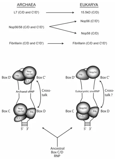
X. Evolutionary Origins of the RNA-guided Nucleotide Modification Complexes ... 23
A. Origins of the Trans-acting Nucleotide Modification Machines ... 23
B. Model for Evolution of the Asymmetric, Eukaryotic Box C/D snoRNP From a Symmetric, Ancestral Box C/D RNP ... 25
XI. Summary of Dissertation Work ... 26
REFERENCES ... 27
CHAPTER I. Archaeal Ribosomal Protein L7 is a Functional Homolog of the Eukaryotic 15.5kD/Snu13p snoRNP Core Protein ... 34
ABSTRACT ... 35
INTRODUCTION ... 36
MATERIALS AND METHODS ... 38
Archaeal L7 Protein Binds the Box C/D Core Motif with the Same Affinity
As Eukaryotic 15.5kD Protein ... 43
L7 Requires the Same Box C/D Sequence and Structural Elements for Binding as Does the 15.5kD Protein ... 44
DISCUSSION ... 49
REFERENCES ... 54
CHAPTER II. Efficient RNA 2'-O-methylation Requires Juxtaposed and Symmetrically Assembled Archaeal Box C/D and C'/D' RNPs ... 66
ABSTRACT ... 67
INTRODUCTION ... 68
RESULTS ... 72
SRNP Core Proteins L7, Nop56/58, and Fibrillarin Bind Both the Terminal And Internal C'/D' Motifs to Assemble Symmetric RNP Complexes ... 72
Nop56/58 and Fibrillarin Associate Through Protein: Protein Interactions And Can Bind the C'/D' Motif in the Absence of L7 ... 74
The Terminal Box C/D RNP is the Minimal Methylation Complex but Efficient Methylation Requires that the Box C/D and C'/D' RNPs are Juxtaposed in the Full-length RNA ... 76
Mutations in Conserved Box Elements Impair Methylation from Both Guide Regions ... 78
Archaeal L7 Binds Cooperatively to the Box C/D and C'/D' Motifs ... 79
DISCUSSION ... 82
MATERIALS AND METHODS ... 88
REFERENCES ... 93
CHAPTER III. Evolutionary Origins of the RNA-Guided Nucleotide Modification Complexes: From the Primitive Translational Apparatus? ... 105
ABSTRACT ... 106
Nucleotide Modification Guide-RNAs are Not Limited to Eukarya ... 107
The Guide RNAs Function as Ribonucleoprotein Complexes ... 108
The Kink Turn or "K-Turn" of the Box C/D RNAs is a Widespread and Evolutionarily Conserved RNA Motif ... 109
Archaeal Box C/D and H/ACA sRNAs Share a Common RNP Core ... 110
A Common Ancestral RNP Complex Derived from the Primitive Translation Apparatus ... 111
Continuing Evolution of the RNA-Guided Nucleotide Modification Complexes ... 113
Concluding Remarks ... 115
BIBLIOGRAPHY ... 117
CONCLUDING REMARKS ... 126
Remaining Questions ... 127
LIST OF TABLES
CHAPTER I.
Table I. Relative binding affinity of L7 protein for selected box C/D
LIST OF FIGURES
LITERATURE REVIEW
Figure 1. Schematic representation of the rRNA primary transcript ... 1
Figure 2. Processing pathways (major and minor) for intronic snoRNAs ... 5
Figure 3. Secondary structure of the eukaryotic box H/ACA snoRNA ... 7
Figure 4. Secondary structure of the eukaryotic box C/D snoRNA ... 8
Figure 5. Alignment of Methanocaldoccus jannaschii ribosomal protein L7 with human Nhp2p and 15.5kD ... 16
Figure 6. Secondary structure of the K-turn consensus sequence and the tertiary structure of the 15.5kD-binding site in U4 ... 17
Figure 7. Comparison of the box C/D core motif with the ribosomal binding site of L7 in the ancestral 23S rRNA illustrates that L7 recognizes variants of the K-turn structure ... 18
Figure 8. Crystal structure of Nop56/58p: Fibrillarin heterodimer ... 22
Figure 9. Model for evolution of the methylation guide RNAs from cis-acting elements in the primitive translation apparatus ... 24
Figure 10. Model for the evolution of the symmetric, archaeal box C/D RNP versus the asymmetric, eukaryotic box C/D RNP ... 25
Figure 4. The box C/D core motif exhibits a highly ordered RNA structure ... 60-61
Figure 5. L7 requires specific sequence and structural elements of the box C/D core
motif for RNA: protein interaction ... 62
Figure 6. Comparison of the U4 snRNA 5' stem loop and the KT-15 K-turn of 23S
rRNA ... 63
Figure 7. Comparison of human 15.5kD, H. marismortui L7Ae and M. jannaschii
L7 protein ... 64
CHAPTER II
Figure 1. Archaeal sR8 sRNP assembly requires a defined order of core protein
addition and forms symmetric RNP complexes on the terminal box C/D core and internal
C'/D' motifs ... 97
Figure 2. Nop56/58 and Fibrillarin interact via protein: protein interactions and can bind
the internal C'/D' motif in the absence of core protein L7 ... 98
Figure 3. in vitro assembled sR8 sRNP guides site-specific 2'-O-methylation from both
terminal box C/D and internal C'/D' RNPs: Efficient methylation requires
juxtapositioning of both RNP complexes in the full-length sRNA ... 99-100
Figure 4. Mutation of either the terminal box C/D core or internal C'/D' motifs in
full-length sR8 affects guided 2'-O-methylation of both RNP complexes ... 101
Figure 5. Core protein L7 exhibits cooperative binding to archaeal sR8 sRNA ... 102
Figure 6. Archaeal L7 binds both the C/D and C'/D' motifs: Eukaryotic 15.5kD protein
binds only the terminal box C/D core motif ... 103
Figure 7. Evolution of the box C/D RNP core proteins: Archaeal and eukaryotic box
C/D RNP complexes exhibit symmetric versus asymmetric distribution of the RNP
CHAPTER III
Box I. Eukaryotic and archaeal RNA-guided nucleotide modification complexes ... 121-122
Figure 1. The L7 protein family ... 123
Figure 2. The kink-turn (K-turn): An evolutionarily conserved RNA motif ... 124
LITERATURE REVIEW
Ribosomes are ubiquitous translational machines that are essential for cell growth and maintenance.
Ribosome biogenesis is an active process involving the transcription of precursor RNA molecules,
cleavage of the pre-ribosomal RNA (pre-rRNA), modification of nucleotides, and assembly with
ribosomal proteins [1-3]. Active cell growth requires maintaining ribosome numbers at an operational
level and thus requiring a large input of cellular resources for any given cell. For example, an actively
growing human cell may require up to 1 million ribosomes, synthesizing approximately 7500 subunits
per minute [4]. Synthesis of such large numbers of ribosomes requires that the steps in ribosome
biogenesis be highly orchestrated.
I. Eukaryotic Ribosome Biosynthesis
A.
B.
Eukaryotic ribosome biogenesis begins with transcription by RNA Polymerase I (RNA Pol I) of the
47S precursor in metazoa or 35S in yeast (see Figure 1A or B, respectively, see [3] for review). The
precursor transcript contains the 18S, 5.8S and 28S (25S in yeast) rRNAs as well as intergenic (ITS)
and extragenic (ETS) transcribed spacer regions (see Figure I). (5S rRNA is also included in the
mature ribosome (60S subunit) but is transcribed by RNA polymerase III outside of nucleolus.) The
nascent precursor rRNA (pre-rRNA) must undergo a variety of post-transcriptional processing events
including cleavage, folding, and nucleotide modification. The vast majority of the nucleotide
modifications are of two types, sugar methylation at the 2'-O-ribose position and pseudouridylation of
uridine residues. These modifications are numerous with upwards of 200 (100 of each type) present in
mature, human rRNAs [5, 6].
A. Nucleotide Modifications in rRNA
Currently, the function of these rRNA nucleotide modifications is unknown. It is commonly believed,
however, that the cumulative effect of these modifications influences ribosome biogenesis and/or
function. The fact that these nucleotide modifications are important stems from several lines of
evidence. First, modifications do not occur in the ITS or ETS regions of the pre-rRNA [3], regions that
are not retained in the mature ribosome RNA. Second, recent positioning of modification sites on
modeled yeast ribosomal subunits revealed that these modifications are clustered in functionally
important regions, including the peptidyl transferase center and the ribosomal subunit interface [7].
Additionally, rRNA regions involved in ribosomal protein binding are virtually devoid of
modifications.
Ribose methylation increases base stacking and pseudouridylation enhances base rotational freedom
center are dispensable for function [10]. The collective effect of nucleotide modifications could
contribute significantly to the overall structure of the folded rRNA particle.
B. Ribosome Biosynthesis
Ribosome biogenesis in eukaryotes occurs in a dense, fibrillar region within the nucleus called the
nucleolus. The nucleolus was first reported in 1898 and was one of the first organelles identified by
microscopists [11]. However, the function of the nucleolus remained elusive until the 1960's, when
two different research groups identified 18S and 28S ribosomal DNA (rDNA) transcriptional units
located within this subcellular compartment [12, 13]. It is now known that the nucleolus is the site of
ribosome biogenesis and is often referred to as the "ribosome factory". The nucleolus is organized
around 50-1000 actively transcribed rDNA repeats of the primary transcript gene. Since this organelle
is not membrane-bound, it is this high level of transcription that is believed to give the nucleolus its
dense appearance in the electron microscope. It is also believed that the nucleolus is formed by a high
concentration of rRNA processing factors transiently associated with multiple sites of activity [14].
II. Small Nucleolar RNAs
The small nucleolar RNAs comprise a large class of stable cellular RNAs that function in various
aspects of ribosome biogenesis. Approximately 100 species of snoRNAs have been identified to date,
but estimates indicate that the full cellular complement numbers around 150 [15, 16]. SnoRNAs are
typically between 75 and 200 nucleotides in length, although, yeast snoRNAs are often considerably
longer due to a lack of 5' end processing (see section IIB). One such example is yeast snR30, which is
approximately 600 nucleotides [17]. Some snoRNAs function as pre-rRNA chaperones, base-pairing
to the pre-rRNA and either preventing rRNA misfolding or facilitating proper folding of the rRNA
snoRNA species results in accumulation of rRNA precursors. However, the vast majority of snoRNAs
guide rRNA nucleotide modifications using rRNA-complementary regions within the snoRNA
molecule.
A. A Brief History of SnoRNAs
In 1966, Busch and coworkers published a study of small nuclear and nucleolar RNAs [18]. They
observed a fraction of nucleolar RNA sedimenting between the 4 and 6S range in a sucrose density
gradient that had not been previously identified. Base composition analysis demonstrated that this low
molecular weight RNA had a higher uridine content (U) with respect to the higher molecular weight
RNA fraction (i.e. rRNA). Two years later, this group published a report that further analyzed this
small nucleolar RNA fraction and revealed that it did not exhibit amino acid acceptor activity (i.e. not
transfer (t) RNA) and had low template activity (i.e. not messenger (m) RNA). They concluded that
this low molecular weight fraction contained unique small RNAs, which were later termed small
nucleolar or snoRNAs [19]. Individual species of snoRNAs are currently named U3, U14, U22, etc.
based on the original base composition analysis and the order in which they were discovered.
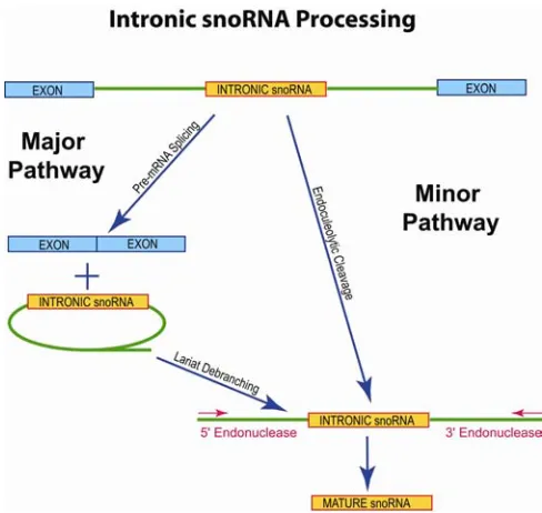
B. SnoRNA Processing in Eukaryotes
SnoRNAs exhibit several genome organizations depending on the eukaryotic organism. Most
snoRNAs in yeast are monocistronic but some (and most in plants) are polycistronic. In metazoa, a
few snoRNAs are individually transcribed (U3, U8, U13), but the vast majority are encoded in introns
of protein-coding genes ("intronic snoRNAs") and transcribed by RNA polymerase II (Pol II) [20].
U14 was the first snoRNA discovered to be intron-encoded and, in mouse, resides within the coding
It is now evident that Pol II transcripts containing intronic snoRNAs ("snoRNA host transcripts")
preferentially code for proteins involved in various aspects of ribosome biosynthesis and translation,
including ribosomal proteins and translation factors [20]. This organization may reflect a regulatory
mechanism important for maintenance of ribosome numbers at operational levels. There are, however,
examples of snoRNA host transcripts whose exonic sequences do not encode protein products and,
instead, act only as snoRNA expression vehicles. These include the gas5 gene, which encodes ten
different snoRNAs in ten different introns [24]. Four such non-protein coding snoRNA host genes
have been identified to date [24-27]. Interestingly, these genes are members of the 5'-terminal
oligopyrimidine (5' TOP) gene family [24, 27]. The 5'-TOP family is associated with gene products
that are translationally regulated by changes in growth conditions [28]. Since this type of snoRNA
host transcript is not translated, it is possible that expression of the 5'-TOP gene family is regulated at
the level of transcription [20].
Figure 2. Processing pathways (major and minor) for intronic snoRNAs.
involves exon-intron splicing, lariat debranching, and finally exonucleolytic trimming. This pathway
is utilized by the vast majority of intronic snoRNAs. The minor pathway is used when snoRNAs are
found in poorly spliced introns (e.g. U16 and U18) [29]. This pathway involves endonucleolytic
excision of the intronic snoRNA followed by exonucleolytic trimming of the ends.
Processing of snoRNAs via either pathway is believed to involve initial packaging of the intronic
snoRNA into a ribonucleoprotein complex (see below). Evidence for snoRNP assembly includes the
finding that intronic snoRNA processing can be inhibited by addition of excess, unprocessed
transcripts indicative of titration of an essential, trans-acting, processing factor(s) [30]. In addition,
precursor snoRNAs can be immunoprecipitated with antibodies against snoRNP core proteins [30].
Recent studies have provided evidence that splicing and snoRNA processing are tightly linked.
SnoRNAs are preferentially positioned 70 to 80 nucleotides upstream of the 3' splice site in humans
and alteration of this spacing is detrimental to snoRNA processing [31, 32]. This conservation of
intronic snoRNA spacing may indicate binding of additional, unidentified snoRNA-processing
factor(s).
III. Classes of SnoRNAs
There are two major classes of snoRNAs based on conserved sequence elements: the box H/ACA
snoRNAs and the box C/D snoRNAs. Both snoRNA classes guide nucleotide modifications through
an antisense base-pairing mechanism. Both the box H/ACA and box C/D snoRNAs contain
complementary sequences that pair with the target rRNA and designate the specific nucleotide for
A. Box H/ACA SnoRNAs
The box H/ACA snoRNAs characteristically fold into a bipartite hairpin-hinge-hairpin tail structure as
seen in Figure 3 [33, 34].
Figure 3. Secondary structure of the eukaryotic box H/ACA snoRNA (reproduced from [35]). Box elements are indicated in bold while the targeted rRNA molecule (green) is shown base-paired to the snoRNA within the pseudouridylation pocket. The target pseudouridylation nucleotide (Ψ) is shown in red.
This snoRNA family is named for conserved box H (sequence ANANNA) and ACA-triplet nucleotide
sequences. The majority of box H/ACA snoRNAs guide pseudouridylation and they do this through
rRNA antisense regions located within an internal loop in one or both of the hairpin structures [36].
These antisense regions base pair to ribosomal RNA, creating short helices (4-10 base pairs in length)
within the loop (see [37] for review). The target uridine lies in an unpaired region at the base of the
proximal helix within the hairpin structure. The internal loop region is called the pseudouridylation
pocket and modification occurs approximately 15 nucleotides from the box H or ACA sequence within
the snoRNA [34]. Only a few members of the box H/ACA family participate in rRNA processing .
The best characterized is yeast snR30 (or U17 in humans) which functions in rRNA precursor cleavage
impaired cell growth [37, 38]. Sequence comparison of snR30 homologs in various species has
revealed conserved sequence elements m1 and m2, which are both unique to this species of snoRNA
and necessary for rRNA processing [17]. However, the mechanism by which snR30 affects pre-rRNA
processing remains unclear.
B. Box C/D SnoRNAs
The second major class of snoRNAs is the box C/D snoRNAs. Members of this family are defined by
conserved box elements C (sequence RUGAUGA where R is any purine) and D (CUGA) (Figure 4).
Figure 4. Secondary structure of the box C/D snoRNA (reproduced from [35]). Box C, D, C' and D' sequences are shown in bold. The rRNA target (green) is shown base-paired upstream of boxes D and D'. The 2'-O-methylated nucleotide (blue) is located 5 nucleotides upstream of boxes D and D'.
Boxes C and D base pair to form a 5+2 asymmetric, nucleotide bulge flanked by external and internal
stems [40]. Many box C/D snoRNAs also contain internal copies of the consensus elements called
boxes C' and D' [41]. The vast majority of box C/D snoRNAs function in guiding 2'-O-ribose
nucleotides in length and are located immediately upstream of boxes D and D' [42, 25]. Nucleotide
modification occurs at a fixed position within the target RNA paired five nucleotides upstream of box
D and/or D' [43]. Unlike box H/ACA snoRNAs, there is no base-specificity for box C/D-guided
modification: any ribonucleotide can be modified.
A few box C/D snoRNAs function in pre-rRNA processing. U3, U14, and U22 are required for
cleavage events necessary for 18S rRNA production whereas U8 is required for 5.8S and 28S
processing [3, 44, 37, 20]. Since no ribonucleases have been identified in association with these
snoRNAs, it is anticipated that processing is facilitated by these snoRNAs acting as RNA chaperones
to assist pre-rRNA folding. Several lines of evidence support this hypothesis. First, U3 acts by
bridging two regions of rRNA that are not adjacent in the precursor transcript, establishing a pre-rRNA
structure that is essential for ribosomal RNA processing [45, 46]. Second, U106 is a suspected
chaperone because it contains two antisense rRNA elements, both of which are complementary to
rRNA regions devoid of modifications [15]. Third, the guide sequence upstream of box D' in U14
base-pairs to a region of 18S rRNA lacking ribose-methylated nucleotides. Experiments have
demonstrated that the U14: rRNA helix formed is required for pre-rRNA cleavage and subsequent 18S
rRNA production [47].
C. MRP
MRP (mitochondrial RNA-processing) is a unique snoRNA species because it is not a member of
either of the two major snoRNA classes. MRP is required for cleavage of pre-rRNA at an ITS I site
upstream of the 5.8S sequence [48]. Interestingly, the RNA component of the MRP snoRNP complex
resembles that of the ribozyme RNase P, which cleaves the 5' end of tRNA [48]. This suggests that the
IV. SnoRNA-Protein Complexes
The two major classes of snoRNAs (box H/ACA and box C/D) form ribonucleoprotein complexes
(RNPs) by associating with a set of core proteins. Whereas the antisense regions within the snoRNAs
select the site for nucleotide modification, the associated proteins provide the necessary enzymatic
activities.
A. Box H/ACA Core Proteins
Box H/ACA snoRNAs associate with four core proteins that are common to all members of this
snoRNA class. These core proteins are Gar1p, Nhp2p, Nop10p, and Cbf5p [49, 51]. Cbf5p (Dyskerin
in humans or Nap57p in rat) is the pseudouridine synthase. Cbf5p shows significant homology to other
known pseudouridine synthases and Cbf5p mutant strains in yeast abolish pseudouridylation of rRNA
[6, 52]. The remaining three proteins are believed to perform structural roles in the H/ACA snoRNP.
Gar1p is an essential component of the box H/ACA snoRNAs and its depletion in yeast results in
pre-rRNA processing defects [53]. Gar1p contains glycine and arginine rich N-terminal domain and
has been found to bind the H/ACA snoRNA directly [54, 55]. In fact, all four core proteins may
contact the RNA because each protein can be UV-crosslinked to a box H/ACA snoRNA in vivo [56].
B. Box C/D Core Proteins
Box C/D snoRNAs also form ribonucleoprotein complexes by assembling with a set of four core
proteins. These core proteins are Fibrillarin, 15.5kD, Nop56p, and Nop58p. Fibrillarin is the
methylase enzyme, utilizing S-adenosyl methionine (SAM) as the methyl donor [57-59]. Interestingly,
supporting RNA-binding activity for this core protein [60]. Experiments have demonstrated that the
association of Fibrillarin with the box C/D snoRNA is dependent on binding of other core proteins to
the snoRNA, indicating that Fibrillarin is only weakly associated with the RNA. However, at least for
one snoRNA species, Fibrillarin associates with the RNA in the absence of other core proteins [61].
The snoRNA core protein, 15.5kD, binds directly to the box C/D motif and this binding is believed to
function as the nucleation event for snoRNP assembly. 15.5kD was originally identified as a protein
that associates with the U4 spliceosomal small nuclear RNA (snRNA) [62]. Subsequent experiments
identified 15.5kD as a dual function protein because it also associates with the box C/D snoRNAs [40].
The co-crystal structure of 15.5kD bound to its binding site in U4 snRNA revealed a unique RNA fold
resulting from an asymmetric bulge flanked on either side by helices [63]. This secondary structure
has been applied to the box C/D motif by analogy [40]. Strikingly, analysis of RNA-folding motifs in
the H. marismortui 50S ribosomal subunit identified a new RNA motif, termed the kink-turn (K-turn)
[64]. Both 15.5kD-binding sites in U4 snRNA and the box C/D snoRNAs exhibit classical features of
this motif and are, therefore, members of this newly identified K-turn family (see section VIIB).
Interestingly, the box H/ACA snoRNA core protein, Nhp2p, is similar to 15.5kD in sequence,
exhibiting 31% identity or 61% similarity in yeast. However, no kink-turn secondary structure has
been identified in the eukaryotic box H/ACA snoRNAs.
The Nop56p and Nop58p core proteins constitute a pair of related proteins, exhibiting 45% sequence
identity in yeast [65]. Like the box H/ACA core proteins Nhp2p and Nop10p, Nop56p and Nop58p are
predicted to be structural components of the box C/D snoRNP. These proteins were originally
identified in synthetic lethal screens using temperature-sensitive Fibrillarin mutants [65]. Interestingly,
only in the presence of Fibrillarin [66, 67]. Additionally, depletion of either Nop58p or Fibrillarin (but
not Nop56p) leads to co-depletion of box C/D snoRNAs [67]. In 2002, Steitz and coworkers utilized
in vivo crosslinking to reveal a differential distribution of Nop56p and Nop58p on a box C/D snoRNA
[60]. This biochemical data supported prior genetic analyses indicating different roles for these two
proteins. More specifically, this study revealed that Nop56p binds to the internal box C'/D' motif
whereas Nop58p binds the terminal box C/D core motif [60]. In another report from the same
laboratory, nucleotide interference analog mapping demonstrated that 15.5kD binds only to the
terminal C/D motif and not the internal C'/D' motif [68]. Thus, the methylase Fibrillarin is the only
core protein bound to both RNA motifs.
V. Archaea: A Separate Domain of Life from Eukaryotes and Bacteria
In 2000, homologs of eukaryotic snoRNAs (termed snoRNA-like or sRNAs) were discovered in
Archaea [69, 70]. This finding indicated that RNA-guided nucleotide modification is an evolutionarily
ancient mechanism. Archaea are prokaryotic organisms, which constitute a domain of life distinct
from both Eukarya and Bacteria [71]. Archaeal metabolism and gene organization resembles Bacteria,
but archaeal replication, transcription and translation machinery more closely resembles eukaryotes
[72].
Many archaeal species live in extreme environments including low pH and high temperature. In 1998,
Noon and coworkers demonstrated that the level of rRNA 2'-O-methylation increases proportionally
with increased growth temperature [73]. This suggests that nucleotide modification in the ribosome
may help stabilize thermolabile RNA at high temperatures. Therefore, rRNA modification in Archaea
Archaeal sRNAs exhibit a genomic organization distinct from eukaryotes. DNA sequences encoding
sRNAs are found on both strands and are dispersed around the entire circular chromosome [74]. Most
are found in intergenic regions between protein-coding genes whereas some overlap the adjacent open
reading frame. The mechanism for transcription and processing of sRNAs in Archaea remains
unknown. Currently, there is only one example of an sRNA encoded within an intron. SR3
(Archeoglobus fulgidus) or sR50 (Pyrococcus abyssi) is found in the intron of its target substrate,
precursor-tRNATrp ( pre-tRNATrp) [75]. The mechanism by which this sRNA guides modification was
predicted to occur in cis via an intramolecular modification reaction [75, 76]. In this model,
intron-encoded sR3 guides two, tRNA nucleotide modifications before its removal from the pre-tRNA
by splicing. New results, however, now indicate that these modifications occur in trans via an
intermolecular reaction (R. Gupta, personal comm.). The details of this mechanism are still under
investigation.
VI. Archaeal sRNAs and Core Proteins
The nucleotide-modification guide RNAs of Archaea exhibit similar consensus box elements and
secondary structures as their eukaryotic counterparts. Not surprisingly, homologs to the eukaryotic
snoRNA core proteins are also present in Archaea. Currently, the only characterized function of Box
C/D RNPs in Archaea is 2'-O-methylation. Possible roles in pre-rRNA folding and/or cleavage remain
to be demonstrated.
A. Archaeal Box H/ACA sRNPs
Although both major classes of modification-guide RNAs are present in Archaea, the H/ACA sRNAs
have only recently been discovered and this sRNA class is the least characterized [77]. The presence
pseudouridylation in Archaea is low. Pseudouridylation levels in Archaea more closely resemble
those in Escherichia coli, where this type of nucleotide modification is catalyzed by protein-only
enzymes [74]; [9]. However, homologs of the core proteins Cbf5p, Gar1p, and Nop10p, had been
detected in archaeal genomes [78, 79]. In 2002, this debate ended with the discovery of four H/ACA
sRNAs in Archeoglobus fulgidus [77]. The box H/ACA sRNAs of Archaea exhibit slightly different
structures and sequence elements than those of eukaryotes. Archaeal H/ACA sRNAs have either one
or three hairpin structures and the box H sequence is sometimes absent. Notably, this is also the case
for the box H/ACA snoRNAs of two early branching eukaryotes, Trypanosoma brucei and Euglena
gracialis [80, 81]. This suggests that the single hairpin H/ACA RNA represents an early evolutionary
form of the box H/ACA snoRNAs found in vertebrates and yeast.
B. Archaeal Box C/D sRNPs
The box C/D sRNAs were the first class of guide RNAs discovered in Archaea [69]; [70]. Unlike the
H/ACA sRNAs, the presence of box C/D snoRNA homologs was anticipated due, in part, to the high
level of 2'-O-methylation in archaeal rRNA sequences. Additionally, homologs to core proteins
Fibrillarin and Nop56p/Nop58p had been detected [82, 83]. Interestingly, Archaea contain a single
homolog to both eukaryotic Nop56p and Nop58p (termed Nop56/58p or Nop5p). This is consistent
with the prediction that the two related proteins in eukaryotes arose from a gene duplication event [74].
With knowledge of these core protein homologs, Omer and coworkers identified the first archaeal box
C/D sRNAs from Sulfolobus solfactaricus by co-immunoprecipitating these RNAs from cell extracts
using antibodies against archaeal Fibrillarin and Nop56/58p [59]. Subsequent development of a
computer algorithm to detect additional archaeal box C/D sRNAs has resulted in the detection of many
The box C/D sRNAs exhibit similar sequences and structures as their eukaryotic snoRNA counterparts
with a few exceptions. First, archaeal box C/D sRNAs are slightly smaller with an average size of 75
nucleotides (as compared to 150 nucleotides in eukaryotes). Additionally, the internal C'/D' motif is
typically a perfect match to the consensus box C and D sequences; Eukaryotic C'/D' sequences are
frequently degenerate. Finally, most box C/D sRNAs in Archaea exhibit "dual guide" function: both
the D and D' guide regions contain sequences antisense to targeted RNA molecules [74]. In contrast,
only 20% of box C/D snoRNAs have been identified as "dual guides".
VII. Identification of the Archaeal Homolog of Eukaryotic 15.5kD
To characterize the assembly and function of the archaeal box C/D sRNPs, it was necessary to identify
the full complement of core proteins. Whereas homologs to both Fibrillarin and Nop56p and Nop58p
were evident, no clear homolog to 15.5kD had been identified. Because 15.5kD is the first protein to
bind and recognize the C/D core motif, it was unlikely that archaeal box C/D sRNP assembly did not
require a comparable core protein homolog.
A. Identification of Archaeal Ribosomal Protein L7
In 2002, we began our investigation of the archaeal box C/D sRNP by identifying ribosomal protein
L7 as a putative homolog to 15.5kD ([84]; see Chapter I). Sequence comparison of
Methanocaldooccus jannaschii (M. jannaschii) L7 with human 15.5kD revealed that these protein
Figure 5. Alignment of Methanocaldococcus jannaschii ribosomal protein L7 with human Nhp2p and 15.5kD (adapted from [84]). Amino acid residues outlined in black indicate identity whereas gray shading indicates similarity. Genbank accession numbers are B64450, CAC08452.1, and NP_004999.1 for L7, 15.5kD, and Nhp2p, respectively.
Identification of a putative archaeal box C/D core protein homolog that is also a ribosomal protein
indicated that L7 might also be a dual function protein, reminiscent of 15.5kD, which binds both the
box C/D snoRNAs and U4 snRNA. Utilizing purified, recombinant L7 protein from M. jannaschii and
a minimal box C/D core motif RNA, we demonstrated that L7 binds the terminal box C/D core motif
with high affinity. This association is dependent on consensus box C and D sequences and formation
of a kink-turn secondary structure (see below). This high affinity and specificity correlated well with
15.5kD's association to eukaryotic box C/D snoRNAs and U4 snRNA [62, 40]. Parallel,
electrophoretic mobility-shift analyzes showed that both eukaryotic 15.5kD and archaeal L7 proteins
specifically recognized the minimal box C/D core motif RNA, thus demonstrating that archaeal
ribosomal protein L7 is a functional homolog of eukaryotic 15.5kD core protein (see Chapter I).
B. Identification of the Kink-Turn Motif
Following careful analysis of the ribosomal 50S subunit crystal structure from archaeal species
into a family termed the kink-turn (or K-turn) motif [64]. This motif is characterized by a
helix-bulge-helix structure with a sharp bend between the two helical axes (Figure 6).
Figure 6. Secondary structure of the K-turn consensus sequence (left; [64]) and the tertiary structure of the 15.5kD-binding site in U4 (right; [63]). Stem structures I and II are coordinately colored in green and blue, respectively. Tandem, sheared G•A base pairs appear in red, and the protruded nucleotide of the asymmetric bulge is shown in yellow.
This bend is facilitated by two adjacent, sheared G•A base pairs, which reside at the base of the
terminal bulge. This structure was first identified in the L30-binding site of the L30 pre-mRNA [85].
Ribosomal protein L30 in eukaryotes regulates its own expression at the level of splicing by
associating with its pre-mRNA. The K-turn structure is also evident in the 15.5kD-binding site in U4
snRNA [63]. In the U4 kink-turn, the RNA is bent at approximately 120 degrees and this bend results
in protrusion of a uridine nucleotide that lies in the RNA-binding pocket of 15.5kD. Logic predicted
that the 15.5kD-binding site in the box C/D terminal core motif should also be K-turn, although this
had not yet been demonstrated directly by detailed structural analysis [40]. Consistently, L7, the
C. Characterization of L7 Association with the Box C/D Core Motif
The archaeal box C/D RNP core protein, L7, is a ribosomal protein that binds a K-turn (KT-15) in the
archaeal large ribosomal subunit (Figure 7B and C; [86, 64]).
Figure 7. Comparison of the box C/D core motif with the ribosomal binding site of L7 in the archaeal 23S rRNA illustrates that L7 recognizes variants of the K-turn structure. The box C/D consensus secondary structure (A) and the secondary (B) and tertiary (C) structure of the L7-binding site in the 23S rRNA of the 50S subunit, KT-15 [64].
KT-15 is a unique structural variant of the K-turn because it incorporates a base-triple in place of one
of the G•A pairs. This indicated that structure may play a larger role in L7 binding than sequence.
To further characterize L7 binding to the box C/D core motif, point mutations in the minimal motif
were utilized in a series of binding studies (see Chapter I). Our results confirmed the hypothesis that
structure was a critical factor for high affinity binding [84]. Mutations predicted to disrupt the
helix-bulge-helix secondary structure, e.g. mutation of the critical GA nucleotides, either reduced or
abolished L7 binding. Conversely, mutations that maintained secondary structure had minimal affect
on L7 binding. The exception is the protruded uridine whose identity should have no bearing on
structure since this residue makes no interactions with other portions of the RNA. Mutation of this
tolerated. Coincidently, Klein and coworkers noted that the protruded uridine in KT-15 is found deep
within the binding pocket of L7 and a purine base ring might be too large to be accommodated in the
RNA binding site of this protein [64].
Comparison of the crystal structures of archaeal ribosomal protein L7 and eukaryotic 15.5kD has
revealed that the tertiary structures of these two proteins are virtually superimposible [84]. This
conservation of structure is striking given that the L7 and 15.5kD proteins share only 30% sequence
identity (see Figure 5). Our results are consistent with the identification of L7 and 15.5kD as
functional homologs ([84]; see Chapter I).
The co-crystal structure of L7 bound to the C/D motif has confirmed that this RNA motif is indeed a
K-turn ([87]; Appendix II). In fact, the L7:C/D RNA complex exhibits high structural similarity to that
of the U4:15.5kD co-crystal structure [63]. This finding was anticipated due to the structural
similarities of the two proteins. Interestingly, our UV-melting profiles revealed conformational
changes in the RNA following L7 binding, indicative of an "induced-fit" mechanism. Interestingly, an
"induced-fit" binding mechanism was also predicted for ribosomal protein L30 binding to its
pre-mRNA and may be the common mode of protein association with K-turn motifs [88]. Consistent
with the idea that the K-turn is not a rigid, preformed structure, Lilley and colleagues demonstrated that
the K-turn motif is highly dynamic and requires other factors, such as proteins, to "lock" the K-turn
motif into its characteristically "bent" conformation [89].
VIII. Archaeal Box C/D sRNP Assembly
In 2002, Omer and colleagues established an in vitro assembly system to study archaeal box C/D
Sulfolobus acidocaldarius, L7, Fibrillarin, and Nop56/58p (Nop56a), to a box C/D RNA from S.
solfactaricus resulted in stable formation of a box C/D RNP in vitro.
A. Analysis of Ribosomal Protein L7 Binding to a Full-length Archaeal Box C/D RNA
Following the work Omer and coworkers, we developed an in vitro assembly system using
Methanocaldococcus jannaschii core proteins and a box C/D RNA to analyze the structure and
function of the archaeal box C/D sRNP. Although Omer and coworkers demonstrated that ribosomal
protein L7 is assembled in the box C/D RNP, it was not clear which motifs this protein bound. Using
full-length, archaeal sR8 box C/D sRNA as well as minimal box C/D or C'/D' RNAs, we determined
that L7 associates with both motifs ([90]; see Chapter II). Additionally, we demonstrated that L7 binds
with positive cooperativity to the two binding sites in the full-length sRNA. RNA footprinting
analysis has now revealed that L7 initially binds to either the box C/D or the C'/D' motif, indicating that
the binding is not sequential (Tran et al., unpublished results). These results were surprising in light
of recent work reporting that the eukaryotic homolog, 15.5kD, binds the eukaryotic terminal box C/D
core but not the internal C'/D' motif [68]. Our comparison of L7 and 15.5kD binding to both a
full-length eukaryotic and archaeal box C/D RNA revealed that the difference in distribution of the two
core protein homologs on the box C/D and C'/D' motifs is due to divergence in the RNA-recognition
capabilities of L7 and 15.5kD ([90]; see Chapter II).
B. Identification of Protein: Protein Interactions Between Nop56/58p and Fibrillarin
To characterize further assembly of the sRNP complex, we utilized a series of "pull-down" assays to
study protein: protein interactions between the archaeal box C/D core proteins. These experiments
suggestion is consistent with our observation that Nop56/58p binding to an L7: RNA complex is
greatly enhanced with the addition of Fibrillarin. Additionally, the Nop56/58p-Fibrillarin protein
complex can associate specifically with the C'/D' motif in the absence of L7, albeit inefficiently. This
result indicated that the Nop56/58p-Fibrillarin "dimer" contacts the RNA directly and that the C/D and
C'/D' motifs are structurally distinct.
C. Demonstration of Archaeal Box C/D sRNP Symmetry
Both electrophoretic mobility-shift and co-purification analyses revealed that all three core proteins
(L7, Nop56/58p, and Fibrillarin) bind both the terminal box C/D and internal C'/D' motifs. This work,
therefore, established the archaeal box C/D sRNP as a "symmetric" particle with respect to core protein
distribution ([90]; see Chapter II). This sharply contrasts with the eukaryotic box C/D snoRNP, where
differential binding of the core proteins to the box C/D and C’/D’ motifs is "asymmetric". Eukaryotic
15.5kD protein is uniquely bound to the terminal box C/D core motif [68]. Nop58p and Nop56p bind
to the terminal C/D and internal C'/D' motifs, respectively [60]. Only Fibrillarin is found associated
with both motifs in the eukaryotic snoRNP. From these contrasting RNP structures, we have proposed
a pathway for the evolution of the box C/D RNP complexes ([90];see section X).
IX. RNP-guided Methylation Activity of the In Vitro Reconstituted Archaeal Box C/D sRNP
Our reconstituted box C/D sRNP guides site-specific methylation from both the box C/D and C'/D'
motif, using both antisense regions to select their respective target RNAs for modification ([90]; see
Chapter II). Surprisingly, although both the C/D and C'/D' motifs bind all three of the core proteins,
both "RNPs" must be juxtaposed in the same sRNP complex for efficient guided-methylation activity.
This requirement was revealed using two different experimental approaches. First, methylation
whereas, the box C'/D' "halfmer" RNP was inactive. This requirement for juxtaposed RNPs in the
same sRNP particle was confirmed using full-length sR8 molecules mutated in either the box C/D or
C'/D' motifs. Strikingly, mutations in either motif affected methylation not only of the mutated motif,
but of the non-mutated motif as well. These results clearly demonstrated that efficient guided
methylation requires box C/D and C'/D' RNPs juxtaposed on the same box C/D sRNP particle. From
these observations, we have concluded that "crosstalk" interactions between the box C/D and C'/D'
motifs are important for guided-nucleotide modification activity ([90]; see Chapter II).
Although the nature of this "crosstalk" is presently unknown, the recent Nop56/58p-Fibrillarin crystal
structure from Archeoglobus fulgidus has suggested an interesting possibility [91].
Figure 8. Crystal structure of Nop56/58p-Fibrillarin heterodimer [91]. Nop56/58p molecules are shown in blue or red whereas Fibrillarin molecules are shown in yellow or orange.
This crystal structure confirms the previously reported interaction between Nop56/58p-Fibrillarin [90],
box C/D RNA between the C/D and C'/D' motifs [91]. The importance of this putative protein: protein
interaction for "crosstalk" between the box C/D and C'/D' motif in guided methylation remains to be
demonstrated.
X. Evolutionary Origins of the RNA-guided Nucleotide Modification Complexes
The archaeal and eukaryal domains are estimated to have diverged approximately 2 billion years ago
[92], making the nucleotide modification complexes evolutionarily ancient ribonucleoprotein
machines. Recently, ribosomal protein L7 was shown to be the archaeal homolog of eukaryotic box
H/ACA core protein, Nhp2p ([93]; see Figure 5). Additionally, L7 binds a K-turn motif within the
archaeal box H/ACA sRNA and is a core component of the pseudouridylation guide complexes in
Archaea. Therefore, archaeal L7 bound to a K-turn motif represents an RNP that is common to both
classes of modification guide complexes as well as the ribosome. These observations led us to propose
that these modification complexes may have their origins in the primitive translational apparatus.
A. Origins of the Trans-acting Nucleotide Modification Machines
Currently, there is no evidence for RNA-guided nucleotide modification in Bacteria. There are no
bacterial proteins with significant sequence similarity to archaeal ribosomal protein L7, although there
are reported K-turn motifs in Bacteria [94]. While we cannot rule out the presence of undiscovered
box C/D and/or H/ACA RNA homologs in Bacteria, it is likely that the modification guide RNAs
originated after the branching of Bacteria and Archaea/Eukarya.
Modern day, nucleotide modification-guide RNPs probably originated as K-turn RNP components of
Figure 9. Model for evolution of the modification guide RNAs from cis-acting elements in the primitive translation apparatus (Adapted from [35]) .
K-turn RNPs present within the primordial apparatus may have been retained as necessary folding
elements. This is supported by the presence of K-turn RNPs in the modern ribosome. Additionally,
some of these cis-acting elements may have evolved to guide rRNA nucleotide modification in the
ribosome. Evolution of the trans-acting modification guide-RNPs probably occurred through a
progenitor RNP (common to both the box C/D and H/ACA RNP class) that guided modification as a
cis or trans complex. The box C/D and H/ACA RNPs then evolved from this progenitor complex. It
is has been suggested that the expansion of the number of guide RNPs and the multitude of RNA
targets may have occurred from duplication events (or retrotransposition events) of the box C/D and
H/ACA RNA genes followed by variation of the antisense elements [6, 15]. Continued variation of
the guide regions has enabled additional guide RNPs to now recognize a variety of non-rRNA targets
B. Model for Evolution of the Asymmetric, Eukaryotic Box C/D SnoRNP from a Symmetric,
Ancestral Box C/D RNP
The demonstration that the archaeal box C/D RNP is symmetric with respect to protein binding
contrasts sharply the asymmetric eukaryotic complex [90, 60, 68]. It is likely that the archaeal box C/D
RNP more closely resembles that of the last ancestor common to both Archaea and eukaryotes. We
have suggested that the evolution of the asymmetric, eukaryotic box C/D RNP is a result of gene
duplication and altered RNA-binding capabilities of the core proteins (Figure 10; [90]).
Figure 10. Model for the evolution of the symmetric, archaeal box C/D RNP (left) versus the asymmetric, eukaryotic box C/D RNP (adapted from [90]).
Gene duplication of Nop56/58p has resulted two highly similar core proteins in eukaryotes, Nop56p
terminal box C/D motif due to differential RNA-binding capabilities [60]. It is possible that Nop56p,
in the presence of Fibrillarin, no longer requires an L7-like protein at the C'/D' motif for binding,
therefore, eukaryotic 15.5kD protein is found only at the box C/D motif. It will be interesting to
determine the core protein distribution on the box C/D RNPs in early branching eukaryotes such as
Trypanosomes and Euglena [80, 81]. These box C/D RNPs may represent evolutionary intermediates,
more closely resembling the symmetric, archaeal RNP complex. Characterization of snoRNPs from
these two species may provide additional information necessary to support this model.
XI. Summary of Dissertation Work
This thesis examines the structure and function of the box C/D sRNP in Archaea. Chapter 1 describes
the identification of ribosomal protein L7 as the archaeal homolog of eukaryotic 15.5kD. L7 serves a
dual role in Archaea as both a ribosomal protein and a box C/D core protein. In both the ribosome and
the box C/D sRNP complex, L7 binds a K-turn motif. Chapter 2 reports development of an in vitro
assembly and methylation assay for the archaeal box C/D sRNP. The archaeal box C/D sRNP is
symmetric with respect to core protein binding which contrasts the asymmetric eukaryotic box C/D
snoRNP. Additionally, we reported that efficient methyation requires that both the box C/D and C'/D'
motifs be juxtaposed in the full-length box C/D sRNP. Finally, the demonstration of L7 as a
component of the ribosome as well as the RNA-modification guide complexes suggested that the
modern day sRNPs and snoRNPs may have had their origins as cis-acting elements in the primitive
translational apparatus. Chapter 3 details a proposed pathway for evolution of the trans-acting
REFERENCES
1. Gerbi, S., A. Borovjagin, and T. Lange, The nucleolus: a site of ribonucleoprotein maturation. Curr. Opin. Cell Biol., 2003. 15: p. 318-325.
2. Sollner-Webb, B., K.T. Tycowski, and J.A. Steitz, Ribosomal RNA Processing in Eukaryotes, in Ribosomal RNA: Structure, Evolution, Gene Expression and Function in Protein Synthesis, R.A. Zimmermann and A.E. Dahlberg, Editors. 1996, CRC Press: New York.
3. Eichler, D.C. and N. Craig, Processing of Eukaryotic Ribosomal RNA, in Progress in Nucleic Acids Research and Molecular Biology, K. Moldave and W. Cohen, Editors. 1994, Academic Press: New York.
4. Lewis, J.D. and D. Tollervey, Like Attracts Like: Getting RNA Processing Together in the Nucleus. Science, 2000. 288: p. 1385-1389.
5. Maden, B.E., The numerous modified nucleotides in eukaryotic ribosomal RNA. Prog. Nucleic Acids Res., 1990. 39: p. 241-303.
6. Lafontaine, D. and D. Tollervey, Birth of the snoRNPs: The evolution of the modification-guide snoRNAs. Trends Biochem. Sci., 1998. 23: p. 383-388.
7. Decatur, W. and M.J. Fournier, rRNA modifications and ribosome function. Trends Biochem. Sci., 2002. 27: p. 344-351.
8. Davis, D., Biophysical and conformational properties of modified nucleotides in RNA (nuclear magnetic resonance studies), in Modification and editing of RNA, H. Grosjean and R. Benne, Editors. 1998, American society for microbiology: Washington, D.C. p. 85-102.
9. Ofengand, J., Ribosomal RNA pseudouridines and pseudouridine synthases. FEBS Letters, 2002. 514: p. 17-25.
10. King, T., et al., Ribosome structure and activity are altered in cells lacking snoRNPs that form pseudouridines in the peptidyl transferase center. Mol. Cell, 2003. 11: p. 425-435.
11. Montgomery, T., Comparative cytological studies, with expecial regard to the morphology of the nucleolus. J. Morphol., 1898. 15: p. 265-565.
12. Ritossa, F. and S. Spiegelman, Localization of DNA complementary to ribosomal RNA in the nucleolus organizer region of Drosophila melonagaster. Proc. Natl. Acad. Sci. USA, 1965. 53: p. 737-745.
13. Birnstiel, M. and M. Chipchase, The nucleolus: pacemaker of the cell. Sci. J., 1970. 6: p. 41-48.
15. Vitali, P., et al., Identification of 13 novel human modification guide RNAs. Nucleic Acids Res., 2003. 31: p. 6543-6551.
16. Terns, M. and R. Terns, Small nucleolar RNAs: versatile trans-acting molecules of ancient evolutionary origin. Gen. Express., 2002. 10: p. 17-39.
17. Atzorn, V., P. Fragapane, and T. Kiss, U17/snR30 is a ubiquitous snoRNA with two conserved sequence motifs essential for 18S rRNA production. EMBO J., 2004. 24: p. 1769-1778.
18. Muramatsu, M., J. Hodnett, and H. Busch, Base composition of fractions of nuclear and nucleolar ribonucleic acid obtained by sedimentation and chromatography. J. Biol. Chem., 1966. 241: p. 1544-1550.
19. Nakamura, T., A. Prestayko, and H. Busch, Studies on nucleolar 4 to 6S ribonucleic acid of Novikoff hepatoma cells. J. Biol. Chem., 1968. 243: p. 1368-1375.
20. Filipowicz, W., et al., Biogenesis, structure and function of small nucleolar RNAs. Acta Biochim Pol., 1999. 46: p. 291-302.
21. Liu, J. and E.S. Maxwell, Mouse U14 snRNA is encoded in an intron of the mouse cognate hsc70 heat shock gene. Nucleic Acids Res., 1990. 18: p. 6565-6571.
22. Leverette, R., M. Andrews, and E.S. Maxwell, Mouse U14 snRNA is a processed intron of the cognate hsc70 heat shock pre-messenger RNA. Cell, 1992. 71: p. 1215-1221.
23. Xia, L., et al., Intronic U14 snoRNAs of Xenopus laevis Are Located in Two Different Parent Genes and Can Be Processed From Their Introns During Early Oogenesis. Nucleic Acids Research, 1995. 23: p. 4844-4849.
24. Smith, C.M. and J.A. Steitz, Classification of gas5 as a Multi-Small-Nucleolar-RNA (snoRNA) Host Gene and a Member of the 5'-Terminal Oligopyrimidine Gene Family Reveals Common Features of snoRNA Host Genes. Molecular and Cellular Biology, 1998. 18: p. 6897-6909.
25. Tycowski, K., et al., A Small Nucleolar RNA Requirement for Site-Specific Ribose
Methylation of rRNA in Xenopus. Proc.of the Natl.Acad. Sci. USA, 1996. 93: p. 1480-1485.
26. Tycowski, K., M. Shu, and J.A. Steitz, A mammalian gene with introns instead of exons generating stable RNA products. Nature, 1996. 379: p. 464-466.
27. Pelczar, P. and W. Filipowicz, The Host Gene for Intronic U17 Small Nucleolar RNAs in Mammals Has No Protein-Coding Potential and Is a Member of the 5'-Terminal
Oligopyrimidine Gene Family. Mol. Cell. Biol., 1998. 18: p. 4509-4518.
29. Caffarelli, E., et al., Processing of the intron-encoded U16 and U18 snoRNAs: the conserved C and D boxes control both the processing reaction and the stability of the mature snoRNA. EMBO J., 1996. 15: p. 1121-1131.
30. Watkins, N., et al., Elements essential for processing intronic U14 snoRNA are located at the termini of the mature snoRNA sequence and include conserved nucleotide boxes C and D. RNA, 1996. 2: p. 118-133.
31. Hirose, T. and J.A. Steitz, Position within the host intron is critical for efficient processing of box C/D snoRNAs in mammalian cells. Proc. Nat. Acad. Sci. USA, 2001. 98: p. 12914-12919.
32. Hirose, T., M.-D. Shu, and J. Steitz, Splicing-dependent and -independent modes of assembly for intron-encoded box C/D snoRNPs in mammalian cells. Mol. Cell, 2003. 12: p. 113-123.
33. Balakin, A., L. Smith, and M. Fournier, The RNA World of the nucleolus: two major families of small RNAs defined by different box elements with related functions. Cell, 1996. 86: p. 823-834.
34. Ganot, P., M. Caizergues-Ferrer, and T. Kiss, The family of box H/ACA small nucleolar RNAs is defined by an evolutionarily conserved secondary structure and ubiquitous sequence elements essential for RNA accumulation. Genes Dev., 1997. 11: p. 941-956.
35. Tran, E., J. Brown, and E.S. Maxwell, Evolutionary origins of the RNA-guided nucleotide-modification complexes: from the primitive translation apparatus? Trends Biochem. Sci., 2004. 29: p. 343-350.
36. Ni, J., A. Tien, and M.J. Fournier, Small nucleolar RNAs direct site-specific synthesis of pseudouridine in ribosomal RNA. Cell, 1997. 89: p. 565-573.
37. Peculis, B., RNA processing: pocket guides to ribosomal RNA. Curr. Biol., 1997. 7: p. R480-R482.
38. Bally, M., J. Hughes, and G. Cesareni, SnR30: A New, Essential Small Nuclear RNA from Saccharomyces cerevisiae. Nucleic Acids Res., 1988. 16: p. 5291-5303.
39. Tollervey, D., A Yeast Small Nuclear RNA Is Required for Normal Processing of Pre-Ribosomal RNA. EMBO J., 1987. 6: p. 4169-75.
40. Watkins, N., et al., A common core RNP structure shared between the small nucleolar box C/D RNPs and the spliceosomal U4 snRNP. Cell, 2000. 103: p. 457-466.
41. Kiss-Laszlo, Z., Y. Henry, and T. Kiss, Sequence and structural elements of methylation guide snoRNAs essential for site-specific ribose methylation of pre-rRNA. EMBO J., 1998. 17: p. 797-807.
43. Kiss-Laszlo, Z., et al., Site-specific ribose methylation of pre-ribosomal RNA: a novel function for small nucleolar RNAs. Cell, 1996. 85: p. 1077-1088.
44. Maxwell, E.S. and M. Fournier, The small nucleolar RNAs. Ann. Rev. Biochem., 1995. 35: p. 897-934.
45. Borovjagin, A. and S. Gerbi, Xenopus U3 snoRNA GAC-box A' and box A sequences play distinct functional roles in rRNA processing. Mol. Cell. Biol., 2001. 21: p. 6210-6220.
46. Borovjagin, A. and S. Gerbi, Xenopus U3 snoRNA docks on pre-rRNA through a novel base-pairing interaction. RNA, 2004. 10: p. 942-953.
47. Liang, W. and M.J. Fournier, U14 base-pairs with 18S rRNA: a novel snoRNA interaction required for rRNA processing. Genes Dev., 1995. 9: p. 2433-2443.
48. Schmitt, M.E. and D.A. Clayton, Nuclear RNase MRP Is Required for Correct Processing of Pre-5.8S rRNA in Saccharomyces cerevisiae. Mol. Cell. Biol., 1993. 13: p. 7935-41.
49. Bousquet-Antonelli, C., et al., A Small Nucleolar RNP Protein Is Required for
Pseudouridylation of Eukaryotic Ribosomal RNAs. EMBO J., 1997. 16: p. 4770-4776.
50. Watkins, N., et al., Cbf5p, a potential pseudouridine synthase, and Nhp2p, a putative RNA-binding protein, are present together with Gar1p in all box H/ACA-motif snoRNPs and constitute a common bipartite structure. RNA, 1998. 4: p. 1549-1568.
51. Henras, A., et al., Nhp2p and Nop10p are essential for the function of H/ACA snoRNPs. EMBO J., 1998. 17: p. 7078-7090.
52. Zebarjadian, Y., et al., Point Mutations in Yeast CBF5 Can Abolish In Vivo Pseudouridylation of rRNA. Mol. Cell. Biol., 1999. 19: p. 7461-7472.
53. Girard, J.P., et al., GAR1 is an essential small nucleolar RNP protein required for pre-rRNA processing in yeast. EMBO J., 1992. 11: p. 673-682.
54. Shaw, P. and E. Jordan, The nucleolus. Annu Rev Cell Dev Biol., 1995. 11: p. 93-121.
55. Bagni, C. and B. Lapeyre, Gar1p Binds to the Small Nucleolar RNAs snR10 and snR30 in Vitro through a Nontypical RNA Binding Element. J. Biol. Chem., 1998. 273: p. 10868-10873.
56. Dragon, F., F. Pogacic, and W. Filipowicz, In vitro assembly of human H/ACA small nucleolar RNPs reveals unique features of U17 and telomerase RNAs. Mol. Cell. Biol., 2000. 20: p. 3037-3048.
58. Wang, H., et al., Crystal structure of a fibrillarin homologue from Methanococcus jannaschii, a hyperthermophile, at 1.6 A resolution. EMBO J., 2000. 19: p. 7-17.
59. Omer, A., et al., In vitro reconstitution and activity of a C/D box methylation guide ribonucleoprotein complex. Proc. Nat. Acad. Sci. USA, 2002. 99: p. 5289-5294.
60. Cahill, N., et al., Site-specific cross-linking analyses reveal an asymmetric distribution for a box C/D snoRNP. EMBO J., 2002. 21: p. 3816-3828.
61. Fatica, A., et al., Fibrillarin binds directly and specifically to U16 box C/D snoRNA. RNA, 2000. 6: p. 88-95.
62. Nottrott, S., et al., Functional interaction of a novel 15.5kD [U4/U6.U5] tri-snRNP protein with the 5' stem-loop of U4 snRNA. EMBO J., 1999. 18: p. 6119-6133.
63. Vidovic, I., et al., Crystal structure of the spliceosomal 15.5kD protein bound to a U4 snRNA fragment. Molecular Cell, 2000. 6: p. 1331-1342.
64. Klein, D., et al., The kink-turn: a new RNA secondary structure motif. EMBO J., 2001. 20: p. 4214-4221.
65. Gautier, T., et al., Nucleolar KKE/D repeat proteins Nop56p and Nop58p interact with Nop1p and are required for ribosome biogenesis. Mol. Cell. Biol., 1997. 17: p. 7088-7098.
66. Lafontaine, D. and D. Tollervey, Nop58p is a common component of the box C+D snoRNPs that is required for snoRNA stability. RNA, 1999. 5: p. 455-467.
67. Lafontaine, D. and D. Tollervey, Synthesis and assembly of the box C+D small nucleolar RNPs. Mol. Cell. Biol., 2000. 20: p. 2650-2659.
68. Szewczak, L.W., et al., Exclusive interaction of the 15.5kD protein with the terminal box C/D motif of a methylation guide snoRNP. Chem. Biol., 2002. 9: p. 1095-1107.
69. Gaspin, C., et al., Archaeal homologs of eukaryotic methylation guide small nucleolar RNAs: lessons from the Pyrococcus genomes. J. Mol. Biol., 2000. 297: p. 895-906.
70. Omer, A., et al., Homologs of small nucleolar RNAs in Archaea. Science, 2000. 288: p. 517-522.
71. Woese, C., O. Kandler, and M. Wheelis, Towards a Natural System of Organisms: Proposal for the Domains Archaea, Bacteria, and Eucarya. Proc. Natl. Acad. Sci. USA, 1990. 87: p. 4576-4579.
72. Dennis, P., Ancient ciphers: translation in archaea. Cell, 1997. 89: p. 1007-1010.
74. Dennis, P., A. Omer, and T. Lowe, A guided tour: small RNA function in Archaea. Mol. Microbiol., 2001. 40: p. 509-519.
75. d'Orval, B.C., et al., Box C/D RNA guides for the ribose methylation of archaeal tRNAs. The tRNATrp intron guides the formation of two ribose-methylated nucleosides in the mature tRNATrp. Nucleic Acids Res., 2001. 29: p. 4518-4529.
76. Bortolin, M.-L., J.-P. Bachellerie, and B. Clouet-d'Orval, In vitro RNP assembly and methylation guide activity of an unusual box C/D RNA, cis-acting archaeal pre-tRNATrp. Nucleic Acids Res., 2003. 31: p. 6524-6535.
77. Tang, T., et al., Identification of 86 candidates for small non-messenger RNAs from the archaeon Archaeoglobus fuldidus. Proc. Nat, Acad. Sci. USA, 2002. 99: p. 7536-7541.
78. Bult, C., et al., Complete genome sequence of the methanogenic archaeon, Methanococcus jannaschii. Science, 1996. 273: p. 1058-1072.
79. Watababe, Y. and M. Gray, Evolutionary appearance of genes encoding proteins asociated with box H/ACA snoRNAs: Cbf5p in Euglena gracilis, an early diverging eukaryote, and candidate Gar1p and Nop10p homologs in archaebacteria. Nucleic Acids Res., 2000. 28: p. 2342-2352.
80. Liang, X., L. Liu, and S. Michaeli, Identification of the first Trypanosome H/ACA RNA that guides pseudouridine formation on rRNA. J. Biol. Chem., 2001. 276: p. 40313-40318.
81. Russell, A., M. Schnare, and M. Gray, Pseudouridine-guide RNAs and other Cbf5p-associated RNAs in Euglena gracilis. RNA, 2004. 10: p. 1034-1046.
82. Amiri, K., Fibrillarin-like proteins occur in the domain Archaea. J. Bacteriol., 1994. 176: p. 2124-2127.
83. Wu, P., et al., Nop5p is a small nucleolar ribonucleoprotein component required for pre-18 S rRNA processing in yeast. J. Biol. Chem., 1998. 273: p. 16453-16463.
84. Kuhn, J., E. Tran, and E.S. Maxwell, Archaeal ribosomal protein L7 is a functional homolog of the eukaryotic 15.5kD/Snu13p snoRNP core protein. Nucleic Acids Res., 2002. 30: p. 931-941.
85. Mao, H., S.A. White, and J.R. Williamson, A novel loop-loop recognition motif in the yeast ribosomal protein L30 autoregulatory RNA complex. Nat. Struct. Biol., 1999. 6: p. 1139-1147.
86. Ban, N., et al., The complete atomic structure of the large ribosomal subunit at 2.4 A resolution. Science, 2000. 289: p. 905-920.
88. Mao, H. and J.R. Williamson, Local Folding Coupled to RNA Binding in the Yeast Ribosomal Protein L30. J. Mol. Biol., 1999. 292: p. 345-359.
89. Goody, T., et al., The kink-turn motif in RNA is dimorphic, and metal ion dependent. RNA, 2004. 10: p. 254-264.
90. Tran, E., X. Zhang, and E.S. Maxwell, Efficient RNA 2'-O-methylation requires juxtaposed and symmetrically assembled archaeal box C/D and C'/D' RNPs. EMBO J., 2003. 22: p. 3930-3940.
91. Aittaleb, M., et al., Structure and function of archael box C/D sRNP core protiens. Nat. Struct. Biol., 2003. 10: p. 256-263.
92. Runnegar, B. Proterozoic eukaryotes: evidence from biology and geology. in Nobel Symposium. 1994. New York: Columbia U. P.: p.287-297.
93. Rozhdestvensky, T.S., et al., Binding of L7Ae protein to the K-turn of archaeal snoRNAs: a shared RNA binding motif for C/D and H/ACA box snoRNAs in Archaea. Nucleic Acids Res., 2003. 31: p. 869-877.
![Figure 1. Schematic representation of the rRNA primary transcript (adapted from [3])](https://thumb-us.123doks.com/thumbv2/123dok_us/1729008.1220747/15.612.163.458.391.596/figure-schematic-representation-rrna-primary-transcript-adapted.webp)
![Figure 3. Secondary structure of the eukaryotic box H/ACA snoRNA (reproduced from [35])](https://thumb-us.123doks.com/thumbv2/123dok_us/1729008.1220747/21.612.220.414.148.338/figure-secondary-structure-eukaryotic-box-aca-snorna-reproduced.webp)
![Figure 4. Secondary structure of the box C/D snoRNA (reproduced from [35]). Box C, D, C' and and D'](https://thumb-us.123doks.com/thumbv2/123dok_us/1729008.1220747/22.612.232.401.277.463/figure-secondary-structure-box-c-snorna-reproduced-box.webp)
![Figure 5. Alignment of Methanocaldococcus jannaschii ribosomal protein L7 with human Nhp2p and 15.5kD (adapted from [84])](https://thumb-us.123doks.com/thumbv2/123dok_us/1729008.1220747/30.612.99.524.73.204/figure-alignment-methanocaldococcus-jannaschii-ribosomal-protein-human-adapted.webp)
![Figure 6. Secondary structure of the K-turn consensus sequence (left; [64]) and the tertiary structure of the 15.5kD-binding site in U4 (right; [63])](https://thumb-us.123doks.com/thumbv2/123dok_us/1729008.1220747/31.612.190.449.149.264/figure-secondary-structure-consensus-sequence-tertiary-structure-binding.webp)

![Figure 8. Crystal structure of Nop56/58p-Fibrillarin heterodimer [91]. Nop56/58p molecules are shown in blue or red whereas Fibrillarin molecules are shown in yellow or orange](https://thumb-us.123doks.com/thumbv2/123dok_us/1729008.1220747/36.612.171.465.352.523/figure-crystal-structure-fibrillarin-heterodimer-molecules-fibrillarin-molecules.webp)
![Figure 9. Model for evolution of the modification guide RNAs from cis-acting elements in the primitive translation apparatus (Adapted from [35])](https://thumb-us.123doks.com/thumbv2/123dok_us/1729008.1220747/38.612.156.480.71.275/figure-evolution-modification-elements-primitive-translation-apparatus-adapted.webp)