Advancement of Brain Tumor Detection Using
SOM-Clustering and Proximal Support
Vector Machine
S.G.Hate1, A.D.Vidhate2
Assistant Professor, Dept. of E&TC, G.H.Raisoni Engineering College, Pune, Maharashtra, India1
PG Student [VLSI], Dept. of E&TC, G.H.Raisoni Engineering College, Ahmednagar, Maharashtra, India2
ABSTRACT: In recent days, image processing is an interesting research field and mainly the medical image processing is increasingly challenging field to process various medical image types. In this paper we have find out advanced technique for detection of brain tumor using SOM-clustring for image segmentation & PSVM is used to automatically detect the tumor from MRI brain image the result is compared with existing a hybrid technique based on the support vector machine (SVM) and fuzzy c-means. The result shows the accuracy comparison result of existing SVM and proposed PSVM algorithm, it is well known that the proposed system works better than existing SVM system with the high accuracy, high precision rate, and high recall rate as it has less execution time than the SVM algorithm.
KEYWORDS: SVM; PSVM; Fuzzy c-means; MRI; image processing;.
I.INTRODUCTION
II.SYSTEM MODEL AND ASSUMPTIONS
The existing methodology consists of a set of stages starting from collecting brain MRI images. The main steps are shown in Figure 1. This hybrid technique involves the following main steps such as enhancement, skull striping, segmentation, feature extraction and training the SVM classifier using MRI images with GLRLM features, storing the database and testing. All the above said steps are involved in testing phase, using the new MRI images with GLRLM features to SVM and brain MRI images are classified. This study used dataset of 120 patients MRI brain images and classified them as normal and abnormal. The image is processed through:
Attainment of images Enhancement of MRI images Skull striping
Fuzzy c-means Feature Extraction SVM Classifier
In Attainment of images Brain MRI images were collected from different medical centers. These brain MRI images were converted into two dimensional matrices using MATLAB (R2013a). The qualities of images are improved using enhancement technique. It is essential to improve the image information for human viewers, so that accurate outcomes are achieved. The first step is enhancement of MRI. Here only the brightness of the images was increased to enhance perceptibility. This was complete to improve the quality of the brain MRI images
Fig.1. Existing Classification System
Mid-range Stretch- this is also an enhancement technique. In this method, the middle range MRI image intensity values are stretched. So it improves the quality of brain MRI images. In this technique, gray scale image pixels are mapped between 0 and 1value by dividing 255 intensity values as shown in(4).
Xij= Imagei/255 (4)
Here i for row index of brain image matrix and j for column.
Fig.2. (a) Enhanced Non-tumor image (b) Enhanced Tumor image
In Skull stripping is a significant step. The steps involved are
Double thresholding- it is a segmentation technique. This technique, convert the image into binary form, that is gray scale image to binary image. This technique generate the mask by setting each pixel in the range of 0.1*255-0.88*255 to 1 means white and remaining pixels to 0 means black. [7].
Erosion- in this stage unwanted pixels are removed from MRI image after thresholding. Thus the skull portions are removed. Here disk of radius 3 was taken as a structuring element for removing all unwanted pixels which are contributing to the brain MRI images.
In Fuzzy C-Means
Segmentation is the technique of separating an image into multiple slices and object region. The skull stripes images are used in image segmentation. The fuzzy c-means clustering algorithm was used in MRI image segmentation. Fuzzy C-Means (FCM) algorithm is used to find out the suspicious region from brain MRI image. Feature extraction is a technique to find the relevant features from images, which are used to understand the images easier. Here the GLRLM feature extraction technique is used. GLRLM is used after the fuzzy c-means algorithm. Here feature extraction is isolating the relevant features which lead to understand the brain MRI images well.
Fig.3. SVM classification (the separating margin between the two classes)
technique. Classification is done to identify the tumor class present in the image. The use of SVM involves two basic steps of training and testing.
In the SVM the classes are assumed to be identified as ± 1, and the decision boundary is estimate as y=0. So using the equation:
= + +
Where xi is the input patterns, w is the weight vector, b the offset. Since the classes are defined as±1 the equation for the line dividing the classes will be:
xi w + b ≥1when y = +1 xi w + b ≥ 1when y = -1
The distance from the hyper-plan ( xi+ b = 0) to the origin is , where w is the norm of w. The distance from the hyper-plane to the origin is:
M =
Where M is the margin. So the maximum margin is obtained by minimizing w .
II.ADVANCED SOM CLUSTRING AND PSVM SYSTEM
In Advanced Brain tumor detection system following operations performed,
A. Image preprocessing
As soon as MR image has been acquired, a pre-processing is performed to remove noise and clean-up the image background. Several algorithms have been constructed for the purpose of Brain tissue extraction from the undesired structure such as Brain Surface Extractor (BSE), Brain Extraction tool (BET), Minneapolis Consensus Strip (McStrip) or Hybrid Watershed Algorithm (HWA) [9]. These structures are already removed from the ISBR 1.0 database. However, images are provided by IBSR 2.0 which is distributed without the scalp/skull already removed. In the latter database, the brain has been extracted in the pre-processing stage using BET.
In order to remove background noise uses a binary mask that built by detecting the greatest contiguous object in the image. After multiplying the binary mask (which contains 0 at the background voxels and 1 otherwise) by the original image, gets the background in black.
B. An Automated MRI Brain Image Segmentation
i) Histogram Equalization for Feature Extraction
Histogram equalization technique is used to increase the dynamic range of the histogram of an image. This technique [10] assigns the input image with their intensity values of pixels. In such a way, there is uniform distribution of intensities in the output image. This improves the contrast of an image. It is known that the good feature set will increase the classification accuracy result and it is very difficult too. As the tissues exists in brain are tedious to classify using only shape features or texture features or shape which defines the intensity level of information. Most of the works done in this area is utilized only the texture features or the shape and texture combination feature for MRI bran classification. By considering this fact and to improve the performance of the system color, texture and shape features which have been extracted in this work and considered for diagnosis. To achieve this goal Mean, intensity, number of occurrences and variance values are calculated to each MRI brain images.
ii) Clustering using SOM
The first step in the system is presented for isolating the tumor from the image. Since the tumor appears dark on the image, the detection of the edge of the tumor becomes confusing. Histogram Equalization is used to overcome this problem. The fast volume segmentation algorithm (HFSSOM) method is used for effective segmentation of brain image. The method is based on image histogram and the features are generated from the computed histogram. Tumor regions are effectively segmented by SOM clustering algorithms and thus the tumor portion from MRI image is detected.
C. An Automated MRI Brain Image Classification for TumorDetection i)Feature Extraction
In this module, Texture feature is defined by using Gray Level Co-occurrence Matrix (GLCM) [11]. Grayscale image from the segmentation phase is obtained from the color image, and then the image co-occurrence matrix is generated. As already known the features are the unique characteristics of in an image or object. To extract these features, various feature extraction techniques is proposed in such a way that the within-class similarity is maximized and between-class similarity is minimized. In this work, the GLCM feature extraction is utilized. The work involves extraction of the important features for brain tumor recognition. The features extracted gives the property of the texture, and are stored in knowledge base and further compared with the features of unknown sample image for classification. Thus, texture features are used to distinguish between normal and abnormal brain tumors. The important texture features are Autocorrelation, Contrast, Correlation, Cluster Prominence, Cluster shade, Dissimilarity, Energy, Entropy, Homogeneity, Maximum probability , Sum of squares, Sum average, Sum variance, Sum entropy, Difference variance, Difference entropy, Information measure of correlation, Inverse difference moment.
ii)Feature Reduction using PCA
In feature reduction stage, which have been applied Principal Component Analysis(PCA) in order to reduce dimensionality of data to get most favorable features from entire data set [4]. PCA converts input feature space to high dimensional feature space wherever they are linearly distinguishable. The reduced Principal Components are then sorted in ascending order. The reduced matrix of PCA features has been arranged as in (1);
PC1≥PC2≥PC3 … … … PCL (5)
PCA features have selected first L columns of matrix M. Although, we have chosen first few columns of PCA reduced feature that have high variations. Where L is the number of columns in above equation. Detail of PCA method is given in [12]. Finally, PSVM classification is performed to identify brain tumor.
III. EXPERIMENTAL RESULTS AND DISCUSSIONS
segmentation and other existing clustering methods such as K-means, FCM etc. The segmentation result for MRI brain image is shown in Fig.5.
A. Performance measures
Classification, the sensitivity, specificity and accuracy were calculated using below formulas: • True Positive (TP): Abnormal brain correctly identified as abnormal.
• True Negative (TN): Normal brain correctly identified as normal. • False Positive (FP): Normal brain incorrectly identified as abnormal. • False Negative (FN): Abnormal brain incorrectly identified as normal. 1) Sensitivity = TP/ (TP+FN) *100%
2) Specificity = TN/ (TN+FP) * 100%
3) Accuracy = (TP+ TN)/ (TP+ TN+FP+FN)* 100 %
All these three parameters are used to check the classifiers performance.
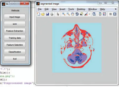
Fig.5. Result Analysis of tumor detection
The overall accuracy percentage details are shown in fig 6. And the comparative analysis is shown in Table 1.
TABLE I. ACCURACY RESULTS COMPARISON WITH OTHER TECHNIQUES
SVM- Fuzzy C-means 91.66
SVM- HFS-SOM 92
SVM- HFS-SOM-GLCM-PCA 94
PSVM- HFS-SOM-GLCM-PCA 97
The accuracy comparison result of existing SVM and proposed PSVM algorithm. From the Fig.7, it is well known that the proposed system works better than existing SVM system with the high accuracy result of 92%.
The values are tabulated in Table.2.
Table II. Accuracy Result Comparison
Technique Accuracy%
SVM 82
PSVM 92
TABLE III. PRECISION RESULTS COMPARISON Technique Precision Rate
SVM 0.82
PSVM 0.93
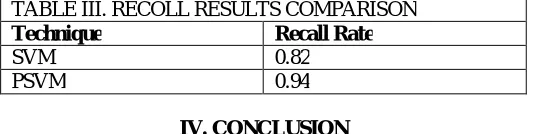
The proposed system has high recall rate as it has less execution time than the SVM algorithm. The values are tabulated in Table.4.
TABLE III. RECOLL RESULTS COMPARISON
Technique Recall Rate
SVM 0.82
PSVM 0.94
IV. CONCLUSION
In this proposed work, effective segmentation and classification is proposed using HFS-SOM and PSVM. After segmentation, the resultant image is given as input to the PSVM classifier followed by feature extraction and selection using GLCM-PCA. At the training phase of PSVM, the texture features are utilized which can reduce the computation complexity of PSVM classifier. The experimental result shows that the proposed system shows a high accuracy rate and less error rate. The proposed system is highly effective for classification to classify normal or abnormal brain with high sensitivity, specificity and accuracy rate.
For future work, to get better accuracy rate and less error rate a hybrid SVM algorithm is to be proposed. In future work, different data mining techniques can be used to train using different kernel functions in order to improve the performance of the classifiers and the data sets can also be increased. In future the system can be improved to support other types of cancer images with few modification either in segmentation and classification stage.
REFERENCES
[1] Prakash Mahindrakar and Dr. M. Hanumanthappa, “Data Mining In Healthcare: A Survey of Techniques and Algorithms with Its Limitations and Challenges”, Int. Journal of Engineering Research and Applications, ISSN : 2248-9622, Vol. 3, Issue 6, Nov-Dec 2013, pp.937-941.
[2] Kailash Sinha, G.R.Sinha, “Efficient Segmentation Methods for Tumor Detection in MRI Images”, 2014 IEEE Student’s Conference on Electrical, Electronics and Computer Science, 978-1-4799-2526- 1/14/$31.00 ©2014 IEEE
[3] R.S.RajKumar and G.Niranjana, “Image Segmentation and Classification of MRI Brain Tumor Based on Cellular Automata and Neural Networks”, IJREAT International Journal of Research in Engineering & Advanced Technology, Volume 1, Issue 1, March, 2013 ISSN: 2320 – 8791.
[4] Parveen, Amritpal singh, “Detection of Brain Tumor in MRI Images, using Combination of Fuzzy C-Means and SVM” 2nd International Conference on Signal Processing and Integrated Networks (SPIN),India, 2015
[5] K.B.Vaishnavee, K.Amshakala, “An Automated MRI Brain Image Segmentation and Tumor Detection using SOM-Clustering andProximal Support Vector Machine Classifier” IEEE International Conference on Engineering and Technology (ICETECH), 20th March 2015, Coimbatore, TN, India.
[6] J. C. Bezdek, (1981), “Pattern Recognition with Fuzzy Objective Function Algorithms”, Plenum Press, New YorkT. Kohonen, Selforganizing maps, Springer,2001.
[7] T. Logeswari, M. Karnan, Hybrid self-organizing map for improved implementation of brain MRI segmentation, in: International Conference on Signal Acquisition and Processing, 2010.
[8] Jian, Y. U. "Texture Image Segmentation Based on Gaussian Mixture Models and Gray Level Co-occurrence Matrix." In Information Science and Engineering (ISISE), 2010 International Symposium on, pp. 149- 152. IEEE, 2010.
[9] Mubashir Ahmad, Mahmood ul-Hassan, Imran Shafi, Abdelrahman Osman, Classification of Tumors in Human Brain MRI using Wavelet and Support Vector Machine, IOSR Journal of Computer Engineering (IOSRJCE), Volume 8, Issue 2 (Nov. - Dec. 2012), PP 25-31
[10] Fung, Glenn, and Olvi L. Mangasarian. "Proximal support vector machine classifiers." In Proceedings of the seventh ACM SIGKDD international conference on Knowledge discovery and data mining, pp. 77-86. ACM, 2001.
[11] O.P. Verma, M. Hammandlu, S. Susan, M. Kulkami and P.K. Jain, "A simple single seeded region growing algorithm for color image segmentation using adaptive thresholding," 2011 International Conference on Communication Systems and Network Technologies, ©2011 IEEE.
[12] G.V. Kumar and Dr G.V. Raju, “Biological early brain cancer detection using artificial neural network”, International Journal on Computer Science and Engineering Vol. 02, No. 08, 2010, 2721-2725.