Lipid Membrane Based Biosensors
Georgia-Paraskevi Nikoleli 1, Dimitrios P. Nikolelis 2,*,
Christina G. Siontorou 3, Marianna-Thalia Nikolelis 2,
Stephanos Karapetis 1
1Laboratory of Inorganic & Analytical Chemistry, School of Chemical
Engineering, Dept 1, Chemical Sciences, National Technical University of Athens, 9 Iroon Polytechniou St., Athens 157 80, Greece
2 Laboratory of Environmental Chemistry, Department of Chemistry, University
of Athens, Panepistimiopolis-Kouponia, GR-15771 Athens, Greece
3 Laboratory of Simulation of Industrial Processes, Department of Industrial Management and Technology, School of Maritime and Industry, University of Piraeus
*To whom correspondence should be addressed
Email: [email protected]
Abstract: The exploitation of lipid membranes in biosensors has provided the ability to reconstitute a considerable part of their functionality to detect trace of
food toxicants and environmental pollutants. Nanotechnology enabled sensor
miniaturization and extended the range of biological moieties that could be
immobilized within a lipid bilayer device. This chapter reviews recent progress
in biosensor technologies based on lipid membranes suitable for
environmental applications and food quality monitoring. Numerous biosensing
applications are presented, putting emphasis on novel systems, new sensing
techniques and nanotechnology-based transduction schemes. The range of
analytes that can be currently detected include, insecticides, pesticides,
herbicides, metals, toxins, antibiotics, microorganisms, hormones, dioxins,
etc. Technology limitations and future prospects are discussed, focused on
the evaluation/ validation and eventually commercialization of the proposed
sensors.
1. Introduction
A biosensor is a device that transforms the chemical information into
an analytically useful signal. Biosensors usually contain two basic
components connected in series: a biological recognition element (“receptor”,
i.e., enzyme, antibody, natural or artificial receptor, etc) and a physical
transducer. The recognition system provides the chemical information (i.e.,
concentration of the analyte) into a physical output signal. The physical
transducer alters the signal into electrical, optical, or piezoelectric, etc. output.
The biological recognition element is in direct contact with a transduction
element. Biosensors have made a large impact in the area of food and
environmental applications and offer advantages in comparison to standard
analytical techniques such as minimal sample preparation, real time detection,
rapid response times, portability, high sensitivity and selectivity, etc. The term
multiple-use biosensor is a device suitable for monitoring both the increase
and decrease of the analyte concentration, whereas a single-use biosensor is
a device which cannot be regenerated and is used only for one analysis test.
Recent progress in nanotechnology has given the opportunity to mass
produce affordable devices and to integrate these into systems for market
applications for environmental or food toxicants monitoring. These
applications include a wide range of food toxicants and environmental
pollutants, such as toxins, insecticides, pesticides, herbicides,
microorganisms, bacteria, viruses and other microorganisms, polycyclic
aromatic hydrocarbons (PAHs), hydrazines, phenolic compounds, allergens,
Lipids are amphiphilic molecules, which possess both hydrophilic head
groups and hydrophobic chains. Since the discovery of model bilayer lipid
membranes (BLM) by Mueller et al. [1], there have been various attempts to
use lipid films to construct biosensor devices for applications in food toxicants
detection or environmental pollutants monitoring. However, free standing
BLMs were very fragile and therefore not suitable for long-term repeated use.
They had low mechanical and electrical stability which was the main obstacle
to their practical applications. However, recent advances in the preparation of
stabilized lipid bilayer have resulted to preparing lipid membrane based
devices for detection of a large diversion of toxicants and pollutants in real
samples. Lipid membranes based biosensors represent an appropriate
biocompatible structure with fast response times (on the order of a few
seconds), high sensitivity (i.e., nanomolar detection limits) and selectivity,
small size, and offers many advantages compared with the bulky analytical
instrumentation such as liquid chromatographic units. Most of these
biosensors are cost efficient, easy-to-use, fast responding portability, and are
good alternative to the expensive, bulky, time consuming standard analytical
methods (i.e., chromatographic techniques). The new generation of lipid
membrane nanosensors has the potential for the development of site-specific
monitors with respect to analytical performance, operational stability and
response.
This work reviews the status of the various nanostructure lipid
membrane based biosensors that are used to monitor food toxicants and
environmental pollutants. The chapter provides the state of art of design and
rapid in the field detection of food toxicants and environmental pollutants and
the challenges that lie ahead.
2. Methods for preparation biosensors based on lipid films
A number of techniques have been described in the literature the last
two decades for techniques for the preparation of stabilized lipid membranes
that are not prone to electrical or mechanical breakage and provide devices
that can be used for practical applications. Most of these techniques provide
lipid membranes with a size is less than 1 µm and therefore can be
considered as nanosensors. These biosensors have been used for
electrochemical experimentation and belong therefore in electrochemical
biosensors. An exception is the development of stabilized polymerized lipid
films on a filter paper that switch on and off their fluorescence and therefore
belong to optical biosensors. Below we provide the various techniques for the
preparation of biosensors based on lipid membranes and mainly those that
are mini- or nanosensors.
2.1. Metal supported lipid membranes
A simple system for the preparation of stabilized bilayer lipid
membrane (sBLM) at the freshly cut tip of Teflon coated metallic wire was
based on the interaction of an amphiphatic lipid molecule with a nascent
metallic surface. One end of a Teflon-coated stainless steel metal wire (with
diameter of 0.1-0.5 mm) was immersed in a lipid solution in chloroform and
then, while still immersed in chloroform, the tip is cut off with a miniature
guillotine. The fresh tip of the wire becomes coated with the lipid membrane
and was placed in the electrolyte (usually 0.1 M KCl), whereupon the lipid film
spontaneously thinned, forming a self-assembled lipid bilayer membrane
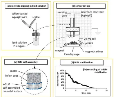
(sBLM). A Schematic of the sensor, measurement setup, and lipid
self-assembly process is shown in Figure 1.
Figure 1. Schematic of the sensor, measurement setup, and lipid self-assembly
process (not drawn to scale): (a) the sensing electrode is tipped with a scalpel and
The electrochemical setup consists of a 20-mL cell and a two-electrode configuration,
i.e., the sensing electrode and a Ag/AgCl reference electrode, placed in a grounded
Faraday cage; an external DC potential of 25 mV is applied between the electrodes
and the ionic current through the BLM is measured with a digital electrometer; the
cell is stirred using a magnetic stirrer. (c) Upon immersion, the lipid droplet attached
to the wire is self-assembled into a bilayer that has one layer adsorbed on the metal
surface and the other facing the aqueous solution. (d) Recording of the ion current
decrease during the self-assembly process; recording started at the immersion of the
sensing electrode in the electrolyte solution [reprinted from ref. 3]..
sBLMs have been fully characterized [2,4,5]. The diameter and
composition of the wires that were used were found to play an important role
in the time for device stabilization, and on the magnitude and noise of the
background ion current [4,5]. The use of a wire of 0.25 mm diameter and of
decane as a solvent should be avoided because the lipid membrane is so
called “black lipid membrane” and these films do not provide reproducible
results. Silver wires of 0.5 and 1.0 mm diameter provide BLMs which were
mechanically and electrical stable for over 48 hours.
Previous studies have provided a model of a potential profile across
sBLMs and have evaluated the structure of the inner lipid layer (facing the
silver wire support). It has been suggested that the lipid headgroups bind to
the electrode by interactions of oxygen atoms of the phosphate groups of the
lipids with silver ions in the metal lattice [6,7]. Note that the silver metal is not
fully insulated from the chloride ion by a BLM. Furthermore, chloride ions can
process and chloride would react with the silver metal to form silver chloride
[4,5]. Potentiometric (against a Ag/AgCl reference electrode) experiments [5]
show only small voltages (relative to a silver wire against a Ag/AgCl reference
electrode) when using the surface of a silver∫ wire after BLM stabilization and
BLM removal with an organic solvent rinse. These results suggest that the
surface of the metal is likely coated with a thin layer of silver chloride and that
the sBLM consists from smaller BLMs on the order of nm which provides a
nanostructure in these devices [8].
2.2. Stabilized lipid films formed on a glass fiber filter
The preparation of stabilized lipid membranes supported on ultrafiltration
glass fiber filters has been reported in the literature [9]. These supported lipid
films on ultrafiltration glass fiber filters have allowed the practical application in
real samples, e.g. for the determination of aflatoxin M1 in milk and milk preparations [10]. The lipid membrane is formed on a microporous filter glass
fiber disk [9,10]. The filters and (nominal) pore size used were GF/F glass
microfiber, 0.7 µm (Whatman ScientificLtd., Kent, U.K.).
The experimental set up which is used for the formation of these
stabilized BLMs consists of two plexiglas chambers separated by a
Saran-Wrap partition (thickness of ca. 10 µm). This plastic partition was cut to more
than twice the size of the contact area of the faces of the chambers and was
folded in half; then, a hole (with diameter 0.32 mm) was punched through the
GF/F microfiber disk (diameter of ca. 0.9 cm and nominal pore size of 0.7 µm)
is placed between the two plastic layers, centered on the 0.32 mm orifice. The
partition containing the filter membrane was then clamped between the two
plexiglas chambers. One of the chambers consisting an electrochemical cell,
had a circular shape (diameter 1.0 cm and depth 0.5 cm); this chamber was
connected with plastic tubing which was used for the flow of the carrier
solution. An Ag/AgCl reference electrode was immersed in the waste of the
carrier electrolyte solution. The second chamber was cylindrical and had its
longitudinal axis perpendicular to the flow of the carrier solution. The upper
hole of this cell was circular (surface area of about 0.2 cm2) and the lower was elliptical (with diameters 0.5 and 1.4 cm parallel and vertical to the flow of the
carrier electrolyte solution, respectively). The lower hole was facing the
opposing cell. An Ag/AgCl reference electrode was positioned at the center of
the cylindrical cell. An external voltage of 25 or 50 mV d.c. is applied between
the two reference electrodes. A Keithley digital electrometer is used as a
current-to-voltage converter. A peristaltic pump is used for the flow of the
carrier electrolyte. Injections of the samples are made with a Hamilton
repeating dispenser. The electrochemical cell and electronic equipment were
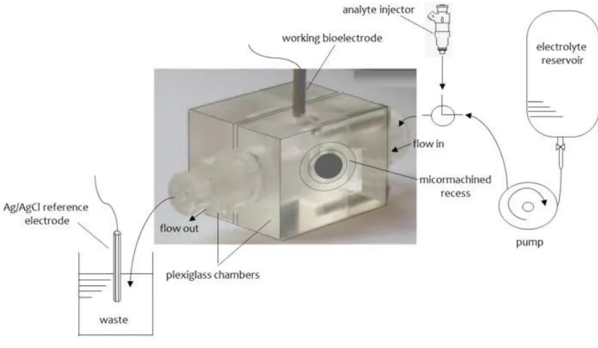
isolated in a grounded Faraday cage. A simple scheme of the apparatus used
is presented in Fig. 2. The procedure for the formation of the stabilized BLMs
is as follows: [9,10]: Lipid solution (ca. 10 µL) is added dropwise from a
microliter syringe to the water surface in the cylindrical cell near the plastic
partition. The level of the electrolyte solution is dropped below the the 0.32
few seconds. The formation of the BLMs has to be verified by the ion current
magnitude and by the electrochemical characterization using gramicidin D.
Figure 2. Schematic of the experimental set-up; the micromachined chambers are
separated by a thin (12.5 μm thick) polyvinylidene chloride wrap and enclose the
microfiber disk. For more details, see text.[from ref. 3]
2.3. Polymer-supported bilayer lipid membranes
The preparation of polymer stabilized was recently described in the
literature’ The construction of these lipid membrane based biosensors took
place by using UV irradiation instead of heating the lipid mixture to 60 °C
[11,12] Thus this process retaines the activity of an enzyme (i.e.,
acetylcholinesterase), whereas heating can deactivate it. The results indicated
methods such as DSC, IR or Raman spectrophotometry. The preparation of
these devices makes possible the practical use of biosensors based on lipid
membranes for chemical sensing, because it allows incorporation of a
“receptor” molecule such as enzyme, antibody or ion-channel receptor in
these lipid films and more importantly these devices are stable outside of the
solution in the air for more than 48 h.
The preparation of these stabilized lipid films is as follows [11,12]: Briefly,
0.8 mL of a mixture containing 4% w/v egg phosphatidylcholine (PC) in
n-hexane (this solvent evaporates so that these films do not retain the solvent in
their structure) were mixed with 0.07 mL of methacrylic acid, 0.8 mL of ethylene
glycol dimethacrylate, 8 mg of 2,2’-azobis-(2-methylpropionitrile) and 1.0 mL of
acetonitrile. The mixture was sparged with nitrogen for about 1 min and
sonicated for 30 min. For the preparation of the stabilized lipid films, 0.15 mL of
this mixture was spread on a microfilter (microporous glass GF/F microfiber
disk with a diameter of ca. 0.9 cm and nominal pore size of 0.7 µm). The filter
with the mixture was then irradiated using the UV deuterium lamp. Raman
spectrometry and differential scanning calorimetry (DSC) were used to monitor
the kinetics of the polymerization process. The measuring set up was similar to
that presented on Fig. 2. These membranes were stable in storage in air for
2.4. Polymer lipid films supported on graphene microelectrodes
Graphene nanomaterials have received tremendous interest in the
field of basic research and in technological applications due to their unique
physicochemical properties, i.e., good sensing ability, and excellent
mechanical, thermal and electrical properties; other advantages include large
surface-to-volume ratio, excellent biocompatibility, high electron-transfer
rates, non-toxicity and bio-safety. The development of biosensors is an
interesting application of this field of nanotechnology. Their implementation in
electrochemical biosensing is quite beneficial as the large
surface-area-to-volume ratio enables miniaturization, increases speed of response and
allows for lower detectabilities while solving the biocompatibility and
biofouling problems. Several examples in the development of
nanobiosensors by integrating enzymes and antibodies were recently
described in literature. Stabilized lipid films were wrapped around a copper
wire containing graphene nanosheets [13,14]. These nanosensors have
been implemented in the rapid detection of food toxicants, environmental
pollutants and toxins in real samples, such insecticides [14], naphthalene
acetic acid [15], cholera toxin [16], and saxitoxin [17].
The preparation of graphene microelectrodes was as follows [13-17]: A
homogeneous graphene dispersion (∼0.4 mg/mL) has been obtained in N
-methyl-pyrrolidone (NMP) through mild sonication for 180 hours and
centrifugation at 700 rpm for 2 h. This suspension has been poured onto a
the organic solvent has been carried out using a fan heater. This copper wire
has been utilized to establish the connection for the extraction of voltage
signals for the calibration curve. Thus, a simplistic approach of drop wise
dispersion of graphene suspended in NMP solution has been utilized to
scatter the graphene nanosheets on the copper wire. The extended sonication
time results in a good fraction of monolayer sheets but with smaller lateral
sizes.
The procedure of construction of these devices is in brief as follows
[13-17]: Stabilized lipid films were prepared by polymerization with a
procedure similar to that previously described [11,12]. Briefly, 0.15 mL of a
mixture containing 5 mg of a mixed lipid powder composed of 35 (w/w) DPPA
and 65 (w/w) of DPPC (1.75 mg DPPA and 3.25 mg DPPC) were mixed with
0.070 mL of methacrylic acid, 0.8 mL of ethylene glycol dimethacrylate, 8 mg
of 2,2′-azobis-(2-methylpropionitrile) and 1.0 mL of acetonitrile. DPPC is used
as lipid and not PC which can be oxidized by air and does not provide
reproducible results The mixture was spumed with nitrogen for about 1 min
and sonicated for 30 min. This mixture could be stored in the refrigerator. For
the preparation of the stabilized lipid films, 0.15 mL of this mixture was spread
on the glass filer microfilter . The filter with the mixture was then irradiated
using a UV deuterium lamp. Raman spectrometry was used to monitor the
kinetics of the polymerization process [11,12].
The enzyme, antibody or receptor (“receptor”) were incorporated in
these BLMs prior polymerization by spreading 15 µL of the “receptor”
stabilized lipid films, 0.15 mL of the polymerization mixture and 15 µL of
“receptor” suspension were spread on the microfilter). The preparation of the
potentiometric biosensor has been finalized by encapsulation of the
filter-supported polymerized lipid film onto the copper wire containing graphene
nanosheets.
3. Applications of lipid film based biosensors in food analysis and environmental monitoring
The stabilized supported lipid membranes biosensors were used for the
flow injection analysis (FIA) of pesticides [18]. Carbofuran was chosen as a
typical pesticide. The determination of the pesticide is based on the degree of
inhibition and reactivation of enzyme by injections of substrate. Carbofuran
was determined at concentration levels of 10−7 to 10−9 M..The investigation of the effect of interferences included compounds usually found in foods,
proteins and lipids. The results have shown no interferences from these
compounds. The technique was applied in various real samples of fruits,
vegetables and dairy products. The recovery ranged between ca. 96 and
106% which shows no interferences from the matrix effects.
A paper was reported in the literature using a synthetic “receptor”
immobilized on supported lipid films on glass fiber filters. The supported lipid
films were modified by calixarenes and permitted sensitive and rapid
determination of insecticides in fruits and vegetables [19]. Other applications in
fast detection of food hormones (i.e., naphthalene acetic acid) in fruits and
vegetables [20] and of zinc in waters [21].
A potentiometric urea lipid film based minisensor on graphene
nanosheets in which a polymeric lipid membrane was deposited has been
recently reported in the literature [22]. The structural characteristics of
graphene nanosheets have been studied through atomic force microscopy
(AFM) and transmission electron microscopy (TEM) measurements. UV-Vis
and Fourrier transform IR (FTIR) spectroscopy have been utilized to study the
pre- and postconjugated surfaces of graphene nanosheets. The presented
potentiometric urea biosensor (Figure 3) exhibits good reproducibility,
reusability, selectivity, fast response times (∼4 s), long shelf life and high
sensitivity having a slope of ca. 70 mV/decade over the urea logarithmic
concentration range from 1×10⁻⁶ M to 1×10⁻³ M.
Figure 3. Schematic of a lipid membrane based biosensor on graphene electrode. This device was used for the potentiometric determination of urea
The electrochemical interactions of naphthalene acetic acid (NAA) with
stabilized lipid films supported on a methacrylate polymer on a glass fiber filter
with incorporated auxin-binding protein 1 receptor were investigated with a
purpose to develop a nanosensor for the rapid determination of NAA in fruits
and vegetables [24]. A FIA technique was used; NAA was injected into the
flowing streams of a carrier electrolyte solution, the flow of the electrolyte
solution stops and an ion current transient was obtained; the peak height of
which was correlated to the hormone concentration (with µM detection limits).
The response times were rapid (on the order of 5 min). The effect of
interferences included a wide range of compounds. The results showed no
interferences from these compounds in concentration levels usually found in
real samples. The method was applied for the determination of NAA in fruits
and vegetables and the reproducibility of the method was satisfactory. Thus, a
quantitative method for the detection of NAA in fruits and vegetables that can be
complimentary to HPLC methods was attained.
A potentiometric carbofuran minisensor on graphene nanosheets with
incorporated lipid membranes has been described in the literature [25]. The
graphene electrode was used to develop a selective and sensitive chemical
sensor for the detection of carbofuran by incorporating an artificial selective
receptor (resorcin[4]arene receptor) on stable lipid films. This chemical
minisensor can determine carbofuran concentrations at nM concentration
range, with rapid response times of ca. 20 s, easy to construct and exhibits
good reproducibility, reusability, selectivity, long shelf life and high electrode
slope of ca. 59 mV/decade over the carbofuran logarithmic concentration
A work that explores the interactions of atrazine with bilayer lipid films
that can be used for the direct electrochemical determination of this herbicide
has been described in the literature [26]. The interactions of atrazine with
solventless bilayer lipid membranes (BLMs) were found to be
electrochemically transduced by these films in the form of a transient current
signal with duration of s and reproducibly appeared within 1 min after
exposure of the membranes to atrazine. The sensitivity of the response was
maximized by the use of BLMs composed of 35% (w./w.) DPPA, and by
alteration of the phase distribution within membranes by the introduction of
calcium ions in bulk solution. The hydrogen bonding between atrazine and the
carbonyl group of the lipid was investigated by the addition of
platelet-activating factor (PAF; an ether analog of PC) in BLMs composed of
phosphatidylcholine. The peak height (i.e., current) was linearly related to the
concentration of this pesticide in bulk solution with µ𝑀 detection limits.
A work that describes a method for the rapid and sensitive
electrochemical flow injection monitoring (FIA) and analysis of mixtures of the
triazine herbicides simazine, atrazine and propazine using stabilized systems
of filter-supported BLMs consisted of egg PC and DPPA has been described
in the literature [27]. Injections of these herbicides were made into flowing
streams of a carrier electrolyte solution and a transient current signal with a
duration of seconds reproducibly appeared in less than two min after
exposure of the lipid membranes to the herbicides. The magnitude of the peak
height was linearly related to the concentration of the herbicide, which could
be determined at µ𝑀 range. Repetitive cycles of injection of herbicides have
the transient signal was different for each triazine and increased to the order
of simazine, atrazine and propazine which has allowed selective detection
and analysis of these triazines in mixtures.
A strategy was described in the literature that was based on monitoring
of changes of ion current through a lipid film with immobilized DNA probes
caused by interaction of these lipid membranes with hydrazine compounds
[28]. A s-BLM that was consisted of egg PC was deposited on a silver metal
electrode. The oligomers used were single stranded deoxyribonucleic acids:
thymidylic acid icosanucleotide terminated with a C-16 alkyl chain to assist
incorporation into s-BLMs (dT20-C16), and deoxyadenylic acid icosanucleotide (dA20). These s-BLMs with incorporated DNA interact with hydrazines, and it is possible to monitor ppb levels of hydrazine compounds (i.e., hydrazine,
methylhydrazine, dimethylhydrazine and phenylhydrazine). This BLM/DNA
biosensor offers a highly sensitive, selective, fast, and portable biosensor for
monitoring these environmentally and toxicologically significant compounds.
A paper appeared in the literature that describes the investigations of
electrochemical interactions of cholera toxin with stabilized lipid films on a
polymer over a glass fiber Whatman GF/F filter with incorporated ganglioside
GM1; this has lead to the development of a minisensor for cholera toxin [29].
The analyte was injected into the flowing streams of a carrier electrolyte
solution, the flow of the solution stopped for 5 min and an ion current transient
was obtained, the peak height of this transient was correlated to the cholera
directed to investigate the rapid detection of other toxins used in bioterrorism
and uses this novel ultrathin film technology.
A potentiometric cholera toxin minisensor on graphene nanosheets
with incorporated lipid films has been described in the literature [30].
Ganglioside GM1 (a natural cholera toxin receptor) was incorporated on
stabilized lipid films on graphene electrodes, providing adequate selectivity for
the detection of cholera toxin over a wide range of concentrations, rapid
response time of ca. 5 min, and detection limits of 1 nM. The proposed sensor
is easy to construct and exhibits good reproducibility, reusability, selectivity,
long shelf life and having a slope of ca. 60 mV/decade of toxin concentration.
The method was evaluated, implemented and validated in lake water
samples. This novel ultrathin film technology is currently adapted to the rapid
detection of other toxins and could be used as a weapon against bioterrorism.
A novel electrochemical biosensor based on a supported polymeric
lipid membranes with immobilized Sheep anti-PCB antibody for the rapid
determination of arochlor 1242 in flowing solution streams (FIA systems) has
been described in the literature [31]. This antibody was immobilized in the lipid
membrane during polymerization of the film; the injections of antigen were
made into flowing streams of a carrier electrolyte solution. The
experimentation was made in a stopped-flow mode; the lipid mixtures were
composed of 15 % (w/w) PA and 85% of DPPC to provide only one and only
single transient current signal with a peak height that was related to the
antigen concentration. Lipid films that were composed of 35 % DPPA were
used to investigate the regeneration of the active sites of antibody after
carrier electrolyte solution. Repetitive cycles of injection of antigen have
exhibited that the maximum number of cycles is ca. 5.
A potentiometric saxitoxin minisensor based on graphene nanosheets
with incorporated lipid films and immobilized anti-STX (which is the natural
saxitoxin receptor) on stabilized lipid films was recently reported in the
literature [32]. A good selectivity and sensitivity for the detection of saxitoxin,
fast response times of ca. 5–20 min, and detection limits of 1 nM were
observed. The proposed minisensor is easy to construct and exhibits good
reproducibility, reusability, selectivity, long shelf life and having a slope of ca.
60 mV/decade over saxitoxin concentration. The method was evaluated and
validated in lake water and shellfish samples. This novel ultrathin film
technology is currently adapted to the rapid detection of other toxins that
could be used as weapons against bioterrorism.
An electrochemical biosensor that is suitable for the rapid and sensitive
screening of the sweetener sucralose based on surface-stabilized bilayer lipid
membranes (s-BLMs) composed of PC was recently described in the
literature [33]. The interactions of sucralose with s-BLMs provided an ion
current increase, that appeared within a few seconds after exposure of the
membranes to the sweetener. The mechanism of signal generation was
explored by DSC studies. The mechanism was found to be related to changes
of the electrostatic fields of the lipid membrane. These studies have shown
that there is an increase of the molecular area of the lipids at the membranes
and a stabilization of a gel phase structure; this was due to adsorption of the
sweetener in the membrane surface. The current signal increases were
concentration range. The present lipid film based biosensor has provided a
rapid response (order of seconds) to alterations of sucralose concentration
(5-50 µM) in the bulk solution. The electrochemical transduction of interactions of
this sweetener with s-BLMs was evaluated by its determination in granulated
sugar substitute products.
A method that reports the FIA and analysis of mixtures of the artificial
sweeteners acesulfame-K, cyclamate, and saccharin using stabilized systems
of filter-supported BLMs was described in the literature [34]. Injections of
artificial sweeteners were made into flowing streams of a carrier electrolyte
solution, and a transient current with duration of seconds appeared in less
than 1 min after exposure of the lipid membranes to the artificial sweeteners.
The peak height of this signal was linearly related to the concentration of
artificial sweeteners, which could be determined at µM concentration range.
The repetitive cycles of injectionsprior to signal degradation was 30. The time
of appearance of the transient response was different for each artificial
sweetener and increased in the order of cyclamic acid, acesulfame-K, and
saccharin. The difference in time of response has allowed selective detection
and analysis of these artificial sweeteners in mixtures. The effect of
interferences usually found in foods, proteins, and lipids was investigated. The
results showed no interferences from these constituents of real food samples.
The method was applied in real food samples (i.e., artificial sweetener tablets,
diet soft drinks, wines, and yogurts) that contain mixtures of these artificial
sweeteners with aspartame and other compounds. A comparison of results
using the present method and that of an Official Method of Analysis showed
Investigation of the transport phenomena through channels/pores it is
very important for various biological, medical, and technical applications. The
scope of a paper appeared in the literature is the development of nanofluidics
for the creation of biosensors capable of detecting single molecules and
manipulating them [35]. The detection of molecules was based on the
measurement of the current through a channel when a molecule enters the
channel, which has a diameter comparable with the molecule size, the
current reduces. In order to improve transport properties of such channels,
their walls are often coated with a lipid bilayer, which behaves as
two-dimensional liquid and thus is capable of supporting transport phenomena.
Presently, this property of lipid membranes was utilized for the development
of a technique for detecting and controlling transport of single-stranded DNA
through channels formed by membrane cylinders with the luminal radii of 5–7
nm. It was demonstrated that in the conditions of small ion strength, the
appearance of a DNA molecule inside such channel is accompanied by an
increase of its ion conductivity and can be controlled by the polarity of the
applied voltage. The peak height of the current increase permits to evaluate
the number of DNA molecules inside the channels. It was also demonstrated
that upon adsorption of DNA molecules on the lipid bilayer surface, the
membrane cylinder behaves as a voltage-sensitive selective ion channel.
Eggshell was used as a biomembrane for immobilization of urease for
the development of a potentiometric urea biosensor [36]. Eggshell membrane
was treated with polyethyleneimine (PEI) to impart polycation characteristics.
Urease was immobilized on the PEI treated eggshell membrane
in surface morphology after immobilization. FTIR study of membrane was
carried out to observe the changes in IR spectra after immobilization of the
enzyme. The biosensor has shown a sigmoidal response for the urea
concentration range from 0.5 to 10 mM. The response time of the biosensor
was 120 s. One membrane could be was reused for 270 reactions without
loss of activity. The urease–eggshell membranes were stable for 2 months
when stored in buffer even at room temperature.
Bilayer lipid membranes (BLMs) can be successfully formed on
polymers electrodeposited on a solid metallic support. Avidin–biotin
interactions were employed for immobilisation of the enzyme on the surface of
the BLM. Improved long-term stability and better selectivity towards certain
interfering electroactive species were observed for a BLM glucose biosensor
based on glucose oxidase immobilised on a platinum support modified with
any of several polymers [37]. However, the best results were obtained for the
mediated system in which the BLM was formed on a Pt support covered with
a layer of evaporated Nafion with incorporated ferrocene. The stable and
sensitive response with significant elimination of the influence of electroactive
interferences on the signal magnitude should allow a practical application of
such miniaturised biosensors.
A novel chemiluminescence biosensor based on a supported lipid layer
incorporated with ganglioside GM1 was reported for the detection of cholera
toxin (CT). The planar supported lipid membrane was prepared as biosensing
interface via spontaneous spread of ganglioside-incorporated phospholipid
vesicles on the octadecanethiol-coated gold surface [38]. The specific
biosensor to be implemented via a sandwiched format using a liposome probe
functionalized with GM1 and horseradish peroxidase (HRP). Then, the
presence of the target CT could be determined via the HRP-catalyzed
enhanced chemiluminescence reaction. The developed strategy offers several
unique advantages over conventional biosensors in that it allows for an easy
construction and renewal of the sensing interface, a small background signal
due to low non-specific adsorption of serum constituents on the lipid
membrane, and effective immobilization of multiple biocatalytic amplifiers and
recognition components via common phospholipid reagents. The developed
biosensor was shown to give chemiluminescence signal in linear correlation to
CT concentration within the range from 1 pg mL−1 to 1 ng mL−1with readily achievable detection limit of 0.8 pg mL−1.
A work that reports a novel bilayer lipid membranes (BLMs) nucleic
acid biosensor supported by modified patch-clamp pipette electrode was
developed to detect staphylococcus enterotoxins B (SEB) gene [39].
Hydrophobic dodecane tail (C12) modified 18 bp single-stranded DNA (ssDNA) probe was immobilized on BLMs. The electrochemical currents
versus the different concentration of ssDNA probe immobilized on BLMs
indicated linear correlation. The BLMs nucleic acid biosensor was fabricated
by selecting the ssDNA probe as the signal sensing element with the
concentration of 273.65 ng/mL. The electrochemical performance of the
biosensor for the detection of SEB was explored. The result showed that a
linear relationship existed between the current and ln(concentration) from 20
to 5000 ng/mL and the detection limit was 20 ng/mL. In addition, the
current alteration in electrolyte which containing no SEB gene. Atom Force
Microscope (AFM) images could be observed and used to evaluate the
superficial microstructure of BLMs, ssDNA immobilized on BLMs and BLMs
after hybridization. The BLMs nucleic acid biosensor supported by modified
patch-clamp pipette electrode will become a highly sensitive, rapid, selective
analytical tool for detection of Staphylococcus aureus, which produce SEB.
A nanostructure electrochemical biosensor was developed to directly
detect and screen estrogenic substances based on estrogen receptor (ER)
binding without the use of radio- or enzyme-labeled compounds [40]. The
biosensor was fabricated by immobilization of ERs in s-BLM modified with Au
nanoparticles, and the properties of the modified electrodes were
characterized by cyclic voltammetry and impedance spectroscopy. The results
have shown that the biosensor was able to detect the natural estrogen
17β-estradiol with an acceptable linear correlation ranging from 5 to 150 ng/L and
a detection limit of 1 ng/L. The biosensor could also detect other known
xenoestrogens such as bisphenol A and 4-nonylphenol with satisfied
sensitivity and quantitative results. The biosensor had good reliability and
repeatability, and the Au nanoparticles greatly enhanced the sensitivity and
stability of the biosensor. Moreover, estrogenic activity of water samples
determined by this biosensor was in good agreement with that determined by
4. Conclusions and future prospects
The present paper provides various ways for the preparation of nanosensors
based on a lipid film technology for food and environmental analytical
applications. Recent technological advances are the construction of stabilized
supported lipid film on graphene nanoelectrodes with an incorporated “receptor”
(enzyme, antibod or natural or artificial receptor) stable in air that can be
portable for in the field applications. These sensors reveal detection limits in
the nM concentration range. The most important aspect of the present efforts
is to provide a portable unit that can be used for in-field and market
applications and that also can be commercialized
The results have shown that a diversity of lipid film based nanosensors
can be reused after storage in air even after a period of a couple of months, and
can be reproducibly fabricated with simplicity and low cost. These nanosensors
have fast response times, are easy to construct and have a lower cost than that
based on chromatographic techniques; they also can be used as rapid portable
detectors complimentary to these methods for in-field and market
measurements in foods and for environmental monitoring.
The present review describes biosensors based on lipid film technology
that can be used for the rapid detection of food toxicants and environmental
pollutants such as toxins, carbamates, hormones, polycyclic aromatic
hydrocarbons, etc and highlights their advantages which are high sensitivity
and selectivity, rapid response times, portability, etc. It is of common sense
that the use of nanotechnology to construct lipid membrane based biosensors
Author Contributions
All authors contributed equally to this work.
Conflicts of Interest
The authors declare no conflict of interest.
References
1. Mueller, P., Rudin, D.O., Tien, H.T., Wescott, W.C. Reconstitution of cell
membrane structure in vitro and its transformation into an excitable system,
Nature 1962, 194, 979-980.
2. Tien, H.T.; Salamon, Z. Formation of self-assembled lipid bilayers on solid
substrates. J. Electroanal. Chem. Interfacial Electrochem.1989, 22, 211–
218.
3. Nikoleli, G.-P., Nikolelis, D., Siontorou, C.G., Karapetis, S. Lipid
membrane nanosensors for environmental monitoring: The art, the
opportunities, and the challenges. Sensors2018, 18(1), 284;
4. Nikolelis, D.P.; Siontorou, C.G.; Krull, U.J.; Katrivanos, P.L. Ammonium
ion minisensors from self-assembled bilayer lipid membranes using
gramicidin as an ionophore. Modulation of ammonium selectivity by
platelet-activating factor. Anal. Chem.1996, 15, 1735–1741.
5. Siontorou, C.G.; Nikolelis, D.P.; Krull, U.J.; Chiang, K.L. A triazine
herbicide minisensor based on surface-stabilized bilayer lipid
6. Hianik, T.; Dlugopolsky, J.; Gyepessova, M. Electrostriction of lipid
bilayers on a solid support. Influence of hydrocarbon solvent and d.c.
voltage. Bioelectrochem. Bioenerg. 1993, 31, 99–111.
7. Hianik, T.; Passechnik, V.I.; Sargent, D.F.; Dlugopolsky, J.; Sokolikova, L.
Surface potentials and solvent redistribution may explain the dependence
of electrical and mechanical properties of supported lipid bilayers on
applied potential and bilayer history. Bioelectrochem. Bioenerg. 1995, 37,
61–68.
8. Passechnik, V.I.; Hianik, T.; Ivanov, S.A.; Sivak, B. Specific capacitance
of metal supported lipid membranes. Electroanalysis 1998, 10, 295–302.
9. Nikolelis D.P., Siontorou C.G., Andreou V.G., Krull U.J. Stabilized
bilayer-lipid membranes for flow-through experiments. Electroanalysis. 1995, 7,
531–536.
10. Andreou V.G., Nikolelis D.P. Flow injection monitoring of aflatoxin M1 in milk and milk preparations using filter-supported bilayer lipid
membranes. Anal. Chem.1998, 70, 2366–2371.
11. Nikolelis D.P., Raftopoulou G., Nikoleli G.-P., Simantiraki M. Stabilized
lipid membrane based biosensors with incorporated enzyme for repetitive
uses. Electroanalysis.2006, 18, 2467–2474.
12. Nikolelis D.P., Raftopoulou G., Chatzigeorgiou P., Nikoleli G.-P., Viras K.
Optical portable biosensors based on stabilized lipid membrane for the
13. Nikoleli, G.-P.; Israr, M.Q.; Tzamtzis, N.; Nikolelis, D.P.; Willander, M.;
Psaroudakis, N. Structural characterization of graphene nanosheets for
miniaturization of potentiometric urea lipid film based
biosensors. Electroanalysis2012, 24, 1285–1295.
14. Bratakou, S.; Nikoleli, G.-P.; Nikolelis, D.P.; Psaroudakis, N.
Development of a potentiometric chemical sensor for the rapid detection
of carbofuran based on air stable lipid films with incorporated
calix[4]arene phosphoryl receptor using graphene
electrodes. Electroanalysis2015, 27, 2608–2613.
15. Bratakou, S.; Nikoleli, G.-P.; Siontorou, C.G.; Nikolelis, D.P.; Tzamtzis,
N. Electrochemical biosensor for naphthalene acetic acid in fruits and
vegetables based on lipid films with incorporated auxin-binding protein
receptor using graphene electrodes. Electroanalysis2016, 28, 2171–
2177.
16. Karapetis, S.; Nikoleli, G.-P.; Siontorou, C.G.; Nikolelis, D.P.; Tzamtzis,
N.; Psaroudakis, N. Development of an electrochemical biosensor for
the rapid detection of cholera toxin based on air stable lipid films with
incorporated ganglioside GM1 using graphene
electrodes. Electroanalysis2016, 28, 1584–1590.
17. Bratakou, S.; Nikoleli, G.-P.; Siontorou, G.C.; Nikolelis, D.P.; Karapetis,
S.; Tzamtzis, N. Development of an electrochemical biosensor for the
rapid detection of saxitoxin based on air stable lipid films with
incorporated Anti-STX using graphene
18. Nikolelis, D. P., Simantiraki, M., Siontorou, G. C., Toth, K. Flow injection
analysis of carbofuran in foods using air stable lipid film based
acetylcholinesterase biosensor, Anal. Chim. Acta2005,537, 169-177 19. Nikolelis, D. P., Raftopoulou, G., Simantiraki, Μ., Psaroudakis, N.,
Nikoleli, G.-P., Hianik, T. Preparation of a selective receptor for
carbofuran for the development of a simple optical spot test for its rapid
detection using stabilized in air lipid films with incorporated receptor,
Anal. Chim. Acta2008, 620, 134-141
20. Nikolelis, D. P., Ntanos, N., Nikoleli, G.-P., Tampouris, K. Development
of an electrochemical biosensor for the rapid detection of naphthalene
acetic acid in fruits by using air stable lipid films with incorporated
auxin-binding protein 1 receptor, Protein and Peptide Lett.2008,15, 789-794 21. Bratakou, S., Nikoleli, G-P. Siontorou, C.G., Karapetis, S., Nikolelis, D.P,
Tzamtzis, N. Electrochemical biosensor for naphthalene acetic acid in
fruits and vegetables based on lipid films with incorporated auxin-binding
protein receptor using graphene electrodes, Electroanalysis 2016, 28,
2171-2177
22. Nikoleli, G.-P.; Israr, M.Q.; Tzamtzis, N.; Nikolelis, D.P.; Willander, M.;
Psaroudakis, N. Structural Characterization of Graphene Nanosheets
for Miniaturization of Potentiometric Urea Lipid Film Based
Biosensors. Electroanalysis2012, 24, 1285–1295.
23. Nikoleli, G.-P., Siontorou, C.G., Nikolelis, D.P., Bratakou, S., Karapetis,
S., Tzamtzis, N. Biosensors based on lipid modified graphene
24. Nikolelis, D. P., Raftopoulou, G., N. Psaroudakis, Nikoleli, G.-P.
Development of an electrochemical chemosensor for the rapid detection
of zinc based on air stable lipid films with incorporated calix4arene
phosphoryl receptor. Int. J. Environ. Anal. Chem.2009,89, 211-222 25. Bratakou, S.; Nikoleli, G.-P.; Nikolelis, D.P.; Psaroudakis, N.
Development of a potentiometric chemical sensor for the rapid
detection of carbofuran based on air stable lipid films with incorporated
calix[4]arene phosphoryl receptor using graphene
electrodes. Electroanalysis 2015, 27, 2608–2613.
26. Nikolelis, D.P.; Andreou, V.G. Electrochemical transduction of
interactions of atrazine with bilayer lipid
membranes. Electroanalysis 2005, 8, 643–647.
27. Nikolelis, D.P.; Siontorou, C.G. Flow injection monitoring and analysis
of mixtures of simazine, atrazine, and propazine using filter-supported
bilayer lipid membranes (BLMs). Electroanalysis 1996, 8, 907–912.
28. Siontorou, C.G.; Nikolelis, D.P.; Tarus, B.; Dumbrava, J.; Krull, U.J.
DNA biosensor based on self-assembled bilayer lipid membranes for
the detection of hydrazines. Electroanalysis 1998, 10, 691–694.
29. Nikoleli, G.-P., Nikolelis, D.P., Tzamtzis, N. Development of an
electrochemical biosensor for the rapid detection of cholera toxin using
air stable lipid films with incorporated ganglioside GM1. Electroanalysis 2011,23(9), 2182-2189.
30. Karapetis, S.; Nikoleli, G.-P.; Siontorou, C.G.; Nikolelis, D.P.;
Tzamtzis, N.; Psaroudakis, N. Development of an electrochemical
lipid films with incorporated ganglioside GM1 using graphene
electrodes. Electroanalysis 2016, 28, 1584–1590.
31. Michaloliakos, A.I.; Nikoleli, G.-P.; Siontorou, C.G.; Nikolelis, D.P.
Rapid flow injection electrochemical detection of arochlor 1242 using
stabilized lipid membranes with incorporated sheep anti-PCB
antibody. Electroanalysis 2012, 24, 495–501.
32. Bratakou, S.; Nikoleli, G.-P.; Siontorou, G.C.; Nikolelis, D.P.; Karapetis,
S.; Tzamtzis, N. Development of an electrochemical biosensor for the
rapid detection of saxitoxin based on air stable lipid films with
incorporated Anti-STX using graphene
electrodes. Electroanalysis2017, 29, 990–997.
33. Nikolelis D.P., Pantoulias S. A minisensor for the rapid screening of
sucralose based on surface-stabilized bilayer lipid membranes. Biosens Bioelectron. 2000, 5(9-10), 439-44.
34. Nikolelis D.P., Pantoulias S. Selective continuous monitoring and
analysis of mixtures of acesulfame-K, cyclamate, and saccharin in
artificial sweetener tablets, diet soft drinks, yogurts, and wines using
filter-supported bilayer lipid membranes. Anal. Chem. 2001, 73(24),
35. Chekashkina, K.V., Galimzyanov, T.R., Kuzmin, P.I. Akimov, S.A., Romanov, S.A., Pozmogova, G.E., Klinov, D.V., Bashkirov, P.V. Detection of DNA molecules in a lipid nanotube channel in the low ion strength conditions, Biochemistry (Moscow), Supplement Series A: Membrane and Cell Biology, 2017, 11(3), 217–224.
36. D'Souza, S.F., Kumar, J., Jha, S.K., Kubal, B.S., Immobilization of the
urease on eggshell membrane and its application in biosensor, Mat. Sci.
& Engin. C2013,33, 850–854.
37. Trojanowicz, M., Miernik, A. Bilayer lipid membrane glucose biosensors
with improved stability and sensitivity, Electr. Acta, 2001, 46(7),
1053-1061.
38. .Chen, H., Zheng, Y., Jian, C.-Y., Wu, H.-L., Shen, G.-L., Yu, R.-Q. An
ultrasensitive chemiluminescence biosensor for cholera toxin based on
ganglioside-functionalized supported lipid membrane and liposome,
Biosens. & Bioelectr., 2008,24(4), 684-689.
39. Liu, N., Gao, Z., Zhou, H.Y., You, M. Detection of SEB gene by bilayer
lipid membranes nucleic acid biosensor supported by modified
patch-clamp pipette electrode, Biosens. & Bioelectr., 2007, 22(9-10),
2371-2376.
40. Xia, W., Li, Y., Wan, Y., Chen, T., Wei, J., Li, Y., Xu, S. ,Electrochemical
biosensor for estrogenic substance using lipid bilayers modified by Au