S.Devasenan1,2*, N. Hajara Beevi2*, S.S. Jayanthi3
1
Department of Chemistry, Sri Ganesh College of Engg and Tech, Puducherry, India.
2
Departments of Chemistry, B.S. AbdurRahman University, Vandalur, Chennai, India.
3
Department of Chemistry, Guru Nanak College, Chennai, India.
*Corresponding author’s E-mail:[email protected]
Accepted on: 18-05-2016; Finalized on: 30-06-2016.
ABSTRACT
In this research paper, we discussed on the Synthesis and characterization of zinc Nanoparticles by green synthesis method. It attempt was made to zinc Nanoparticles is prepared by using a medicinally plant Andrographis paniculata (Family: Acanthaceae). Zinc sulphate as used to synthesis the zinc Nanoparticles by using leaf extract of Andrographis paniculata. The structural characterization of synthesized Nanoparticles was carried out using X-RD, EDAX, and SEM. The optical characterization was carried out using UV – Vis and FT – IR analysis. The results showed that the leaf extract is optimum for the synthesis of Zinc Nanoparticles and it is also known to have the ability to inhibit the growth of various pathogenic microorganisms. The synthesized Zinc Nanoparticles can be used for various applications due to its eco – friendly, non-toxic and compatibility for pharmaceutical and other applications.
Keywords: Zinc Nanoparticles, Andrographis paniculata, characterization, antimicrobial activity.
INTRODUCTION
n recent years, Nanomaterials are being used in a wide variety of applications due to its varying properties on scaling down from bulk size to nanometre size (10-9m). The surface area to volume ratio plays an important role in Nanoparticles, due to which they become more reactive. Nanotechnology and Nanoparticles based product and application are increased now a days due to various fields like biotechnology, physics, chemistry, material sciences, engineering, and medicine. Zinc Nanoparticles are being widely under use in a variety of fields due to its uniqueness and attractiveness in their properties like electrical, optical, dermatological and anti-bacterial1,2. This makes them to be a promising element the widely distributed fields like automobiles, electronics, optoelectronics, textiles, medicine, drug delivery and cosmetics.
Most commonly, Zinc Nanoparticles are produced through chemical methods3,4, like sol-gel processing5, precipitation and electro deposition method6. Zinc has been found highly attractive because of its remarkable application potential in solar cells, piezoelectric devices, UV absorbers, pharmaceutical and cosmetic industries7,8. Potentially, Zinc removed all the dyes and water pollutants from textile effluent under UV light have been proved9,10. Nanoparticles exhibit completely new or improved properties with larger particles of the bulk materials and these novel properties are derived due to the variation in specific characteristics such as size, distribution and morphology of the particles11. The properties of materials change as their size approaches the nanoscale and as the percentage of atoms at the surface of a material becomes significant12. The growing
need of environmental friendly nanoparticles, researchers are using green methods for the synthesis of various metal nanoparticles for pharmaceutical applications13. Although different biological based synthetic methods are known for Zn are sought by researchers. Biological process has led to the development of an eco-friendly approach for the synthesis of nanoparticles. The use of non-toxic materials like plant extract & bacteria for synthesis of zinc nanoparticles offers numerous benefits of pharmaceutical application14.
Biological methods of nanoparticles synthesis using microorganisms15, enzymes16, fungus17 and plants or plant extracts18 have already shown to be possible.
In recent, green synthesis of Zinc nanoparticles was achieved by using microorganisms, plant extract. Zinc nanoparticles show potential antimicrobial effects against infectious organisms such as Escherichia coli, Bacillus subtilis, Vibriocholerae, Pseudomonas aeruginosa, Syphilis typhus, and Staphylococcus aureus19,20.
In the present work was carried out to synthesize and characterize the Zinc Nanoparticles using Andrographis paniculata leaf extracts. Further Zinc nanoparticles were optical characterization using UV-VIS and FT-IR spectrometer, structural characterization using scanning electron microscopy (SEM), EDAX, X – RD and antimicrobial activities.
MATERIALS AND METHODS
Materials
The following analytical grade materials were used without further purification: Zinc Sulphate (ZnSO4). A.C.S. reagent (Sigma – Aldrich, 99% purity by wt).
Green Synthesis and Characterization of Zinc Nanoparticle Using
Andrographis paniculata
Leaf Extract
I
Plant Materials
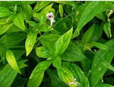
The plants Andrographispaniculata leaf were collected from the cuddalore area, Tamilnadu, during March2016.
Image of Andrographis Paniculata plant
Methods
Preparation of the Leaf Extract
Andrographis paniculata leaf were collected and used to prepare the aqueous extract. Leaf weighing 30gm were thoroughly washed in distilled water, dried, cut into fine pieces and were crushed into 100ml distilled water was added and boiled to 60°C – 70°C for about 15mins. Then the resulting crude extracts filtered through Whatman No.1 filter paper (pore size 25µm). The filter was further filtered through 0.6µm sized filters.
Synthesis of Zinc Nanoparticles
One millimole aqueous solution of Zinc sulphate (ZnSO4) was prepared and aqueous extract of leaf of Andrographis paniculata used for the synthesis of zinc Nanoparticles. 20ml of Andrographis paniculata leaf extract was added into 80ml of aqueous solution of 1mM zinc sulphate. It is kept in magnetic stirrer for 2hours at room temperature.
Characterization of Zinc Nanoparticles
Optical Characteristics of Zinc Nanoparticles
Synthesis of Zinc Nanoparticles by reducing the zinc ions solutions with andrographis paniculata leaves extract to optical characterized by using UV – Visible spectrometer, the absorption spectra was recorded using Perkin Elmer LS 45 spectrophotometer. Zinc Nanoparticles sample is dispersed in UV – Vis methanol with the help of the sonicator. The spectrum was recorded under room temperature. The FT-IR spectra of zinc nanoparticles of andrographis paniculata powder were recorded in SHIMADZU-8400 spectrometer using pellet method.
Structural characteristics of Zinc Nanoparticles
The surface morphology of the Zinc Nanoparticles was characterized using SEM analysis. The Scanning Electron Microscopy (SEM) used for this purpose is a Jeol – JSM – 3.5 CF – Japan. The EDAX analysis spectrum shows the formation of pure Zinc Nanoparticles. The particles size and nature of the zinc nanoparticles were determined
using X – RD. The powdered X – ray diffraction was performed using Scifert X – ray diffractometer with a CuKα radiation.
Antimicrobial Test
The antimicrobial activity of the synthesized zinc Nanoparticles is investigated against different types of pathogenic bacteria such as Escherichia coli, Staphylococcus aureus and Pseudomonas aeruginosa that were cultured on agar plates added with same concentration of zinc Nanoparticles by disc diffusion method.
RESULTS AND DISCUSSION
Structural Characterization and Morphology
The structural characterization was carried out using powder X – RD analysis. Fig: 1 shows typical X – RD pattern of the as-obtained Zinc Nanoparticles using anandrographis paniculata leaf extract.
All the diffraction peaks can be well indexed to the characteristics of cubic centred of zinc Nanoparticles.
The sharp and narrow diffraction peaks appearing at about 2θ of 47.51, 51.20 and 74.83(deg) were assigned to (111), (200) and (220) plane values of Zinc Nanoparticles.
The dry powders of the zinc Nanoparticles were used for X-RD analysis. The diffracted intensities were recorded from 20° to 80° at 2theta angles. The diffraction pattern is corresponds to pure zinc powder.
The X-RD pattern indicates that the zinc Nanoparticles had a spherical structure. The obtained results illustrate that zinc ions had indeed been reduced to Zn by andrographis paniculata plant extract under reaction conditions. To determine the average particles size of the Zinc Nanoparticles, the Debye Sherrer’s equation is used.
D=Kλ / B cosӨ
Where, D is the crystalline size of Nanoparticles, K is the sherrer’s constant, λ is the wavelength of the X – Ray sources used in X – RD, B is the full width at half maximum of the diffraction peak, Ө is the Bragg’s angle.
The structural characterization was carried out using powder EDAX analysis. Fig 2 show the quantitative and qualitative analysis of elements may be concerned is the formation of zinc Nanoparticles. They were identified by EDAX analysis. Due to the surface Plasmon resonance, the zinc Nanoparticles shows the absorption peaks of higher counts. The EDAX analysis spectrum reported is the clearly revels. The composition of zinc is 89.23 percentages. The absence of any extra peak in the EDAX spectrum shows the formation of pure zinc Nanoparticles.
Figure 2: EDAX spectrum for the Zinc Nanoparticles.
The surface morphology and size of the Zinc Nanoparticles was examined using scanning electron microscopy (SEM). Fig 3 shows the scanning electron microscopy of Zinc Nanoparticles synthesized by the plant extract of andrographis paniculata is obtained from the proposed by bio-reduction method. The spherical in shape of the Zinc Nanoparticles was confirmed.
Figure 3 A&B: – SEM micrograph of the Zinc Nanoparticles.
Optical Characterization of Zinc Nanoparticles
The brownish green colour crystalline of Zinc nanopowder was insoluble in water and almost in all organic solvents like methanol. Hence a UV – Visible spectrum was recorded for the Zinc nano dispersed in methanol solution and it is represented in Fig 4. It is the most important method of analysis to detect the surface Plasmon resonance property of zinc Nanoparticles. UV – Visible absorption results confirmed the formation of zinc Nanoparticles prepared in liquid by bio reduction
method. The absorption peak observed at 224nm is the characteristics peak of zinc Nanoparticles.
Figure 4: UV – Vis spectrum of the Zinc Nanoparticles
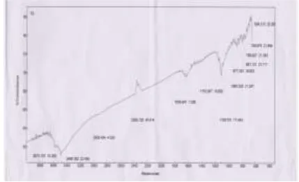
In order to determine the functional groups on andrographis paniculata leaf extract and identify their role in the synthesis of zinc nanoparticles, FT – IR analysis was performed. FT – IR spectrum of andrographis paniculata leaf extract and synthesized zinc nanoparticles are shown in Fig 5. In the zinc nanoparticles, peak values at 699, 750, 799, 851, 877, 1080, 1109, 1153, 1636, 2285, 2926, 3486 and 3573cm-1 was observed. Peak at 699, 750, 799cm-1 corresponds to C – H bending of aromatic, 851cm-1and 877cm-1 corresponds to N–H bending and C– H bending. The peak located at 1080cm-1, 1109cm-1 could be assigned to the C – O stretching vibration. Peak at 1636cm-1,3486cm-1 2926cm-1 corresponds to C=O stretching of amides and O–H stretching of phenolic compound. The FTIR analysis of Zinc nanoparticles suggested that they might surround by the any of these organic molecules. The physicochemical properties of andrographis paniculata leaf extract act as capping agent and prevents the nanoparticles formed from aggregation.
Figure 5: FT – IR spectrum of the Zinc Nanoparticles
Antimicrobial Activity of Zinc Nanoparticles
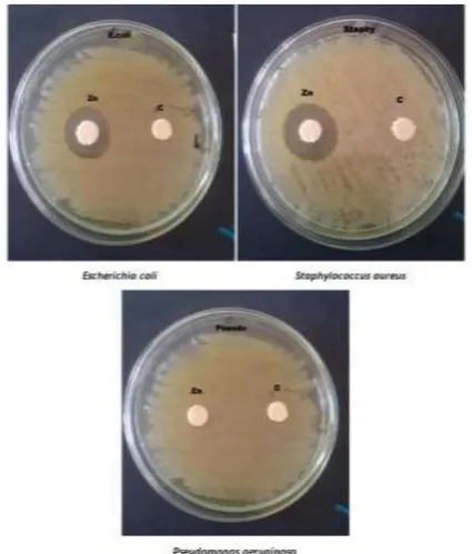
aeruginosa as it shown clear inhibition zone at the same concentration is 50µl/ml. After the incubation period, the growth inhibition zone was measured and the results of the inhibition were measured in millimetre. The maximum zone of inhibition (ZOI) values is observed as 10mm in Escherichia coli, 18mm in Staphylococcus aureus bacteria as shown in the Table 1. But Pseudomonas aeruginosa bacteria are not clear inhibitinga growth in this process by using same concentration of zinc Nanoparticles.
Figure 6: Antimicrobial activity of Zinc nanoparticles withEscherichia coli, Staphylococcus aureus and Pseudomonas aeruginosa.
Table 1: Zone of inhibition of ZnNPs (mm)
S. No Pathogenic Bacteria 50µl
1. Escherichia coli 10±1mm
2. Staphylococcus aureus 18±1mm
3. Pseudomonas aeruginosa No inhibit the growth
CONCLUSION
In conclusion, the field of nanoscience and nanotechnology is the development of eco-friendly processes for synthesis of zinc Nanoparticles. Here we have reported the zinc Nanoparticles were successfully synthesized by using andrographis paniculata leaf extract for the antimicrobial activity. The structural characteristics and morphology of the obtained zinc Nanoparticles were studied using the X – RD, EDAX and SEM techniques. The result is confirmed the zinc Nanoparticles with high stability and without any impurities. The optical characteristics of zinc Nanoparticles were studied using the UV – Vis analysis. The peak in the absorption spectrum is confirmed the
formation of zinc Nanoparticles. The functional group present in the leaf extract was confirmed by FT – IR analysis. Further antimicrobial activity of andrographis paniculata plant extract and synthesized zinc nanoparticles were investigated in the disc diffusion method. From the results it is clear to know that the zinc nanoparticles from andrographis paniculata plant extract also have the ability to inhibit the growth of various pathogenic microorganisms like Escherichia coli, Staphylococcus aureus, and it not produced inhibit the growth of pathogenic microorganism like Pseudomonas aeruginosa.
REFERENCES
1. Tomaszewska-Grzedaa.A, ÃLojkowskia W, Godlewskib M, Yatsunenkob S, Drozdowicz-Tomsiad K, Goldysd E. M and Phillipse M. R Growth and Characterization of ZnO Nanoparticles, International Schoolof Semiconducting Compounds, Proceedings of the XXXIV, (2005).
2. Kamaldeep, Dhirendra Kumar and Kashyap Kumar Dubey, Optimization of Zinc Oxide nanoparticles synthesis to fabricate glucose oxidase sensor, Advances in Applied Science Research.3(5), 2012, 3081-3088.
3. AlessioBecheri, Maximilian Du¨rr, Pierandrea Lo NostroPieroBaglioni Synthesis and characterization of zinc oxide nanoparticles: application to textiles as UV-absorbers, Journal of Nanopart Res, 10, 2007, 679-689. 4. Sheree E. Cross A., Brian Innes B., Michael S., Roberts A.,
Takuya Tsuzuki B., Terry A., Robertson C., Paul McCormick, Human Skin Penetration of Sunscreen Nanoparticles: In-vitro Assessment of a Novel Micronized Zinc Oxide Formulation, Skin Pharmacol Physiol, 20, 2007, 148–154. 5. Surabhisivakumar, Putchavenkateswarlu, Vankarangorao
and Gollapallinageswsararao, synthesis, characterization and optical properties of zinc oxide nanoparticles,
International nano letters, 3, 2013, 30.
6. Grasseta F, Saitoa N, Lia D, Parka D, Sakaguchia I, Ohashia N, Hanedaa H, Roisnelc T, Mornet S, Duguet E. Surface modification of zinc oxide nanoparticles by aminopropyltriethoxysilane, Journal of Alloys and Compounds, 360, 2003, 298-311.
7. Deepti K, Pradeep T J., Precursor-controlled synthesis of hierarchical ZnO nanostructures, using oligoaniline-coated Au nanoparticle seeds, Crystal growth. 2009, 311-3889. 8. Li M, BalaH, Lv X. Ma X, Direct synthesis of monodispersed
ZnO nanoparticles in an aqueous solution Mater.Lett. 2007, 61-690.
9. Xiaoxia Lin, FeiRong, Degang Fu, Chunwei Yuan. Enhanced photocatalytic activity of fluorine doped TiO2 by loaded with Ag for degradation of organic pollutants, Powder Tech. 219, 2012, 173-178.
10. PieqiangLi, Guohua Zhao. An efficient and energy saving approach to photocatalytic degradation of opaque high-chroma methylene blue wastewater by electrocatalytic pre oxidation, Dye and pigments, 92, 2012, 923-928.
and its characterization, Advanced Materials Letters, Vol. 2 No.4, 2011, 313-317.
12. Harish K Handral, Prashanth Kumar Jha, Shruthi SD. Pharmacognostic and phytochemical studies on the leaves of Murrayakoenigii L Spreng. Pharmacophore; 1 3, 2010, 231-238.
13. Akl M. Awwad, Nidà M. Salem, Amany O. Abdeen, Biosynthesis of Silver Nanoparticles using Oleaeuropaea Leaves Extract and its Antibacterial Activity, Nanoscience and Nanotechnology, Vol. 2012, 2(6), 2012, 164-170. 14. Sigh R P, Magesh S, Rakkiyappan C, Self stabilized Ag
nanoparticles from green Murrayakoenigii leaves and a few application, Nanotechnology and application, 6, 2012, 43-51.
15. Klaus T, Joerger R, Olsson E and Granqvist C G., Silver-Based Crystalline Nanoparticles, Microbially Fabricated, J. Proc. Natl. Acad. Sci., 96, 1999, 13611-13614.
16. Konishi Y, and Uruga T, Bioreductive Deposition of Platinum Nanoparticles on theBacteriumShewanella algae, J. Biotechnol., 128, 2007, 648-653.
17. Willner I., Baron R. and Willner B. Growing Metal Nanoparticles by Enzymes, J. Adv.Matter., 18, 2006, 1109-1120.
18. Chandran S. P., Chaudhary M., Pasricha R., Ahmad A. and Sastry M. Synthesis of Goldand Silver Nanoparticles Using Aloe Vera Plant Extract, J. Biotechnol. Prog., 22, 2006, 577-583.
19. Vigneshwaran N., Ashtaputre N. M., Varadarajan P. V., Nachane R. P., Paraliker K. M. AndBalasubramanya R. H., Biological Synthesis of Silver Nanoparticles Using the FungusAspergillusflavus, Mater. Lett., 61, 2007, 1413-1418. 20. Ravindra P. Singh, Vineet K. Shukla, Raghvendra S. Yadav, Prashant K. Sharma, Prashant K. Singh, Avinash C. Pandey. Biological approach of zinc oxide nanoparticles formation and its characterization, Adv. Mat. Lett., 2(4), 2011, 313-317.