THE ROLE OF CBP AND p300 IN CARDIAC
HYPERTROPHY
ROSALIND JANE GUSTERSON
A thesis submitted to the University o f London for the
degree o f Doctor of Philosophy
July 2002
Medical Molecular Biology Unit
Institute o f Child Health
ProQuest Number: 10042804
All rights reserved
INFORMATION TO ALL USERS
The quality of this reproduction is dependent upon the quality of the copy submitted.
In the unlikely event that the author did not send a complete manuscript and there are missing pages, these will be noted. Also, if material had to be removed,
a note will indicate the deletion.
uest.
ProQuest 10042804
Published by ProQuest LLC(2016). Copyright of the Dissertation is held by the Author.
All rights reserved.
This work is protected against unauthorized copying under Title 17, United States Code. Microform Edition © ProQuest LLC.
ProQuest LLC
789 East Eisenhower Parkway P.O. Box 1346
Abstract
The closely related CBP and p300 co-activator molecules play a key role in gene activation by interacting with a number o f different transcription factors, and have been shown to be essential for normal cardiac development and expression o f genes encoding cardiac muscle-specific proteins. Evidence from non-cardiac cells also indicates that these factors are essential for the proper functioning of transcription factors known to be involved in cardiac hypertrophy. Hypertrophy is initially a compensatory response in the heart, defined by enlargement of cells without division and reactivation o f foetal genes. Chronic hypertrophy can be detrimental and may lead to heart failure. The subject o f this thesis is an investigation of the role of CBP and p300 in cardiac hypertrophy.
Phenylephrine (PE) is an a-adrenergic agonist known to induce hypertrophy in cardiac myocytes. Here it is shown that in cardiac cells the activity o f CBP and p300 is stimulated by PE treatment by a mechanism which involves the p42/p44 MAPK pathway, targeting primarily the N-terminus of p300 and the C-terminus of CBP which are not homologous to one another. This suggests that CBP and p300 play an important role in the hypertrophic effect of PE. To further support this theory, data here shows that inhibition o f either CBP or p300 with antisense or dominant negative mutant constructs inhibits PE- induced hypertrophy as assayed by atrial naturetic protein production (ANP), cardiac cell protein:DNA ratio and cell size.
Acknowledgements
I would like to thank Professor David Latchman for giving me the opportunity to work in his laboratory, and for his ideas, support and patience throughout the project. I would also like to thank Dr Bhawanjit Brar for her continual help and advice during the initial stages o f my project. I would like to thank Dr David Faulkes for his help, and in particular for teaching me new techniques.
A special thanks to Jo Buddie for her assistance with the FACS machine, and Vanita Shah for her time and guidance helping the development of the radioimmunoassay technique. I would also like to thank all members o f the Medical Molecular Biology Unit for their assistance and friendship over the last 3 years.
I am very grateful to the British Heart Foundation for their generous funding of the project, and for travel grants provided for both national and international conferences.
Finally, I would like to thank my parents for their support and encouragement over the last 3 years, and Jon Lovick to whom I am indebted for his daily support, patience and advice.
Declaration
Publications
1) Gusterson, R. J., Brar, B. K., Faulkes, D., Giordano, A., Chrivia, J. C., and Latchman, D. S. (2002). The transcriptional co-activators CBP and p300 are activated via phenylephrine through the p42/44 MAPK cascade.
Journal o f Biological Chemistry 111, 2517-2524.
CONTENTS
A bstract...2
Acknowledgements...4
D eclaration...4
Publications...5
C ontents...6
CHAPTER 1
INTRODUCTION
...161.1 Transcription F actors...17
1.1.1 Introduction...17
1.1.2 Mechanisms o f Action o f Transcription Factors... 17
1.1.2.1 DNA-binding Domains... 18
1.1.2.1.1 Helix-Tum-Helix M otif... 18
1.1.2.1.2 Zinc Finger M otif...18
1.1.2.1.3 Leucine Zipper and Basic DNA Binding Domain... 19
1.1.2.1.4 Helix-Loop-Helix M otif... 19
1.1.2.2 Activation o f Transcription...20
1.1.2.3 Inhibition o f Transcription...24
1.1.3 Regulation of Transcription Factors... 28
1.1.4 Chromatin Structure and the Control of Gene Expression... 29
1.1.4.1 Modification o f Histones by Acétylation... 29
1.1.4.2 Histone Acetyltransferases... 34
1.1.4.2.1 p55 and yGCN5...36
1.1.4.2.2 P/CAF...36
1.1.4.2.3 SRC-1 and ACTR...37
1.1.4.2.4 hTAFii250 and its homologs... 38
1.1.4.2.5 ESAl and Tip60...38
1.2 CBP and p300 Transcriptional A ctivity... 41
1.2.1 Introduction...41
1.2.2 CBP and p300 Structural Features...47
1.2.3 Acetylation-Dependent Functions o f CBP and p300...51
1.2.4 Acetylation-Independent Functions o f CBP and p300... 54
1.2.5 Regulation of CBP and p300 by Phosphorylation... 55
1.3 Biological Functions of CBP and p300...57
1.3.1 CBP and p300 in Cell Cycle Regulation and Differentiation 57 1.3.2 CBP and p300 in Embryogenesis and Development... 57
1.3.3 CBP, p300 and Disease...62
1.3.4 Functional Differences Between CBP and p300...69
1.4 C ardiac H y p ertro p h y ... 71
1.4.1 Definition... 71
1.4.2 Cardiac Hypertrophy and Ageing... 73
1.4.3 Characteristics of the Hypertrophic Response...74
1.4.4 Hypertrophic Stimuli... .76
1.4.5 Hypertrophic Regulation by Catecholamines...77
1.4.6 Hypertrophic Regulation by the Sympathomimetic Drug Phenylephrine...79
1.4.7 Hypertrophic Regulation by Cardiotrophin-1...80
1.4.8 Hypertrophic Regulation by Endothelin-1...80
1.4.9 Hypertrophic Regulation by Angiotensin II... 80
1.4.10 Hypertrophic Regulation by Urocortin... 81
1.5 Signalling Pathways in Cardiac H y p ertro p h y ...82
1.5.1 Ras and the ERK Cascade... .84
1.5.2 Stress Response and p38 Protein Kinases... .88
1.5.3 Protein Kinase C Pathway... 90
1.5.4 Calcineurin and NF-AT3... .91
1.6 Induction of C ardiac H ypertrophy by Gene A ctivation...92
CHAPTER 2
MATERIALS AND M ETHODS
...972.1 L aboratory Reagents... 98
2.1.1 General Suppliers... 98
2.1.2 Bacterial Reagents...98
2.1.3 Molecular Reagents... .98
2.1.4 Western Blotting Reagents... 98
2.1.5 Tissue Culture Reagents... 99
2.1.6 Assay Reagents...99
2.1.7 Equipment...100
2.2 Cell C u ltu re...101
2.2.1 Primary Neonatal Rat Cardiac Cell Culture... 101
2.2.2 Transient Transfection...101
2.2.3 Stimulation o f Cells... 108
2.3 Propagation, Purification and M anipulation of Plasm id DNA...109
2.3.1 Transformation of E.coli... 109
2.3.2 Large Scale Plasmid DNA Extraction from E.coli...110
2.3.3 Small Scale Plasmid DNA Extraction from E.coli...110
2.3.4 Examination o f DNA by Restriction Digest...I l l 2.3.5 Isolation of DNA Fragments from Agarose Gels... I l l 2.3.6 Ligation of DNA... 112
2.3.7 Bacterial Lysate Electrophoresis... 112
2.3.8 Site-Directed Mutagenesis... 112
2.4 Analysis of Protein Levels... 114
2.4.1 Western Blotting... 114
2.4.2 Immunofluorescence...115
2.4.3 Protein Assay...115
2.5 Assessment of Prom oter Activity... 117
2.5.1 Luciferase Assay... 117
2.5.2 P-Galactosidase (P-Gal Assay)... 117
2.6 M easurem ent of H ypertrophy ...119
2.6.1 Measurement o f Cell Size...119
2.6.2 Measurement o f Protein and DNA Content... 119
2.6.3 ANP Measurement...120
2.7 Statistical Analysis...121
CHAPTER 3
The Effect of Hypertrophic Stimuli on CBP and p300
A ctivity
... 1223.1 In tro d u ctio n ... 123
3.2 Effect of Different E xtracellular Stimuli on the Transcriptional Activity of C B P... 124
3.3 Activation of a-A drenergic R eceptors...127
3.4 Investigation of the Signalling Pathw ay Involved in the H ypertrophic Effect of Phenylephrine on CBP Activity...129
3.4.1 Involvement o f the MAPK Pathway in Regulating CBP Activity Using Chemical MAPK Inhibitors...129
3.4.2 Involvement o f the MAPK Pathway in Regulating CBP Activity Using DNA Plasmids...133
3.5 Localisation of the CBP Region Involved in Up-Regulation by Phenylephrine...136
3.5.1 Effect o f Phenylephrine on the Transcriptional Activity of a Series o f Smaller CBP Chimera Constructs... 136
3.5.2 Dependence of C B P l961-2039 Up-Regulation by Phenylephrine on MAPK Pathway...136
3.6 Activation of Endogenous CBP by Phenylephrine...140
3.7 Effect of Phenylephrine on p300 T ranscriptional Activity...142
3.8 Effect of M APK Inhibition on p300 Activity...144
3.9 Putative Phosphorylation Sites within CBP and p300... 148
3.9.1 CBP Phosphorylation Site within the C-terminus...148
3.9.2 p300 Phosphorylation Site within the N-terminus...157
CHAPTER 4
The Effect of CBP and p300 Inhibition on Phenylephrine
Induced Cardiac Hypertrophy
... .1624.1 In troduction ... 163
4.2 Cloning CBP Antisense C onstruct...163
4.3 CBP Antisense Inhibits Endogenous CBP Expression... 167
4.4 Effect of Antisense CBP and Dominant Negative p300 on CBP and p300 Activity... 169
4.5 Effect of Antisense CBP and Dom inant Negative p300 on H ypertrophy Induced by Phenylephrine...173
4.5.1 Inhibition o f Increased ANP Release...173
4.5.2 Inhibition o f Increased Cell Protein to DNA Content... .178
4.5.3 Inhibition o f Increased Cell Size...181
4.6 Effect of E l A on Increased Cell Size and Up-Regulation of ANP by Phenylephrine...185
4.7 Discussion... 188
CHAPTER 5
Involvement of CBP and p300 Histone Acetyltransferase
Activity in Hypertrophy
... .1905.1 In troduction...191
5.2 Role of CBP HAT Activity in the Enhanced Activity of CBP Induced by Phenylephrine...191
5.3 Ability of CBP b ut not CBP HAT M utant to Induce H yp ertro p h y .. .193
5.4 Ability of p300 b u t not p300 HAT M utants to Induce H ypertrophy.. 197
5.5 Induction of H ypertrophy by Increased HAT Activity... 201
5.6 Involvement of CBP and p300 in the H ypertrophic Effect of Trichostatin A ... 205
CHAPTER 6
DISCUSSION... 212
LIST OF FIGURES AND TABLES
Figure 1.1 Assembly of the general transcription factors required for the
initiation of transcription... 22
Figure 1.2 Inhibition of gene expression by transcription factors...25
Figure 1.3 Activation and repression o f thyroid hormone receptor... 27
Figure 1.4 Histone acétylation as a reversible mechanism... 31
Figure 1.5 Model o f histone acétylation and histone deacetylation... 33
Table 1.1 Known histone acetyltransferases... 35
Table 1.2 Known histone deacetylases...40
Figure 1.6 Activation of the cAMP-CREB signalling pathway... .42
Figure 1.7 Alignment of human CBP and p300 molecules... 44
Figure 1.8 CBP/p300 organisation and interactions...46
Figure 1.9 CBP/p300 co-ordinate transcription with chromatin remodelling... .48
Figure 1.10 Structural features of CBP/p300 gene relative to their function...50
Figure 1.11 Chromosomal translocations involving CBP/p300... 64
Figure 1.12 Models for E l A inhibition of CBP/p300...67
Figure 1.13 Chemical structure o f epinephrine and phenylephrine...79
Figure 1.14 Signal transduction pathways implicated in the hypertrophic response... 83
Figure 1.15 The MAPK cascades... 85
Table 2.1 DNA Plasmids... 102
Table 2.2 Primers for Site-Directed Mutagenesis... 113
Table 2.3 Antibody Conditions... 115
Figure 3.1 Effect o f different extracellular stimuli on the transcriptional activity of CBP in cardiac myocytes...125
Figure 3.2 Effect o f PE on the transcriptional activity o f Gal4DBD in cardiac myocytes... 126
Figure 3.4 Effect o f specific inhibitors on the up-regulation of CBP activity
by PE in cardiac m yocytes...130
Figure 3.5 PE induces p42/p44 MAPK phosphorylation in cardiac myocytes and is blocked by PD ... 331
Figure 3.6 Effect o f protein kinase inhibitors on the up-regulation of CBP activity by PE in cardiac myocytes... 132
Figure 3.7 Effect o f over-expression o f the MAPK dominant negative mutant (P42Y185F) and wild type p42 (P42WT) on the up-regulation o f CBP activity by PE in cardiac myocytes... 134
Figure 3.8 Effect o f over-expression o f a constitutively active MEK-1 mutant on the up-regulation of CBP activity by PE in cardiac myocytes...135
Figure 3.9 Effect o f PE on the transcriptional activity o f different N-terminal and C-terminal regions of CBP in cardiac myocytes...138
Figure 3.10 Activation o f p42/44 MAPK pathway mediates up-regulation of CBP (1961-2039) activity in cardiac myocytes... 139
Figure 3.11 Ability o f PE to stimulate transcriptional activation via the C R E Bd i e d m l construct...141
Figure 3.12 Effect o f PE on the transcriptional activity o f different N-terminal and C-terminal regions o f p300 in cardiac myocytes... 343
Figure 3.13 Effect of the specific MAPK inhibition on the up-regulation of p300 activity by PE in cardiac myocytes...145
Figure 3.14 Effect o f overexpression of the MAPK dominant negative mutant and wild type p42 on the up-regulation o f p300 activity by PE in cardiac myocytes... 146
Figure 3.15 Effect o f protein kinase inhibitors on the up-regulation of p300 activity by PE in cardiac myocytes... 147
Figure 3.16 Structure o f CBP (amino acids 1961-2039)... 151
Figure 3.17 Screening for CBP S1994A and CBP S2010A...152
Figure 3.18 Screening for CBP S2015/2016A...353
Figure 3.21 Effect o f serine 2015 and 2016 CBP mutations on CBP 1961-2039
up-regulation by PE ... 156
Figure 3.22 Effect o f serine 89 mutations on p300 up-regulation by PE...158
Figure 4.1 Bacterial lysate electrophoresis to screen for CBP antisense clones... 165
Figure 4.2 Mini-prep digests to confirm CBP antisense... 166
Figure 4.3 Effect of CBP antisense on endogenous CBP levels by immunofluorescence...168
Figure 4.4 Effect of antisense CBP and dominant negative p300 on CBP and p300 activity...170
Figure 4.5 Effect of CBP antisense on CBP C-terminal activity in the absence o f P E ... 171
Figure 4.6 Effect of CBP antisense on promoter activation in cardiac myocytes...172
Figure 4.7 Effect of CBP antisense on ANP promoter activation in cardiac myocytes... 175
Figure 4.8 Effect o f a dominant negative p300 mutant on the up-regulation of ANP promoter activity by PE in cardiac m yocytes...176
Figure 4.9 Effect o f CBP antisense and p300 dominant negative on ANP release by PE in neonatal cardiac myocytes... 177
Figure 4.10 Use of FACS analysis to measure cell cycle and protein:DNA content... 179
Figure 4.11 Effect of antisense CBP and dominant negative p300 on the hypertrophic response to PE as measured by cell protein:DNA content... 180
Figure 4.12 Effect o f antisense CBP on PE induced cell size...182
Figure 4.13 Effect o f antisense CBP on PE induced cell size...183
Figure 4.14 Effect o f dominant negative p300 on PE induced cell size... 184
Figure 4.15 Effect o f E l A on enhanced ANP activity by PE... 186
Figure 5.1 Effect o f CBP HAT mutant on up-regulation by PE... 192
Figure 5.2 Effect o f CBP overexpression and CBP HAT mutant on cell size... 194
Figure 5.3 Effect o f CBP overexpression and CBP HAT mutant on cell proteiniDNA... 195
Figure 5.4 Effect o f CBP overexpression and CBP HAT mutant on cell ANP production... 196
Figure 5.5 Effect o f p300 overexpression and p300 HAT activity on cell size... 198
Figure 5.6 Effect o f p300 overexpression and p300 HAT activity on cell protein:DNA... 199
Figure 5.7 Effect o f p300 overexpression and p300 HAT activity on cell ANP production...200
Figure 5.8 Effect o f TSA on cell size... 202
Figure 5.9 Effect o f TSA on cell proteiniDNA... 203
Figure 5.10 Effect o f TSA on cell ANP production...204
Figure 5.11 Effect o f CBP antisense on PE and TSA induced cell size... 206
Figure 5.12 Effect o f p300 dominant negative on PE and TSA induced cell size... 207
Figure 5.13 Effect o f CBP anti sense and p300 dominant negative on PE and TSA induced cell proteiniDNA... 208
CHAPTER 1
1.1
Transcription Factors
1.1.1 Introduction
The regulation o f gene expression is central to the normal development and proper functioning o f all organisms (for review see Latchman, 1997a). Such gene regulation is primarily achieved at the level o f gene transcription, whereby the DNA is copied into an RNA transcript. Thus, different genes are transcribed in different cell types leading to the production o f their corresponding proteins, while a particular stimulus will produce new protein synthesis by activating the transcription o f new genes.
1.1.2 Mechanisms o f Action o f Transcription Factors
Transcription o f a specific gene is dependent upon an array o f regulatory sequences, known as the gene promoter, which determine both the basal transcription level o f the gene and its response to specific stimuli. Transcription itself is controlled by proteins known as transcription factors, that bind to specific DNA sequences in the gene promoter and activate or inhibit transcription. In order to produce their effects, transcription factors generally, but not always, require the ability to bind DNA, and then to affect transcription either positively or negatively (for review see Latchman, 2001). Transcription factors which act by linking the DNA binding transcriptional activator to the basal complex are called co-activators.
1.1.2.1 DNA-binding Domains
The ability of a protein to bind selectively to a particular DNA site in the genome is the foundation upon which transcriptional regulatory pathways are built. As mentioned above, transcription factors contain DNA-binding domains which can be grouped into a few families based on common structural motifs.
1.1.2.1.1 Helix-Turn-Helix M otif
The proteins in this family span a broad range of protein folds that contain a conserved bi-belical motif, termed the helix-tum-helix, and are generally dissimilar in stmcture outside the helix-tum-helix (for review see Garvie and Wolberger, 2001). The two helices are related by a relatively fixed angle, connected by a tight bend, and the length of each helix varies among different subclasses of the family. The second o f the two helices, referred to as the recognition helix, inserts into the major groove of DNA, and its amino acid side chains, which differ from protein to protein, play an important part in recognising the specific DNA sequence to which the protein binds. The first helix, though not embedded in the major groove, in some cases makes additional DNA contacts. An example o f the helix-tum-helix motif is found in the homeobox transcription factors first discovered in Drosophila (Dura and Ingham,
1988).
1.1.2.1.2 Zinc Finger M otif
1995). In these molecules, the zinc finger motif involves an approximately 70 amino acid domain, in which two zinc ions are each co-ordinated by four cysteines.
1.1.2.1.3 Leucine Zipper and Basic DNA Binding Domain
The leucine zipper and basic DNA binding domain does not refer specifically to a DNA binding domain, but to the characteristic structure which allows two a helices to dimerise, facilitating the correct positioning of two basic regions N-terminal to the a helices. This in turn allows the a helices to interact directly with acidic DNA (Kouzarides et a l, 1988, Turner et al, 1989). In helical portions o f the leucine zipper proteins, every seventh amino acid is a leucine. Since there are on average 3.6 amino acids per turn o f an a helix, all o f the leucines appear on the same side of the helix (after every two helical turns), thereby forming a hydrophobic surface and arranging into an a helical parallel coiled coil (O’Shea et al., 1989). The DNA binding portion of the motif is a highly positively charged basic region containing several arginine and lysine residues, and attaches to the major groove of the DNA in a ‘scissors-grip’ manner (for review see Vinson et al., 1989). The proto-oncogene products c-jun and c-fos, which heterodimersie to form the AP I transcription complex, interact with one another through leucine zippers and with the AP-1 response element through N-terminal basic residues. The leucine zipper and basic DNA-binding domain has also been detected in several other transcription factors, including the CCAAT-box binding protein C/EBP, and the yeast factor GCN4.
1.1.2.1.4 Helix-Loop-Helix M otif
1.1.2.2 Activation o f Transcription
Many transcription factors contain, in addition to the DNA binding domain, specific regions which are necessary for the activation o f transcription. Such regions were identified on the basis that they can stimulate transcription when linked to the DNA binding domain of a completely unrelated factor, and are known as activation domains (for review see Mitchell and Tjian, 1989). As with DNA binding domains, a number of distinct types o f activation domain have been identified on the basis that they contain a cluster o f negatively charged (acidic) amino acids, glutamine residues or proline residues.
The activation domains are thought to function by interacting with components of the basal complex. This is a complex o f RNA polymerase II and various transcription factors such as TFIIB and TFIID, which assemble at the gene promoter, and are essential for transcription to occur (Buratowski et al.,
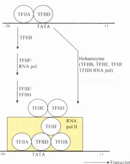
1989; Furukawa et al., 2001; Nikolov et al., 1997; for review see Roeder, 1996). Early studies showed the first clear insights into the stepwise assembly of the basal transcription factors and RNA polymerase II on the promoter template (Fig. 1.1). Initially the TATA-binding protein (TBP) subunit o f TFIID recognises and binds the TATA box in the promoter. This is followed by TFIIA binding to the upstream side of TFIID, stabilising the TFIID/DNA complex. TFIIB then binds the TFIIA/TFIID/DNA complex, and is thought to act as a bridge between TFIIA/TFIID/DNA and RNA polymerase II. Finally, TFIIE, TFIIH and TFIIJ bind the pre-initiation complex. After RNA polymerase II has been tethered to the promoter, it is released from the complex o f general transcription factors to begin transcription. An important step in the initiation of transcription is carried out by TFIIH, a protein kinase subunit that phosphorylates RNA polymerase II, and in the case of some promoters this phosphorylation is thought to disengage the polymerase and activate transcription.
T F I I A I T F I I D
-50 T A T A + 1
T F I I B
F l o l o e n z y m e
( T F I I B , T F I I E , T F I I F
T F I I H R N A p o l ) T F I I F /
R N A p o l
T F I I E /
T F I I H
T F I I E I T F I I H
X R N A
T F I I F ) p o l II
T F I I A I T F I I D I T F I I B
-50 T A T A +1
►Transcription
Figure 1.1 Assembly of the general transcription factors required for the
initiation of transcription
1.1.2.3 Inhibition of Transcription
Although it was originally thought that most eukaryotic transcription factors acted by stimulating transcription, it is now clear that a wide variety of factors act by inhibiting the transcription o f specific genes, and that such inhibitory transcription factors are as important as stimulatory factors (for reviews see Hanna-Rose et a l, 1996; Herschbach et a l, 1993; Latchman, 2002). Repression can be established by proteins that act over a short range, or at a long distance (for reviews see Blackwell and Walker, 2002; Courey and Jia, 2001; Gray and Levine, 1996). The latter is often referred to as silencing because an entire chromosomal locus is inactivated, as opposed to short range repression, which only blocks the function o f nearby DNA-bound activators, and does not interfere with more distantly bound activators.
Short range repressors have been shown to act by interfering with the activity o f a positively acting factor, thereby blocking its stimulatory effect on transcription. This is achieved by DNA binding competition between a repressor and a gene activator protein, whereby the repressor binds to overlapping DNA sites (Fig. 1.2 A), or by the formation o f a complex between the activator and the repressor in solution preventing the activator binding to the DNA (Fig.l.2B). Alternatively, the repressor could bind with the activator to block the interaction between the activation domain and the general machinery, sometimes referred to as quenching (Fig.l.2C) (Alberts et al., 1994; Latchman, 2002).
B
A c tiv a tio n su r fa ce
A c tiv a to r
ÎÏ
R ep resso r for a ctiv a to rX
B in d in g site for a ctiv a to r
X B in d in g site B in d in g s ite for repressor
TATA
R ep resso r
TATA
B in d in g s ite for a ctiv a to r
B in d in g site for repressor
TATA
D
X
x ^ i r c c t R ep ressio n
0
Figure 1.2 Inhibition of gene expression by transcription factors
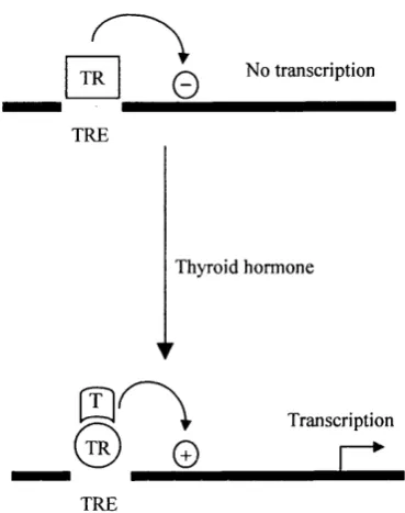
Hence, the balance between binding o f transcriptional activators and transcriptional repressors to the regulatory region o f a particular gene will determine the rate o f its transcription in any particular situation. However, in order for a particular gene to respond to specific signals or to be regulated in a cell type specific manner, the balance between these activating and repressing molecules must change in different situations. It is now recognised that although some factors are pure activators or pure repressors, many can both activate and repress transcription in a way that is dependent on the particular situation. One such example is the thyroid hormone receptor which regulates gene expression in response to thyroid hormone, and is a member o f the steroid hormone receptor family o f transcription factors. This study, and others, have led to the idea that many nuclear receptors switch, in a ligand dependent manner, between binding o f a multi-component co-repressor complex containing histone deacetylase activity, and binding o f a co-activator complex containing factors with histone acetyltransferase activity (for review see Torchia et al., 1998) (see section 1.1.4).
T R
0
N o transcriptionT R E
Th yro id h o r m o n e
©
0
Transcri pt ion
-►
T R E
Figure 1.3 Activation and repression of thyroid hormone receptor
1.1.3 Regulation o f Transcription Factors
Transcription factors can be regulated at two levels, the regulation of transcription factor synthesis and the regulation o f transcription factor activity. In a number of different situations, a transcription factor is regulated by being synthesised in one particular tissue or cell type, and not in other tissues. One such example is the MyoD transcription factor. This transcription factor is synthesised only in skeletal muscle cells, but its artificially induced expression in other cell types, such as undifferentiated fibroblast cells, is sufficient to convert them to skeletal muscle cells. This indicates a critical role for MyoD factor in the induction o f muscle specific gene expression.
1.1.4 Chromatin Structure and Control o f Gene Expression
The genomes o f eukaryotes are highly compacted to allow the DNA molecules to form inside the cell and to be easily managed. The first level of compaction is the wrapping of DNA around histones to form nucleosomes. Each nucleosome consists of a central core complex o f eight basic histone proteins (two of each o f histones H2A, H2B, H3 and H4), surrounded by 146bp DNA (for reviews see Arents and Moudrianakis, 1993; Pruss et al., 1995). Each histone has a high proportion of positively charged amino acids (lysine and arginine). The positive charge helps the histones bind tightly to the DNA (which is negatively charged). Adjacent nucleosomes are connected by linker DNA, in the so called ‘beads on a string’ structure. In a second level o f compaction the nucleosomes are further coiled into a chromatin fibre o f 30nm diameter. The histone HI molecules are thought to be responsible for pulling the nucleosomes closer together to form the 30nm fibre.
1.1.4.1 Modification of Histones by Acétylation
In the resting cell, DNA is tightly compacted into nucleosomes to prevent transcription factor accessibility, and in this way acts as a barrier to the initiation of transcription, by preventing the access of transcriptional factors and RNA polymerase II to their cognate recognition sequences. During activation of the cells, this compact inaccessible DNA is made available to DNA binding proteins by unwinding of the 30nm fibre to the ‘beads on a string’ structure to generate a more open chromatin structure, thus allowing the induction o f gene transcription. Specific lysine resides in the N-terminal tails of core histones can be post- translationally modified by acétylation o f the s-amino group, possibly neutralising the positive charge o f the lysine s-amino group, primarily on histones H3 and H4 (for reviews see Wade et al., 1997; Wade and Wolffe, 1997). This modification is thought to weaken the interaction between the histone tails and the DNA, thus creating an open chromatin structure, more accessible to transcription factors.
C H j
C = 0
S
NU/
I
C \\ 2
I
{ C \ h h
I
-X -L y s-X -H isto n e
SH
I I
C o e n z y m e A C o e n z y m e A
X h d a cN H20
C H ,
I c = o
C H ]
I
c = o
N H
CH] (CH])]
I
-X -L y s -X -H is to n e
OH
Figure 1.4 Histone acétylation as a reversible mechanism
Several transcriptional regulators possess intrinsic HAT and HDAC activities, strongly suggesting that histone acétylation and deacetylation play a role in regulating transcription (Fig. 1.5) (Ito and Adcock, 2002). There is evidence that increased gene transcription is associated with an increase in histone acétylation (hyperacetylation), whereas hypoacetylation o f histones is correlated with reduced transcription or gene silencing (see section 1.1.4.2) (for reviews see Grunstein, 1997; Kuo and Allis, 1998; Wolffe, 1997; Workman and Kingston, 1998). Using DNase I sensitivity as an indicator of differences between active and inactive DNA, the hyperacetylated forms o f histone H3 and H4 have been shown to be localised in active genes exhibiting DNasel sensitivity, whereas hypoacetylated histones H3 and H4 have been shown to exist in transcriptionally inactive regions. Furthermore, treatment of cells with sodium butyrate, which inhibits cellular deacetylase activity and therefore increases histone acétylation, has been shown to result in DNasel sensitivity in some regions o f chromatin, and to activate the expression of some previously silent cellular genes (for review see Reeves and Cseijesi, 1979).
Histone Acétylation
(Gene transcription)
Histone Deacetylation
(Gene silencing)
Polymerase II complex HAT complex
C o r e hist o n e
HDAC complex
u T m f m T ^
D N A
Figure 1.5 Model o f histone acétylation and histone deacetylation
1.1.4.2 Histone Acetyltransferases
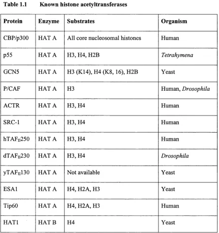
Histone acetyltransferases (HATs) can be grouped into at least two classes (Table 1.1) (for review see Kuo and Allis, 1998). Type A HATs are localised in nuclei and are thought to acetylate nucleosomal histones in reactions closely tied to transcriptional activation. By contrast, type B HAT can be purified from cytoplasmic fractions, and it is thought that these activities are responsible for acetylating newly synthesised histones before chromatin assembly during DNA replication. Several distinct type A HAT families have been characterised to date, including the GCN5 family (Brownell et a l, 1996; Kuo et a l, 1996; Wang et al., 1997) with homologs in Tetrahymena thermophilia, Saccharomyces
cerevisiae. Drosophila and human, including P/CAF (p300/CBP associated
factor) (Yang et al., 1996). Other HAT families include CBP/p300 (Ogryzko et al., 1996), human TAFn250 and its homolog yeast TAFnl30 and Drosophila
TAFii230 (Mencia and Strahl, 2001; Mizzen et al., 1996), the MYST family (named after its founding members, MOZ, YBF2/SAS3, SAS2, Tip60), and members o f the steroid receptor co-activators (SRC-1, ACTR/AIB- l/pCIP/RAC3/TRAM-l, and TIF2/GRIP1) (Chen et al., 1997; Spencer et al.,
Table 1.1 Known histone acetyltransferases
Protein Enzyme Substrates Organism
CBP/p300 H A TA All core nucleosomal histones Human
p55 H A TA H3, H4, H2B Tetrahymena
GCN5 H A TA H3 (K14), H4 (K8, 16), H2B Yeast
P/CAF H A TA H3 Human, Drosophila
A G IR H A TA H 3,H 4 Human
SRC-1 H A TA H 3,H 4 Human
hXAFii250 H A TA H 3,H 4 Human
dTAFii230 H A TA H 3,H 4 Drosophila
yTAFiil30 H A TA Not available Yeast
ESAl H A TA H4, H2A, H3 Yeast
Tip60 H A TA H4, H2A, H3 Human
The known HATs are described below, except CBP and p300 which are discussed in detail in section 1.2
1.1.4.2.1 p55 and yGCN5
The first type A HAT ever cloned and identified was a 55kDa- polypeptide (p55) fi"om the ciliated protozoan Tetrahymena thermophilia
(Brownell et al., 1996), and is highly similar to a known transcriptional co activator, yGCNS, in the budding yeast (Georgakopoulous and Thireos, 1992). Mutations specifically affecting the nucleosomal HAT activity o f yGCNS containing complexes correlate with both a slow growth phenotype and reduced levels o f transcription from specific promoters, supporting the functional role of acétylation in transcription (Candau et al., 1997; Wang et al, 1998a; for review see Gregory and Horz, 1998). The HAT activity o f p55 and yGCNS have been studied extensively in vitro (Brownell et al., 1996; Gregory et al., 1998; Zhang et al., 1998). Both proteins preferentially acetylate histone H3 under certain in vitro
assay conditions, although histones H4 and H2B can also be acetylated when they are purified and presented separately to the enzyme.
The site specificity o f yGCNS was further determined at the amino acid level. Lysine 14 of H3 and lysines 8 and 16 of H4 are efficiently acetylated by yGCNS in vitro (Kuo et al., 1996). It was also shown that Lysine 9 is the preferred position o f acétylation in newly synthesised yeast H3. This finding, along with the fact that lysines S and 12 in H4 are predominant acétylation sites during chromatin assembly in many organisms, indicates that yGCNS acetylates a distinct set of lysines that do not overlap with those sites characteristically used by type B HATs for histone deposition and chromatin assembly (Kuo et al.,
1996).
1.1.4.2.2 P/CAF
since these two proteins bind to an overlapping region of CBP/p300. Displacement o f P/CAF by E lA from CBP/p300 causes cells to enter S-phase; overproducing P/CAF counteracts this E lA mediated mitogenic effect (Wang et al., 1997). Outside the GCN5 homologous region, function o f the N-terminal half of P/CAF is not yet understood. Because the entire P/CAF, unlike the GCN5 related HAT domain alone, is able to acetylate nucleosomal substrates, it has been suggested that this P/CAF specific domain may assist the GCN5 related HAT domain to recognise specific nucleosomal substrates (Korzus et al., 1998; Wang et al., 1997, Yang et al., 1996).
1.1.4.2.3 SRC-1 and ACTR
SRC-1 and ACTR were identified through screening for protein factors that interact with nuclear hormone receptors in a ligand dependent manner. Steroid receptor co-activator-1 (SRC-1) is a steroid hormone receptor interacting co-activator that can facilitate the transcription activities o f multiple nuclear receptors. SRC-1 interacts with P/CAF (Spencer et al., 1997) and CBP/p300 (Yao et al., 1996) and, like P/CAF and CBP/p300, SRC-1 is able to acetylate nucleosomal histones H3 and H4 in vitro (Spencer et al., 1997). The catalytic domain o f SRC-1 has been mapped to an extreme C-terminal region that is not obviously homologous to the catalytic domain o f GCN5 family members, suggesting a potentially different catalytic mechanism.
ACTR is homologous to SRC-1 and TIF2/GRIP1 proteins in several motifs throughout the entire open reading frame, including the putative HAT catalytic domain o f SRC-1 (there is 34-38% identity between these three proteins in this region) (Chen et al., 1997). HAT activity o f ACTR was demonstrated in
vitro, and similar to SRC-1, nucleosomal histones H3 and H4 are preferred
1.1.4.2.4 hTAFii250 and its homologs
hTAFii250, and its homologs, are each the largest subunit o f TBP associated factors (TAFus), and thought to serve as a scaffold for the entry of other TAFs to TFIID. The discovery that hTAFn250, dTAFn230, and yTAFnl30 all possess intrinsic HAT activity in vitro suggests that TFIID may gain access to the promoter via its ability to modify the nucleosomal TATA elements (Mizzen
et al., 1996). Consistent with this theory, nucleosome assembly in vitro represses
transcription of plasmids unless TBP promoter or TFIID promoter complexes are formed prior to nucleosome assembly, suggesting that nucleosomes inhibit binding o f TBP or TFIID to chromatin (Mizzen et a l, 1996). In addition, the TFIID promoter interaction can be facilitated by using tail-less histones for the nucleosome assembly (Godde, 1995), or by adding the Swi/Snf chromatin remodelling complex into the reaction (Imbalzano et al., 1994).
1.1.4.2.5 E SA l andX ip60
The most recently discovered HATs are the essential SAS family acetyltransferase (ESAl) gene product in yeast (Smith et al., 1998), and HIV-1- Tat interactive protein, Tipl60 (Kieman et al., 1999; Yamamoto et al., 1997). Both proteins are members o f the MYST family. Yeast ESA l, first identified by sequence homology, is similar to Drosophila MOF protein, and likely to play an important role in accomplishing dosage compensation in male flies (Hilfiker et al., 1997). In Drosophila, male flies have only one X chromosome while females have two. To compensate for the lower copy number, genes on the males X chromosomes are expressed to a higher level (twice as much as each single X chromosome does in female flies). Consistent with the general correlation between increased transcription and histone acétylation, histone H4 on the male X chromosome is acetylated at Lysine 16 (Bone et al., 1994). This acétylation pattern is specific for the male X chromosome. Male specific lethality and loss o f dosage compensation have been linked to mutations o f several genes, and in all cases the Lysine 16 acétylation pattern is lost.
1.1.4.3 Histone Deacetylases
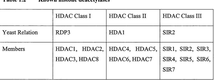
HDAC proteins identified to date fall into three main classes through similarities in their domain organisation, and relation to a yeast founding member (Table 1.2) (for reviews see Khochbin et al., 2001; Narlikar et a l, 2002). The Sin3 complex and NuRD complex contain members o f the class I HDAC family, HDACl and HDAC2. Complexes containing members o f class II HDACs have yet to be purified. Two complexes containing a member of class III HDACs, SIR2, have recently been identified and show distinct deacetylase activities (Yang et a l, 2002).
The histone specificities o f the class I HDAC family complexes are just beginning to be characterised. Recent studies using highly specific antibodies suggest that the yeast homolog o f HDACl, RPD3, deacetylates all sites except lysine 16 on H4. This site is strongly linked to heterochromatic silencing, and its acétylation by dMOF is linked to activation of the Drosophila male X chromosome (see section 1.1.4.2.5) (Suka et al., 2001; for review see Roth et al.,
2001). This is consistent with RDP3 having distinct functions from the SIR2 deacetylases. Furthermore, this suggests that RDP3 does not act antagonistically to ESA l, the yeast homolog o f dMOF.
Table 1.2 Known histone deacetylases
HDAC Class I HDAC Class II HDAC Class III
Yeast Relation RDP3 HDAl SIR2
Members HDACl, HDAC2, HDAC3, HDACS
HDAC4, HDACS, HDAC6, HDAC7
1.2
CBP and p300 Transcriptional Activity
1.2.1 Introduction
Cyclic adenosine 3’,5’-monophosphate (cAMP) response element binding protein (CREB), is a 43kDa leucine zipper transcription factor which was identified and cloned via a study o f cAMP regulated genes (Hoeffier et al.,
1988; for review see Andrisani, 1999). In the last decade, numerous studies have contributed to the current understanding of CREB structure, function, and CREB-mediated transcription. CREB binds to the cAMP-response element (CRE) as a homodimer formed via the leucine zipper motif present at its C- terminus, and its transcriptional activity is regulated by phosphorylation at serine residue 133, located within the N-terminal transactivation domain (Gonzalez and Montminy, 1989; Mayr and Montminy, 2001; Parker et al., 1996; Swope et al.,
1996). Ser 133 is phosphorylated by a number o f kinases including cAMP dependent kinase (PKA) (for review see Lalli and Sassone-Corsi, 1994). In the basal state, PKA resides in the cytoplasm as an inactive heterotetramer o f paired regulatory (R) and catalytic (C) subunits. Induction o f cAMP liberates C subunits, which passively diffuse into the nucleus and induce cellular gene expression by phosphorylating CREB. CREB mediates the activation of cAMP responsive genes by binding as a dimer to CRE, TGACGTCA (Comb et al.,
( D
C R E B
Figure 1.6 Activation of the cAMP-CREB signalling pathway
CBP displays great similarity to another larger nuclear protein, p300, which was isolated separately from CBP, through its interaction with the adenoviral transforming protein E lA (Arany et al., 1995; Eckner et a l, 1994). E lA exerts its biological effects through its ability to modulate the transcriptional activity o f target genes, either directly or indirectly (Nevins, 1992). Two conserved regions within ElA , known as CRl and CR2, together with a less well conserved N-terminal region, are necessary for E lA to exert its wide-ranging effects (Hasegawa et a l, 1997a; Jelsma et al., 1989; Lillie et al.,
1986; Moran et al., 1986; Smith and Ziff, 1988; Subramanian et al., 1988; Whyte
et al., 1988; for review see Shikama et al., 1997). Binding o f the N-terminal
region of E lA together with a sub-domain o f CRl to p300 is sufficient to induce S-phase entry of quiescent cells, and thus DNA synthesis (Liu and Kitsis, 1996). In the same study it was also shown that E lA can re-stimulate DNA replication in terminally differentiated cardiac myocyte cells through its p300 binding domain.
CHI B r omo C H 2 CH 3
C B P
I ■
66%
p300
I ■
86% [
2 4 4 l a a
86% 66% 82%
2414aa
Figure 1.7 Alignment of human CBP and p300 molecules
The recognition that these two proteins have highly conserved sequences and interact with transcription factors involved in different aspects of gene regulation suggested that they have the potential to participate in a variety of cellular functions (for reviews see Janknecht and Hunter, 1996; Goodman and Smolik, 2000). Indeed a variety o f sequence-specific DNA binding factors are found in complex with CBP and p300, including nuclear hormone receptors (Chakravarti et a l, 1996; Kamei et al., 1996), c-Jun (Arias et a l, 1994; Bannister
et a l, 1995), c-Fos (Bannister et a l, 1995), c-Myb (Dai et al., 1996;
Oelgeschlager et a l, 1996), p53 (Avantaggiati et a l, 1997; Wadgaonkar et al.,
1999), Statl and Stat2 (Zhang et al., 1996), MyoD (Yuan et al., 1996), HlF-1 (Arany et al., 1996), and GATA-1 (Blobel et al., 1998) (Figl.S). Further, CBP and p300 interact with TBP, TFllB, TFllD, RNA helicase A, and with RNA polymerase 11 itself (Dallas et al., 1997; Kee et al., 1996; Kwok et al., 1994), suggesting that they are recruited to RNA polymerase 11 holoenzyme (see section 1.1.2.2). Indeed, as mentioned previously, later studies showed that CBP and p300 have the ability to act as a bridge between transcription factors and the basal transcription machinery, thus resulting in transcriptional activation. In addition CBP and p300 are also histone acetyltransferases and therefore capable of enhancing transcriptional activation by acétylation o f histones, allowing gene target promoters to become more accessible (discussed in section 1.2.2.)
CBP/ p300
S t a t - 1
SF -1
N u c l e a r
H o r m o n e
r e c e p t o r s
d
I
C H I
J M Y
d m a d
P y L T
H P V E 6
C I I T A
T at
SF -1
E 2 F
H P V E 2 E t s - I
B R A C I j u n B
P 4 5 / N F - E 2 R N A h e l i c a s e A
T A L I c - J u n C / E B P p
p 7 3 c - m y b G A T A - I
M d m 2 T a x N e u r o - D
T B P S a p I M i c r o p h t h a l m i a
H I F - I Y Y I E l A p 5 3
E t s - I S R E B P T F I I B
R X R A T F - I P / C A F
p 6 5 A T F - 4 T w i s t M y o D Y Y I
Pit-1 C u b i t u s p p 9 0 R S K
H N F - 4 In te rr uptu s A T F - 2 c - F o s p / C I P
S t a t - 2 G l i 3 v I R F S V 4 0 L a r g e T S R C - I
Î
Î
K I X B r o m o - C H 2
Î Î
d o m a i nA T
Î
Î
2441/ 2414aa
C H 3 G l u t a m i n e - r i c h
r e g i o n
Figure 1.8 CBP/p300 organisation and interactions
1.2.2 CBP and p300 Structural Features
The sequence organisation o f CBP/p300 genes isolated from Drosophila,
nematodes, and vertebrates are very similar (Chen et al., 2000, Yoshida et al.,
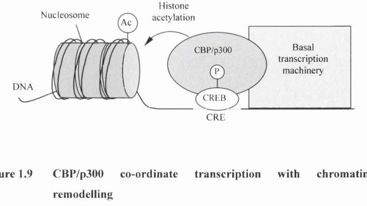
1998). As mentioned, several amino acid domains such as the bromodomain, the CREB binding domain, referred to as KIX, and three cysteine-histidine rich regions (CH), provide binding sites for a variety o f factors. The central portion o f both CBP/p300 genes encodes a relatively large domain possessing intrinsic acetyltransferase activity (Ogryzko et al., 1996). This region alone is sufficient to acetylate lysine residues o f certain target proteins in vitro (see section 1.1.4.1). Although the exact role o f histone acétylation remains uncertain, it is proposed that the loss of positive charge conferred to the histone tail by acétylation weakens the interaction of histones with DNA and facilitates the access of RNA polymerase II and its associated factors to DNA templates (Fig. 1.9) (Giles et al.,
1998).
Histone
N u c l e o s o m e a c é t y l a t i o n A c
B a s a i
t r a n s c r i p t i o n
m a c h i n e r y C B P / p 3 0 0
D N A
C R E B
C R E
Figure 1.9 CBP/p300 co-ordinate transcription with chromatin
remodelling
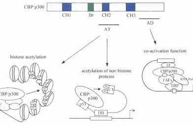
Non-histone proteins are also substrates of acétylation by CBP/p300, for example TFIEF, TFIIE and GATA-1 (Boyes et a l, 1998; for review see Imhof et a l, 1997). Moreover, reports have shown that the tumour suppressor p53 protein is acetylated in vitro by p300, and this in turn stimulates p53 binding to its DNA- binding site (Gu and Roeder, 1997). However, p300 does not acetylate CREB, inspite o f the fact that these two proteins are strongly associated, suggesting the existence o f a high degree o f specificity o f these enzymes (Gu and Roeder, 1997). Although the relevance o f protein acétylation in vivo remains to be addressed, these findings imply that it is a general regulatory event in the modulation o f structure-function relationship o f proteins.
C B P / p 3 0 0
h i s t o n e a c é t y l a t i o n
C B P / p 3 0 0
T F
C H B r C H 2 C H 3
A T
a c é t y l a t i o n o f n o n h i s t o n e
p r o t e i n s
TT
C B P / p 3 0 0
DB
A D
c o - a c t i v a t i o n f u n c t i o n
r
X ü ii.
CBP/p300
Pol
TAFs
I BP
Figure 1.10 Structural feature of CBP/p300 gene relative to their function
1.2.3 Acetylation-Dependent Functions o f CBP and p300
As discussed in section 1.1.4.2, mammalian acetyltransferases belong to a large family o f proteins. Interestingly, CBP is found in association with both ACTR and P/CAF, and the simultaneous recruitment o f these proteins in large multi-protein complexes appears to be necessary for activation o f transcription nuclear receptors (Chen et al., 1997). The finding that transcription co-activators possess intrinsic acetyltransferase activity and are recruited into complexes with DNA-binding sequence-specific activators strongly implies that histone acétylation may be targeted to transcriptionally active chromatin domains in a signal dependent fashion. The purpose o f multiple acetyltransferases, however, still remains unanswered. The lack of any obvious homology between these proteins argues that they exert distinct, non-overlapping effects. In support o f this view is the existence of a P/CAF co-activation complex which excludes CBP and p300 (Ogryzko et al., 1998). This P/CAF complex comprises several histone-like subunits forming a histone octamer-like structure which lacks the regions corresponding to the N-terminal tails o f histones. These findings have led to the proposal that in addition to converting a silent chromatin domain into a potentially active one, acetyltransferases could replace the histone octamer, and by preventing deacetylation o f the histone tail contribute to the maintenance of an active chromatin state.
al., 1998; Lill et al., 1997; Sang et al., 1997; Somasundaran and El-Deiry, 1997). This indicates that the ability of p53 to function as a transcription factor and also to transmit signals to cells, is dependent upon its binding to CBP/p300. It is not clear, however, whether the acetyltransferase activity o f p300 directly contributes to either function of p53. Evidence suggests that p300 enhances the transactivation potential o f p53, regardless o f its acetyltransferase activity, through protein-protein interactions. This is argued because a p300 deletion mutant with an intact acetyltransferase domain, but lacking the binding site for the acetyltransferase P/CAF, is impaired for rescuing E l A mediated repression o f the p53 responsive gene, p21/WAF, compared to the full length protein (Lill et al., 1997) This inhibition is overridden more efficiently when the expression levels of p300, CBP and P/CAF are raised simultaneously (Scolnick et ah, 1997). This indicates that P/CAF is also an important protein in p53 transcription and signalling, a view consistent with the observation that P/CAF acetylates p53 at amino acids residues distinct from those acetylated by p300 (Liu et al., 1999a; Sakaguchi et al., 1998). Yet, similarly to p300, P/CAF increases sequence specific binding o f the protein to DNA.
Thus, there seems to be a degree o f redundancy between certain effects of CBP/p300 and P/CAF. While in the case o f p53 this issue remains to be addressed, in other cases these proteins have been shown to regulate transcription in a unique fashion. In muscle cells, microinjection with anti-p300, anti-CBP, or anti-P/CAF antibodies impairs transcription activation of MyoD regulated genes, implying that these proteins equally contribute to MyoD dependent transcription (Sartorelli et al., 1997; for reviews see Eckner et al., 1996; Puri et al., 1997a). A p300 protein containing a deletion o f the acetyltransferase domain is still able to activate MyoD transcription, whereas deletions of the acetyltransferase domain of P/CAF significantly impair MyoD dependent transactivation (Puri et al.,
Thus the ability of CBP/p300 to modulate gene expression can be either dependent or independent o f their acetyltransferase activity and the requirement o f this function appears to be strictly promoter-activator specific. A more recent study has demonstrated that the intrinsic acetyltransferase domain of Drosophila
1.2.4 Acetylation-Independent Functions o f CBP and p300
Apart from their ability to acetylate histones and non-histone proteins, CBP/p300 are important for the regulation o f transcription by serving a structural role as scaffolds upon which a variety of proteins can efficiently assemble and operate targeted promoters. By providing multiple binding sites for transcription activators, other acetyltransferases and basal components of the transcription apparatus, it is thought that CBP/p300 may increase the relative concentration of these factors where they are needed and provide a nucléation site which facilitates protein-protein and DNA-protein interactions. This type o f multi protein complex may have evolved in eukaryotic cells not only to deal with the complexity o f nucleosomal templates but also to regulate the number o f genes activated in response to a given stimulus.
A model to explain regulation o f transcription by CBP/p300 has been proposed by Maniatis and colleagues (Kim et al., 1998; for review see Carey,
1.2.5 Regulation o f CBP and p300 by Phosphorylation
Most proteins involved in the control o f cell growth are regulated by phosphorylation, and CBP/p300 are examples of this. However, little is known about how phosphorylation affects CBP/p300 functions. Studies first showed that p300 could be phosphorylated by cyclin/Cdc2 and cyclin/Cdk2, and that these phosphorylation events were blocked by E l A (for review see Goodman and Smolik, 2000). Further understanding of the functional implications of CBP/p300 phosphorylation by cyclin dependent kinases (Cdks) has been provided by studies showing that p300 is regulated negatively by cyclinE/Cdk2, thus preventing DNA synthesis (further discussed in section 1.3.1). Recent work has suggested that CBP is phosphorylated at G l/S by cyclinE/Cdk2, and that this phosphorylation increases the histone acetyltransferase activity o f CBP (Ait-Si-
Ali e ta l, 1999).
Several papers have suggested that CBP is regulated by the calcium/calmodulin (CaM) dependent protein kinase CaMKIV (Chawla et al.,
1998), and it has been suggested that this phosphorylation event may be required for CBP mediated transcriptional activation (Hardingham et al., 1999; Hu et al.,
1999). Using constructs in which CBP is artificially recruited to the promoter by means of fusion to the Gal4 DNA binding domain, this provides a model that suggests that CaMKIV, which is a well known activator o f CREB (Dash et al.,
1991; Sheng et al., 1991; Sun et al., 1994), mediates transcription of CREB via a mechanism regulated by CBP. However, mapping o f the critical phosphorylation site on CBP remains to be determined. Moreover, by using a form of CREB that has been mutated so that it interacts with CBP/p300 constitutively, it has been found in F9 teratocarcinoma cells (which contain low levels o f CBP but normally require the addition o f CREB and PKA to activate a CRE reporter), that CBP/p300 recruitment by itself is sufficient for gene activation. Phosphorylation o f CBP is therefore not essential in this system (Cardinaux et al., 2000).
1.3
Biological Functions of CBP and p300
The initial studies developed to characterise the biological activities of CBP/p300 were performed in transient transfection assays by overexpressing the proteins in cells in culture. These investigations highlighted that in many respects the functions of CBP and p300 overlap. CBP/p300 were shown to interact with the same set o f intracellular proteins, to activate similar sets of genes and to participate in common biological processes. As it became possible to study the effects of down-modulation of the expression levels o f these proteins, potentially important differences have been discovered. By using hammerhead ribozymes capable o f cleaving specifically the CBP or p300 mRNAs, a study showed that both differentiation and cell-cycle arrest induced by retinoic acid are dependent upon normal p300 levels and are unaffected by inhibition of CBP (Kawasaki et al., 1998). In contrast, both proteins are required for apoptosis induced by retinoic acid. CBP/p300 were both involved in signalling through the second messenger cAMP, yet mouse cell lines lacking a functional p300 gene respond normally to incoming signals inducing CREB activation (Yao et al., 1998). Furthermore, a recent study has shown that p300, but not CBP, stimulates transcription o f the cyclin D1 promoter. Thus, at physiological concentrations, CBP and p300 control the expression of distinct but overlapping target genes.
1.3.1 CBP and p300 in Cell Cycle Regulation and Differentiation
Until recently there was relatively little evidence for a role for CBP/p300 in cell cycle progression. Initial studies showed that E l A can stimulate DNA synthesis in quiescent neonatal rat kidney cells through its CBP/p300 binding region, implying that CBP/p300 may play a role in cell cycle regulation (Stein et al., 1990). Furthermore, E lA can also re-stimulate DNA replication in terminally differentiated cardiac myocytes through its CBP/p300 binding domain (Liu and Kitsis, 1996). More recently, CBP/p300 have been implicated in differentiation of hematopoietic tissues through their association with GATA-1.
CBP/p300 modulate the expression pattern o f downstream myogenic factors, such as myogenin and MEF-2, and promote cell-cycle withdrawal in myoblasts induced to differentiate (Puri et al., 1997a; Puri et al., 1997b; Sartorelli et al.,
1999; Yuan et al., 1996). The cell cycle arrest induced by MyoD can be overridden by inactivating the function o f either CBP/p300, indicating that these proteins are both required for appropriate cell cycle exit, to commit the cells to GO. Furthermore, during terminal differentiation, p300 transactivates the Cdk inhibitor, p21, in co-operation with the cellular transcription factors Spl and Sp3, or tissue-specific transcription factors, again suggesting that p300 may play a role in keeping cells in GO/Gl (Billon et al., 1996; for review see Goodman and Smolik, 2000). However, fibroblasts isolated from p300 knockout mice are unable to replicate and appear to undergo cell cycle arrest, suggesting that in addition to being involved in cell cycle arrest p300 may also be necessary for cell cycle progression (Yao et al., 1998).
In addition to the involvement o f CBP/p300 in skeletal muscle, these proteins have also been shown to be important in cardiac muscle development (see section 1.6 for further detail). The transcription factors GATA-4/5/6 have been implicated as key regulators of cardiogenesis and the transcription o f many cardiac specific genes (Charron and Nemer, 1999). GATA-4/5 function, in particular have been shown to be regulated by p300, and p300 is therefore likely to be involved in the regulated expression o f cardiac genes itself.
1.3.2 CBP and p300 in Embryogenesis and Development
Many (if not most) developmental pathways in mammals culminate in interactions that involve CBP/p300. Two independent studies have shown that normal CBP and p300 levels are required for the appropriate completion of developmental processes in mice (Tanaka et al., 1997; Yao et al., 1998). Deletion o f either CBP or p300 alleles is lethal and leads to embryonic death between days 9 and 11.5 o f gestation. CBP and p300 nullizygous embryos are much smaller than their littermates, exhibiting severe open neural tube defects and defects in heart development, shown by abnormalities o f cardiac muscle differentiation and trabeculation. Furthermore, the expression o f the myocardial contractile proteins was clearly reduced in mutant hearts compared with the wild type hearts of littermates, thus suggesting p300 is required for proper heart development. Cells removed from the p300 homozygous mutants also displayed poor proliferation properties, implying that p300 is required for growth stimulation, an idea that is contrary to the general opinion that CBP and p300 are tumour suppressor proteins (see section 1.3.3).
Disruption o f one p300 allele is associated with a high incidence of embryonic deaths (Yao et al., 1998). However, once past embryogenesis p300 heterozygous mice are viable and do not display phenotypic abnormalities. Unlike p300, CBP heterozygous mutant mice manifest skeletal and cardiac abnormalities consistent with the human congenital disorder Rubinstein-Taybi Syndrome (RTS) (Rubinstein and Taybi, 1963), in which one CBP allele is inactivated, supporting the idea that loss o f heterozygosity at the CBP locus might be the cause of the disease (Eckner, 1996; Oike et al., 1999; Petrij et al.,
in Drosophila embryos, demonstrating that in the tight regulation of CBP/p300, more is not always better.