Synthesis And Characterization Of Silver
Nanoparticles Using Flower Extract Ofaerva
Lanata And Evaluation Of Its antimicrobial
Activities
D. Devi Priya, J. Thomas Joseph Prakash
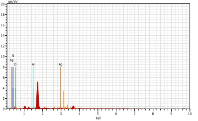
Abstract : Silver nanoparticles (AgNPs) with flower extract of Aerva lanata are successful in the process of synthesis. The characterization of obtained AgNPs are accomplished by the following methods like UV-Visible Spectrometer, Field Emission Scanning Electron Microscopy paired with Energy Dispersive X- ray Spectroscopy, X-ray Diffraction, Fourier Transform Infrared Spectroscopy, Dynamic Light Scattering. The spherical morphologies are confirmed, with the size of range 10 to 50 nm. The Energy Dispersive X-ray Spectroscopy present in the elemental absorption peak at 3 KeV. The existence of elemental silver in the sample is also proved by the planes (111), ( 200), (220) and (311) corresp onds peaks in the X-Ray Diffraction patterns which implies the crystalline nature of the nanoparticles in the face centered cubic structure. The evaluation of size dis tribution of nanoparticles were done using Dynamic Light Scattering measurements. Additionally, nanoparticles show effective antibacterial and antifungal activities. As synthesized AgNPs showed excellent antibacterial activities against pathogenic two gram positive and one gram negative and antifungal activities showed good test organisms, from the result sample candida vulgaris was most effective and the highest activity.
Index Terms : Aerva lanata, Antibacterial activity, Antifungal activity, DLS, FTIR, silver nanoparticles, UV.
—————————— ——————————
1 INTRODUCTION
Nanotechnology is one of the most exciting field in current area. Nanotechnology is used to depict the creation and utilization of materials with structural feature in the nano range between those atoms and bulk materials with at least one dimensions. Synthesis of metal nanoparticles is significant in multidiscipline such as developing medicine, energy, chemistry, electronics, environment etc. So it is always an emerging area. Depending upon their size, distribution and morphology, nanoparticles brandish new or improved properties [1], [2]. Silver nanoparticles (AgNPs) have been extensively experienced in biomedical devices because of their admitted antibacterial, antifungal properties [3], [4], [5], [6], [7], [8], [9]. Having said that, the method of synthesis size, shape, functionalization and stabilizing agents are correlated to these effects [10], [11].
The conventional methods of synthesis consists of physical, chemical and biological methods whereas these processes have various demerits such as high cost and energy demand use of toxic reductant agents and production of toxic waste. Recently, the green methods to synthesis nanoparticles has been proposed over conventional methods having a list of merits such as low cost, utilize less energy, produce less and nontoxic waste. Moreover green methods can control the size, shape and stability and they also offer simplicity and swiftness than other methods [12], [13], [14], [15]. The target of this research paper on the Aerva lanata flower (sirupeelai) which belongs to the family Amaranthaceae and its medicinal purposes. The flower is well known for its medical use since ancient times. In Ayurveda, the indian system of medicine, the Aerva lanata flower has been used as it attributes several medicinal properties. Aerva lanata is an excellent medicine for treating diseases related to stomach and the digestive system, and is a good remedy for stones in urinary bladder and kidney. Also it regulates the blood sugar level so the flower is good for diabetes patients. In this study we have successfully conducted a process of biosynthesis of AgNPs with extract of Aerva lanata. The obtained AgNPs were characterized by UV-Vis Spectrophotometer (UV), Fourier Transform Infrared Spectroscopy (FTIR), X-ray Diffraction (XRD), Dynamic Light Scattering (DLS), Field Emission Scanning Electron Microscopy (FESEM) and Energy Dispersive X-ray Spectroscopy (EDAX). The nanoparticles were evaluated for antibacterial and antifungal activities. The antibacterial activities of the samples were studied on Staphylococcus auerus (MTCC25923), Bacillus subtilis (MTCC 2451) and Escherichia Coli (MTCC 25922) bacterial strains using disc-diffusion method. The antifungal test organisms used for study are Candida albicans (MTCC 3498) and Candida Vulgaris (MTCC 227).
________________________
D. Devi priya is currently pursuing in Ph.D (Research scholar) PG And Research Department of Physics, Government Arts College (Affiliated to the Bharathidasan University), Trichy-620 022, Tamil Nadu, India, Cell: +919486242165
E-mail: [email protected]
J. Thomas Joseph Prakash* is currently work as a Assistant Professor PG And Research Department of Physics, Government Arts College (Affiliated to the Bharathidasan University), Trichy-620 022, Tamil Nadu, India, Cell: 09842470521
4197
2 MATERIALS AND METHOD
Materials
All the reagents which are required in this experiment were procured from Sigma Aldrich. For this process, double distilled water was utilized whereas establishment of filtration using Whatman No.1 paper. Double distilled water were used to wash well and rinse purposes of glass wares which were used for the whole reactions and kept dried. The Aerva lanata flowers were freshly collected from Trichy as shown in Fig.1.
Fig.1 Aerva lanata
Preparation of flower extract
The flowers were completely washed with normal tap water for many times and the same was followed by double distilled water to get rid of the impurities. A mixture of 10 g of Aerva lanata powder and 100 ml of double distilled water were boiled for 15 min. The outcome solution was filtered through Whatman No.1 filter paper initially. The final filtered extract was stored.
Synthesis of silver nanoparticles
In order to synthesize nanoparticles 10 ml of flower extract was added with 90 ml of 1mM silver nitrate solution. After the time period of 1 hour the solution was turned from pale yellow to brown denoting that the silver nanoparticles were formed Fig.2.
3
CHARACTERIZATION
OF
SILVER
NANOPARTICLES
The formation of silver nanoparticles proved by UV-Visible spectroscopy using UV-VISIBLE SPECTROPHOTOMETER LAMBDA 35 PERKIN ELMER
showing ranges between 190 nm to 1100 nm. FTIR analysis was accomplished for the reduction of silver ions with the spectral range of 400-4000 cm-1. The experiment sample was centrifuged at 8000 rpm for 15 min and dried with help of hot air oven. The resultant sample was ground with Kbr to form a pellet. Then the pellet analysed using
FTIR SPECTRUM 1000 PERKIN ELMER
SPECTROMETER instrument. The silver nanoparticles having crystalline structure was determined by X-ray Diffraction analysis with XRDX ―X‖ PERT PRO Diffractrometer. To examine the morphology of silver nanoparticles by employing Field Emission Scanning Electron Microscopy FEI QUANTA-250 FEG equipped with an Energy Dispersive X-ray Spectroscopy instruments. Dynamic Light Scattering analysis were used to determine the size of the silver particles by the help of instrument Nanoplus.
Antibacterial activity
Collection of test organisms:
To examine the antibacterial activity of silver nanoparticles sample, 2 grams of positive bacterial strains such as Staphylococcus aureus (MTCC 25923) and Bacillus strains Escherichia coli (MTCC 25922) were taken and prepared as test organisms. The procurement of required bacterial strains were from the microbial type culture and collection (MTCC) at Chandigarh, India. The cultivation of bacterial strains at 37º C and maintained on nutrient agar (Difco, USA) slant at for 4 º C.
Screening of Antibacterial Activities (disc diffusion method):
The determination of antibacterial activity of silver nanoparticle samples was performed with the help of disc diffusion method. The Muller Hinton Agar were used to prepare petridishes (Diameter 60 mm) and immunized with test organisms. Disc of six mm width were sterilized and impregnated with 10 µl of various samples respectively. At room temperature, the prepared discs were kept on the top layer of the agar plates and left for about 30 minutes for compound diffusion. Positive control was prepared with the help of Amoxicillin as standard antibiotic disc with the quantity of 10 µl. The dishes were incubated for 24 h at 37 º C and the zone of inhibition was recorded in millimetres and the experiment was repeated twice.
Screening of Antifungal Activities:
Candida albicans (MTCC 3498) and Candida vulgaris (MTCC 227) are the fungal test organisms which were used for this study were procured from National Chemical Laboratory (NCL), Pune, Maharashtra, India.
Determination of antifungal activity
prepare positive control. The dishes were incubated for 24 h at 37 º C and the zone of inhibition was recorded in millimetres.
4 RESULTS AND DISCUSSION
UV-Visible Spectral studies
The Aerva lanata flower extract was mixed with aqueous solution of the silver nitrate due to the reduction of silver ion the change of colour takes place from pale yellow to brown. It is found that when the surface plasmon vibrations in silver nanoparticles are charged, the silver nanoparticles displayed some brown colour in the aqueous solution. The existence of nanoparticles was proved by obtaining a spectrum in the Visible range of 200-1000 nm using UV-Visible Spectrophotometer Fig.3. From the analysis absorbance peak as found at around 445 nm which was specific for silver nanoparticles [16].
Fig.3 UV-Visible Spectroscopy
Fourier Transform Infrared Spectroscopy
FTIR measurements was executed to spot the possible biomolecules in Aerva lanata flower extract which are liable for capping leading to efficient stabilization of the silver nanoparticles Fig.4. The IR spectrum of silver nanoparticles shows significant absorption bands situated at 2916 Cm-1 N-H Stretching, 2356 Cm-1 C-O Stretching, 1679 Cm-1 C-H Bending, 1606 Cm-1 C=C Stretching, 1435 Cm-1 O-H Bending, 1001 Cm-1 C-O Stretching, 892 Cm-1 C=C Bending, 760 Cm-1 C-H Bending, 571 Cm-1 C-Br Stretching. The FTIR data confirms that the peak is present in the samples [17].
Fig.4 Fourier Transform Infrared Spectroscopy
X-Ray Diffraction
The XRD pattern of powder sample of AgNPs exhibited various peaks at 38.04 , 44.19 , 64.1 and 77.09 . The 2 values that implied that (111), (200), (220) and (311) faces of silver respectively to face centred cubic (FCC) crystalline structure of metallic silver as shown in Fig.5. The values are in the good agreement with the JCPDS file No. 04-0873. It recommends the prepared silver nanoparticles having crystalline and amorphous organic phases [18].
Fig.5 X-ray diffraction analysis
Field Emission Scanning Electron Microscopy
In FESEM , the morphology of green synthesized AgNPs was viewed. As represented The FESEM image illustrated that the AgNPs formed having sizes ranging from 10 to 50 nm and were well dispersed with spherical shape as shown in Fig.6 (a) [19].
Fig.6 (a) Field Emission Scanning Electron Microscopy
Energy Dispersive X-ray Spectroscopy
4199
Fig.6 (b) Energy dispersive X-ray spectroscopy
Particle Size analysis (DLS)
Dynamic Light Scattering measurements were performed to persuade the size of the formed silver nanoparticles. The particle size distribution curve of the synthesized AgNPs. In the same figure the particles of ranging from 282.0 nm and had an average particle size of 151.7 nm as shown in Fig.7 [21].
Fig.7 Dynamic Light Scattering
Antibacterial activities
By using disc diffusion method, the results of the antibacterial activity of different samples were tested against pathogens are shown in Table.1. The inhibitory activity against positive strains Staphylococcus aureus (7mm) and Bacillus subtilis (8 mm) were shown by sample D whereas at sample D, exhibited the antibacterial activity in all the four bacteria. But as shown in Figure.8 sample C was more susceptible against staphylococcus aureus (7mm), Bacillus subtilis (8mm), Escherichia coli (6mm). Nevertheless, the crude extract and synthesized nanoparticles manifested better inhibitory actions against pathogens.
Table 1: Antibacterial activity of silver nanoparticle sample
Antifungal activities
The experiment represents the outcomes of the antifungal susceptibility test of the various samples and against the test organisms. As shown in the Figure.9, the experiment outcome demonstrates that the sample D was the most effective and the highest activity against Candida vulgaris (4 mm zone of inhibition) as shown in table.2.
Table 2: Antifungal activity of silver nanoparticle sample
Samples
Conc entrat ions (µl/ml )
Organisms/ Zone of Inhibition (mm)
Candida Candida Vulgaris Albicans
A(Silver Nitrate) 10 µl 0 0
B (Amoxicillin) 10 µl 8 8
C (Plant Extract) 10 µl 3 6
D (Nanoparticles) 10 µl 4 7
1 2 3 4 5 6 7 8 9 10 keV 0 2 4 6 8 10 12 14 16 18 20 cps/eV O Ag Ag Ag Al N Samples Con centr ation s (µl/m l) Organisms/ Zone of Inhibition (mm) Staphylo Bacillus Escherihi hia coli Coccus subtilis aureus A(Silver Nitrate) 10 µl 0 0 0
B (Amoxicillin) 10 µl 9 9 9
C(Plant Extract) 10 µl 6 7 4
CONCLUSION
Aerva lanata flower extract was found suitable for the green synthesis of silver nanoparticles. The reduction of silver nanoparticles by the flower extract resulted in the formation of stable nanoparticles with spherical morphologies. The concentration of flower extract and metal ions play an important role in the green synthesis of AgNPs. The spectroscopic characterization using UV-Vis, XRD, FESEM, EDAX and particles size analyzier were useful in proving the formation of nanoparticles and also in confirming their size and shape. FTIR evidenced the formation and stability of the biosynthesized AgNPs which can be studied further to understand the chemical and molecular interaction which could be responsible for nanoparticle synthesis. The results of the antibacterial activities were test against pathogens by two gram positive Staphylococcus aureus (MTCC 25923), Bacillus subtilis (MTCC 2451) one gram negative Escherichia Coli (MTCC 25922) and the antifungal activities showed most effective and the highest activity.
REFERENCES
[1]. Song JY, KimL BS, ―Rapid biological synthesis of silver
nanoparticles using plant leaf extracts‖ Bioprocess Biosyst Eng (2009) 32; 79-84.
[2]. Nalwa HS, ―Handbook of nanostructure mater and nanotechnology‖. Academic press newyork; 2000. [3]. Li Y, Qin T, Ingle T, et al, ―Differential genotoxicity
mechanisms of silver nanoparticles and silver ions‖. Arch Toxicol (2017) 91(1): 509–519.
[4]. Albanese A, Tang PS, Chan WC, ―The effect of nanoparticle size, shape and surface chemistry on biological systems‖ Annu Rev Biomed Eng (2012) 14(1):1–16.
[5]. Ge L, Li Q, Wang M,Ouyang J, Li X, Xing MM, ―Nanosilver particles in medical applications: Synthesis, Performance, and toxicity‖ Int J Nanomed (2014) 9(1): 2399–2407.
[6]. Zhang T, Wang L, Chen Q, Chen C, ―Cytotoxic potential of silver nanoparticles‖ Yonsei Med J (2014) 55(2): 283– 291.
[7]. Moldovan, Bianca; David, Luminita; Vulcu , Adriana; et al, ―in vitro and in vivo anti-inflammatory properties of green synthesized silver nanoparticles using Viburnum opulus L. fruits extract‖ Mater Sci & Eng C- Materials for biological Applications (2017) 79:720–727.
[8]. Prateek Mathur, Swati Jha, Suman Ramteke, Jain NK, ―Pharmaceutical aspects of silver nanoparticles‖. Artif Cells Nanomed Biotechnol (2018) 12:1–12.
[9]. Li J, Wang Y, Liang R, An X, Wang K, Sheng G, Tu Y, Zhu J, Tao J, ―Recent advances in targeted nanoparticles drug delivery to melanoma‖ Nanomedicine (2015) 11(3): 769–794.
[10].Wei L, Lu J, Xu H, Patel A, Chen ZS, Chen G, ―Silver nanoparticles: Synthesis, properties and therapeutic applications‖ Drug Discov Today (2015) 20(5):595–601. [11]. Kausar B. Riaz Ahmed, Amber M. Nagy, Ronald P.
Brown, Qin Zhang, Subhas G. Malghan, Peter L Goering, ― Silver nanoparticles: Significance of physicochemical properties and assay interference on the interpretation of in vitro cytotoxity studies‖ Toxicology in vitro (2017) 38:179–192.
[12]. Mohammed AE, Al-Qahtani A, Al-Mutairi A, Al-Shamri B, Aabed KF, ― Antibacterial and cytotoxic potential of biosynthesized silver nanoparticles by some plant extracts‖ Nanomaterials (2018) 8(6): 1–15.
[13]. Zainal Abidin Ali, Rosiyah Yahya, Shamala Devi Sekaran, R. Puteh, ―Green synthesis of silver nanoparticles using Apple extract and its antibacterial properties‖ Adv in Mater Sci and Eng (2016):6.
[14]. Kummara, S. Patil, M.B., Uriah, ―Synthesis, Characterization, biocompatible and anticancer activity of green and chemically synthesized silver nanoparticles A comparative study‖ Biomed Pharmacother (2016) 84:10–21.
[15]. Kumar R, Ghoshal G, Jain A, Goyal M, ―Rapid green synthesis of silver nanoparticles (AgNPs) using prunus persica plants extract: Exploring its antimicrobial and catalytic activities‖ J Nanomed Nanotechnol (2017) 8:4, 2157–7439.
[16]. C. Krishnaraj, Jagan EG, S. Rajasekar, P. Selvakumar, P.T. Kalaichelvan, N. Mohan, ―Synthesis of silver nanoparticles using Acalypha indica leaf extracts and its antibacterial activity against water bone pathogens‖colloids surfaces B Biointerfaces 76 (1) (2010) 50-56.
[17]. Dubey, S. P. Lahtinen, Sarkka H, Sillanpaa M, ―Bioprospective of Sorbus aucuparia leaf extract in development of silver and gold nanocolloids‖ Colloids surf B Biointerfaces 80, (2010) 26-33.
[18]. Kittel, Charles, ―Introduction to solid state physics‖ John Whiley & Sons INC, Eighted, (1992)
[19]. Hong Wei, Hongxing Xu, ―Nanowire-based plasmonic waveguides and devices for integrated nanophotonic circuits‖ Nanophotonics. 1(2) 2012, 155-169.
[20]. P. Magudapathy, P. Gangopadhyay, Panigrahi BK, Nair BGM, S. Dhara, ― Electrical transport studies of Ag nonoclusters embedded in glass matrix‖ Phy. B. (2001) 299, 142-146.