Original Article
170 | P a g e
Int J Med Res Prof.2016; 2(2); 170-76. P-ISSN:2454-6356; E-ISSN:2454-6364
A Clinical Study of Prognostic Factors of Guillain Barré Syndrome in a
Tertiary Care Hospital of North
-
East India
Zain Fakih
1, Duncan Khanikar
2*, Chitralekha Baruah
3, Marami Das
4 1PG trainee,
2Registrar,
3Professor, Department of Medicine,
4Associate Professor,
Department of Neurology, Gauhati Medical College and Hospital, Guwahati, Assam, India.
Article History
Received: 14 Mar 2016 Revised: 17 Mar 2016 Accepted: 27 Mar 2016
*Correspondence to:
Duncan Khanikar, Registrar, Department of Medicine, Gauhati Medical College and Hospital, Guwahati, Assam, India.
ABSTRACT
Introduction: Guillain-Barré syndrome (GBS) is an acute, frequently severe, and fulminant polyradiculoneuropathy that is autoimmune in nature. Various factors play an important role in the mortality and morbidity of the disease. Aim of the study is to look for the prognostic factors.
Methods: We conducted a hospital based observational, descriptive study comprising of 52 patients of Guillain Barré Syndrome who had been diagnosed based on the criteria laid down by Asbury AK, Cornblath DR (1990), admitted in Gauhati Medical College and Hospital, Guwahati, Assam (India) and fulfilled the inclusion and exclusion criteria. Statistical analysis was performed using GraphPad InStat version 3.00 for Windows 7, GraphPad Software, San Diego California USA.
Results: In our study, 32 were male and 20 were female; 38.46% were in the third decade of life followed by the second decade (21.15%) and sixth decade (13.46%). On follow up at the end of 1 month , 30 out of 52 patients(57.7%) had good prognosis while the rest (42.3%) had a poor prognosis (GBS disability score of >2). Amongst the patients with poor prognosis, 40.91% of the patients were more than 50 years of age (p-value- 0.02), 68.18% had a low MRC sum score (<30) on presentation to the hospital (p-value-0.0002), 54.55% had an antecedent history of diarrhoea value-0.0023), 68.18% showed autonomic dysfunction (p-value-0.023) during hospital stay and 72.72% had cranial nerve involvement. Mortality was 7.69% due to ventilator associated pneumonia.
Conclusion: Early identification of several clinical factors are crucial to predict the prognosis of the disease and early initiation of treatment ensures a better outcome.
KEYWORDS:
Autonomic dysfunction, Guillain-Barré syndrome (GBS), GBS disability score, Polyradiculoneuropathy, Poor prognosis.INTRODUCTION
Guillain-Barré Syndrome is an acquired immunologically mediated inflammatory polyradiculoneuropathy. It initially presents with weakness with or without paresthetic sensory symptoms. The fairly symmetrical weakness of the lower limbs ascends proximally over hours to several days and may subsequently involve arm, facial, and oropharyngeal muscles, and in severe cases, respiratory muscles. Its severity varies from mild, in which patients are still capable of walking unassisted, to a nearly total quadriplegia1. This is the most common cause of acute or subacute generalized paralysis in practice2.
It has a yearly incidence rate between 1.1 and 1.8 per 100000 in western countries. The lifetime likelihood of
any individual acquiring GBS is 1:1000. GBS is equally common in men and women and can occur at any age. In western countries, GBS is common in the 5th decade, but in India it occurs more commonly at a younger age3. There is a male preponderance among the hospitalized population4.
patients whose GBS is otherwise mild. Several subtypes of GBS are recognized.
Guillain-Barré syndrome (GBS) is usually associated with a good prognosis. However, it can have a poor prognosis needing ventilatory support and causing major deficits at discharge6. GBS remains fatal in about 4% of cases; only about 20% of patients are able to walk unaided after 4 weeks. After 1 year only 60% recover full motor strength and 14% are left with a severe disability7.
Regarding clinical prognostic factors, most studies demonstrate higher age (>40 or >50 years) as poor outcome. Also preceding diarrhoea, severe weakness leading to a low MRC score on admission and high early GBS disability grade are all reliable predictors of a poor outcome7. González-Suárez et al. (2013)8 demonstrated cranial nerve involvement and need for mechanical ventilation also leads to a poor prognosis.
MATERIALS AND METHODS
PatientsIn this descriptive, observational study, conducted from June 2014 to May 2015, a total of 52 patients were included. These patients, all above the age of 12 years, had attended Medicine O.P.D./Ward or Neurology O.P.D./Ward in Gauhati Medical College and Hospital, Assam, India; and had been diagnosed based on the criteria laid down by Asbury AK, Cornblath DR (1990)9. Patients with acute neuromuscular weakness due to other causes (e.g., myasthenia gravis, botulism, poliomyelitis, toxic neuropathy, diphtheria, vasculitic neuropathy) and those with major illness like CAD, DM, Hypertension, HIV and tuberculous infection were excluded.
Ethical clearance was obtained from the ethical committee of Gauhati Medical College & Hospital. Assessment
Data was collected by taking proper history from patients and attendants, thorough clinical examination and relevant investigations. Data was recorded in preformed proforma. Clinical data mainly comprised of age, sex, date of onset of disease, date of presentation to the hospital, duration of progression of symptoms, disability at the time of presentation, preceding and concurrent infections, sensory symptoms, bladder and bowel complaints, exposure to toxins, heavy metals and recent vaccination. In clinical examination due importance was given to pulse, blood pressure, respiratory rate, single breath count and neurological examination including cranial nerves especially III, IV, VI, VII, IX, X, tone and power of the muscles, reflexes (both deep and superficial) and sensory system examination. Power of the muscles was assessed by MRC sum score (Annexure 2), a summation of the MRC grades (range 0–5) given in full numbers of the following muscle pairs: upper arm abductors, elbow flexors, wrist extensors, hip flexors, knee extensors and
foot dorsal flexors as given by Kleyweg RP et al (1991)10. The MRC sum score ranged from 0 (“total
paralysis”) to 60 (“normal strength”).
Laboratory data consisted of Complete Blood Count, ESR, RFT, LFT, serum electrolytes, RBS, Urine routine examination and Urine for Porphobilinogen, HIV ELISA, Chest X-Ray, ECG and Nerve Conduction Velocity (NCV) testing.
Patients were followed up after 1 month and were assessed by the GBS Disability Score (Annexure 1) advocated by Hughes RA et al (1978)11. Poor prognosis was defined as the inability to walk unaided 10 meters across an open space (GBS disability score of 3 or higher) as given by Walgaard et al (2011)12. GBS
disability score of ≤2 was taken as good prognosis.
Statistics
All the statistical graphs were prepared using Microsoft Excel 2007 and Microsoft Word 2007. Statistical analysis was performed using GraphPad InStat version 3.00 for Windows 7, GraphPad Software, San Diego California USA. (www.graphpad.com).
RESULTS
Out of the 52 cases, 30 (57.69%) were males and 22 (42.31%) were females. Male to Female ratio was 1.36:1, the age ranged from 12 to 80 years and mean age was 34.02±16.78 years. Majority (38.46%) were in the age group of 21-30 years followed by 12-20 years (21.15%).
The highest incidence of GBS (38.46%) (20 cases) was seen in the summer months from May to July. There were 16 (30.76%) cases in the spring season from February to April, 10 (19.24%) in the winter season from November to January and 6 (11.54%) in the rainy season from August to October. Majority of the patients (38.46%) achieved clinical nadir, which is the maximum disease activity after the onset of the disease, during the first week of presentation. By the end of the 2nd week, 28.84% had achieved nadir. Mean number of days for nadir was 9.71 ± 6.5 days.
Thirty three (33) out of 52 patients (63.46%) of GBS had some antecedent event while 19 (36.54%) reported no such clinical event. Gastrointestinal infection was reported by 16 (30.77%) patients. Most of the patients (93.94 %) developed GBS within 28 days from the antecedent event and 66.67% had a latent period of < 2 weeks. The mean latent period was 14.08 ± 8.21 days. In nerve conduction studies, 38 (73.08%) patients were found to have AIDP. AMAN was diagnosed in 8 (15.38%) patients while AMSAN was diagnosed in 6 (11.54%) of the cases.
Five (9.62%) patients developed aspiration pneumonia and 4 (7.69%) patients had ventilator associated pneumonia during hospital stay. Respiratory muscle weakness was seen in 26.31%, 37.5% and 16.67% of patients with AIDP, AMAN and AMSAN respectively. Sensory Symptoms in the form of pain was seen in 21 patients (40.38%) and 37 patients (71.15%) had parasthesias. Objective sensory deficit was seen in 17 patients (32.69%). Autonomic dysfunctions were seen in 25 out of 52 patients (48.07%) while 27 (51.93%) patients had no evidence of autonomic dysfunctions. Out of the 52 patients studied 4 patients expired. Mortality was 7.69%. The cause of death in all the patients was ventilator associated pneumonia. Two out of the 4 patients who died, also had autonomic dysfunction. Mortality in AIDP patients was 7.9% and in AMAN patients it was 12.5%.
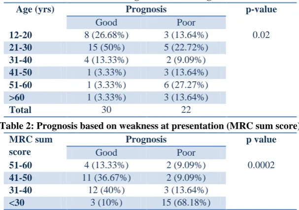
Thirty out of 52 patients had good prognosis at the end of 1 month (57.7%) while the rest had a poor prognosis (42.3%). Maximum numbers of patients with poor prognosis were of age group 51-60 years (27.27%).
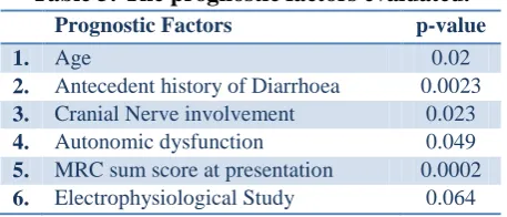
Statistical analysis testing reveals a p-value of 0.02 which is significant. Majority of the patients with poor prognosis (68.18%) had a low MRC sum score (<30) on presentation to the hospital. The p-value is 0.002 on statistical analysis which is significant. There was history of diarrhoea in 54.55% of the patients with poor prognosis. Statistical analysis test reveals a p-value of 0.0023 which is significant. There were autonomic dysfunctions in 68.18% of the patients with poor prognosis. Statistical analysis test shows a p value of 0.023 which is significant. Sixteen out of 22(72.72%) patients with poor prognosis had cranial nerve involvement while cranial nerve was not involved in 6 (27.28%) patients with poor prognosis. Statistical test shows p-value to be 0.49 which is significant. Amongst patients with poor prognosis electrophysiological studies showing axonal neuropathy was found in 40.91% of the patients and demyelinating neuropathy was detected in 59.09% of the patients with poor prognosis. Statistical analysis test shows a p value of 0.064 which is insignificant.
DISCUSSION
Gender DistributionIn our study, we found a male to female ratio of 1.36:1. An Indian study Dhadke SV et al. 201313, McGrogan A et al. 200914, González-Suárez et al. 20138 have also reported small predominance of male gender.
Age Distribution
In this study the youngest age was 13 years and the oldest age was 80 years with a mean age of 34.02±16.78 years. Maximum number of cases were in the third decade of life (38.46%) followed by the second decade (21.15%) and sixth decade (13.46%). Dhadke SV et al. 58%
42%
Prognosis at 1 month
Good Poor
13.3%
86.7%
54.6%
45.5%
Present Absent
ANTECEDENT H/O DIARRHOEA
Good Poor
43.3% 52.7%
72.7% 27.3%
Present Absent
CRANIAL NERVE
INVOLVEMENT
Good Poor
33.3% 67.7%
68.2%
31.8%
Present Absent
AUTONOMIC DYSFUNCTIONS
201313 found maximum number of patients in 4th decade of life (30%) followed by the 3rd decade of life (27.5%). An Indian study by Sharma G et al. 20133 reported a
mean age of 37.5 years. Jiang - Guoxin in their study in Sweden (1996)15 have observed two peaks one between 20 to 24 years and the other between 70-75 years. Table 1: Prognosis based on age.
Age (yrs) Prognosis p-value
Good Poor
12-20 8 (26.68%) 3 (13.64%) 0.02
21-30 15 (50%) 5 (22.72%)
31-40 4 (13.33%) 2 (9.09%)
41-50 1 (3.33%) 3 (13.64%)
51-60 1 (3.33%) 6 (27.27%)
>60 1 (3.33%) 3 (13.64%)
Total 30 22
Table 2: Prognosis based on weakness at presentation (MRC sum score)
MRC sum score
Prognosis p value
Good Poor
51-60 4 (13.33%) 2 (9.09%) 0.0002
41-50 11 (36.67%) 2 (9.09%)
31-40 12 (40%) 3 (13.64%)
<30 3 (10%) 15 (68.18%)
Seasonal Variation
In this study maximum number of patients were seen in the summer season from May to July (38.46%) followed by the spring season from February to April (30.76%). Sharma G et al (2013)3 in India, Zaheer M et al (2008)16 in Pakistan, Akbayram M et al (2011)17 in Turkey found maximum incidence of 41.53%, 64% and 40% respectively during summer season which correlates with our study.
Clinical Nadir
By the end of the second week of onset of disease, 68.3% of the patients achieved clinical nadir. All the patients (100%) had maximum weakness by 4 weeks. Maximum number of days from disease to nadir was 28 days and minimum was 1 day. According to Asbury AK et al, 19909,18 ~50% of patients reach clinical nadir by 2 weeks and more than 90% by 4 weeks. Sejvar JJ et al. 201119 described that the weakness in GBS progresses over a period of 12 hours to 28 days before a plateau is reached.
Antecedent Events
Our study revealed 63.46% to have respiratory tract infection or gastrointestinal tract infection before the development of GBS. Two thirds of cases (66.67%) of GBS are preceded by upper respiratory tract infection or diarrhoea according to Yuki N et al. 201220 which matches our study. Respiratory Tract Infection was present in 32.69% of our cases and gastrointestinal tract infection in 30.77%. González-Suárez et al. 20138 have reported upper respiratory infection in 37.7% of cases and gastrointestinal infection in 27.4%.
Latent Period between the antecedent event and onset of the disease
The mean latent period, 14.08 ± 8.21 days, in our study is consistent with the study carried out by González-Suárez et al. 20138. However, the study carried out by
Dhadke SV et al. 201321, majority i.e. 20 out of 22 (90.90%) patients developed neurologic illness within 4 weeks of antecedent event, while remaining 2 patients developed it between 1-3 months of preceding illness. Subtypes of GBS
In our study the most common subtype of GBS was AIDP which was found in 73.08% of the patients. AMAN was diagnosed in 8 (15.38%) patients while AMSAN was diagnosed in 6 (11.54%) of the cases. We did not find any case of Miller Fisher Syndrome or any other variant of GBS. Kalita et al (2014)22 in Lucknow have reported AIDP in 73.4%, AMAN in 13.4% and AMSAN in 4.6% which is similar to our study. Gupta et al (2008)23 in Thiruvananthapuram, Alexander et al (2001)24 in Vellore and Vengamma (2011)25 in Tirupati have reported the most common subtype of GBS as AIDP in 85.2%, 38.2% and 76.2% respectively followed by AMAN in 10.6%, 30.4% and 3.4% patients respectively.
Cranial Nerve Involvement
Cranial nerves were involved in 53.84% of the patients. According to Löffel et al. 197727 and Winer et al. 1988a28 cranial nerves are involved in 50% of the patients. González-Suárez et al. 20138 have shown cranial nerve involvement in 35.5% of the patients. Most common nerve involvement was the VII cranial nerve bilaterally in our study (38.46%) followed by the IX and X cranial nerves (26.92%).
Respiratory System Involvement
20-30% of patients which is consistent with our study. González-Suárez et al. 20138 have shown respiratory paralysis in 17% of cases. In Indian study by Dhadke SV et al. 201321 respiratory failure was observed in 32.5% patients.
Autonomic Dysfunctions
We found autonomic disturbances in 25 (48.07%) patients. Autonomic dysfunction is common in the Guillain–Barré syndrome, occurring in over 60 % cases according to Zochodne 199432. Flachenecker P et al 199733 and Burns TM et al 200134 have all reported autonomic disturbances in over 50% of patients. Chatterjee A et al 200935 found autonomic signs in 35.3% of patients in their study. However, González-Suárez et al 20138 found autonomic abnormalities in 8.5% in their study which is less than that in our study. Mortality
Four (7.69%) patients died in our study, all due to ventilator associated pneumonia. Winer JB et al. 198836 has reported mortality in the range of 3-13%. Hughes RAC et al. 200737, Rajabally YA et al. 20127 have found mortality in GBS to be 5% and 4% respectively. In northern India, Kalita J et al (2014)22 have reported death in 3.4 % of the patients studied.Lawn and Wijdicks (1999)38 did a comprehensive audit of in-hospital GBS-related deaths and found that deaths most commonly resulted from ventilator-associated pneumonia as found in our study.
Prognosis of the patients
On follow up at the end of 1 month in the study 30 out of 52 patients had good prognosis (57.7%) while the rest had a poor prognosis (42.3%). In the study done by Walgaard et al (2011)39 54% had poor outcome at 4 weeks, 29% at 3 months, and 15% at 6 months after hospital admission.
Prognosis based on Age at one month
In our study after 1 month 40.91% of the patients with poor prognosis were more than 50 years of age. Statistical analysis showed significant p-value of 0.02. The Plasma Exchange Sandoglobulin Trial40 has shown a significant correlation between ages more than 50 years and death or inability to walk at 48 weeks after the disease. Visser et al (1999)41 using data of 147 patients who had participated in the Dutch GBS trial found by multivariate logistic regression analysis that age >50 was a predictor of a poor outcome. Walgaard et al (2011)42 has advocated age more than 60 years as a predictor of poor outcome of GBS.
Prognosis based on weakness
In this study, 15 (68.18%) out of 22 patients with poor prognosis had a low MRC sum score (<30) on presentation to the hospital. Statistical analysis showed p-value of 0.0002 which is statistically significant. Visser et al (1999)41 have shown low MRC score (<40) to be a predictor of a poor outcome. Walgaard et al (2011)42 have reported low MRC score at admission to
the hospital and at day 7 to be a predictor of poor outcome. In India, Verma R et al (2013)43 have shown that MRC sum score <30 on hospital admission is a predictor of poor outcome at 6 months.
Prognosis based on history of diarrhoea
Statistical analysis test revealed a p-value of 0.0023 between the two groups of patients with good and poor prognosis. Arami MA et al (2006)44 and Walgaard et al (2011)42 have shown a significant correlation between history of diarrhoea and worse outcome at 6 months. Visser et al (1999)41 have shown in their study previous gastrointestinal infection being associated with a poor outcome. History of diarrhoea leading to a poor outcome has also be shown by Hadden RDM et al (2001)45 and van Koningsveld et al (2007)46.
Prognosis based on autonomic dysfunction
15 out of 22 patients (68.18%) with poor prognosis showed autonomic dysfunction as against 10 out of 30 patients (33.33%) with good prognosis. Statistical analysis test showed the p-value to be 0.023 which is significant. In India Kalita J et al (2014)22 and Verma R et al (2013)43 have reported autonomic dysfunction to be a marker of poor outcome. However, Singh NK et al (1987)47 have found no association between autonomic dysfunction and clinical outcome of the patient.
Prognosis based on cranial nerve involvement Out of 22 patients with poor prognosis 16 (72.72%) patients had cranial nerve involvement while cranial nerve was not involved in 6 (27.28%) patients. Statistical analysis showed significant p-value. Rajabally et al. 20127 have reported facial and/or bulbar weakness at admission was a strong predictor of mechanical ventilation and poor prognosis. Sundar U et al. 200548 reported that bulbar weakness is a predictor of poor outcome.
Table 3: The prognostic factors evaluated.
Prognostic Factors p-value
1. Age 0.02
2. Antecedent history of Diarrhoea 0.0023
3. Cranial Nerve involvement 0.023
4. Autonomic dysfunction 0.049
5. MRC sum score at presentation 0.0002
6. Electrophysiological Study 0.064
(1998)49 have reported that patients with axonal Guillain-Barré syndrome can show both rapid and slow recoveries.
CONCLUSION
This study concludes that there are several factors which play a key role in the prognosis of the disease. Most of the patients having poor prognosis were of advanced age, had severe disability at presentation, had a previous history of diarrhoea, had some autonomic dysfunctions or had cranial nerve involvement. It is, therefore, crucial that these factors are identified early with an aim to treat and manage them, and subsequently ensure better outcomes.
REFERENCES
1. Katirji B, Koontz D: Bradley’s Neurology in Clinical
Practice 6th Edition 2012; 76; 1955-1963
2. Ropper AH, Samuels MA, Klein JP: Adams and Victor’s Principles of Neurology 10th Edition 2014; 46; 1322-1330 3. Sharma G, Sood S, Sharma S. Seasonal, Age & Gender Variation of Guillain Barre Syndrome in a Tertiary Referral Center in India Neuroscience & Medicine, 2013, 4, 23-28. 4. Sinha S, Prasad KN, Jain D, Pandey CM, Jha S, Pradhan S. Preceding infections and gangliosides antibodies in patients with Guillain–Barré syndrome: A single center prospective case-control study. Clin Microbial Infect. 2007;13:334–7
5. Hauser S.L., Amato AA: Guillain-Barre Syndrome and other immune-related neuropathies. In Kasper DL, Fauci AS, Hauser SL, Longo D, Jameson JL, Loscalzo J et al.
Harrison’s Principles of Internal Medicine. 19th edition,
vol-II. New York: Mc Graw-Hill, 2015; 2694-2698
6. Kaur U, Chopra JS, Prabhakar S, Radhakrishnan K, Rana S. Guillain-Barré syndrome - a clinical electrophysiological and biochemical study. Acta Neurol Scand. 1986;73:394– 402
7. Rajabally YA, Unicini A: Outcome and its predictors in Guillain-barré syndrome. J Neurol Neurosurg Psychiatry 2012, 83:711–718.
8. González-Suárez et al.: Guillain-Barré Syndrome: Natural history and prognostic factors: a retrospective review of 106 cases. BMC Neurology 2013 13:95.
9. Asbury, A. K. and Cornblath, D. R. (1990). Assessment of current diagnostic criteria for Guillain–Barré syndrome. Ann. Neurol., 27, 521–4.
10. Kleyweg RP, van der Meché FGA, Schmitz PIM. Interobserver agreement in the assessment of muscle strength and functional abilities in Guillain–Barré syndrome. Muscle Nerve 1991;14:1103–9.
11. Hughes RA, Newsom-Davis JM, Perkin GD, Pierce JM. Controlled trial prednisolone in acute polyneuropathy. Lancet 1978;2:750–753.
12. Walgaard C, Lingsma HF, Ruts L, et al: Early recognition of poor prognosis in Guillain-Barré syndrome. Neurology 2011, 76:968–975
13. Dhadke SV, Dhadke VN et al. J Assoc Physicians India. 2013 Mar;61(3):168-72.
14. McGrogan A, Madle GC, Seaman HE, et al. The epidemiology of Guillain-Barre ´ syndrome worldwide. Neuroepidemiology 2009;32:150e63.
15. Jiang GX, de Pedro-Cuesta J, Strigard K, Olsson T, Link H. Pregnancy and Guillain-Barré syndrome: a nationwide register cohort study. Neuroepidemiology 1996;15:192-200.
16. Zaheer M, Naeem M and Nasrullah M, “Seasonal Vari-
ation and Sex Distribution in Patients with Guillain-Barre
Syndrome,” Pakistan Journal of Neurological Sciences,
Vol. 3, No. 1, 2008, pp. 6-8.
17. Akbayram S, Dogan M, Akgün C, Peker E, Sayin R
and Aktar F, “Clinical Features and Prognosis with Guil
-lain-Barré Syndrome,” Annals of Indian Academy of Neu- rology, Vol. 14, No. 2, 2011, pp. 98-102.
18. Asbury, A. K. and Cornblath, D. R. (1990). Assessment of current diagnostic criteria for Guillain–Barré syndrome. Ann. Neurol., 27, 521–4.
19. Sejvar JJ, Baughman AL, Wise M, Morgan OW. Population incidence of Guillain-Barré syndrome: a systematic review and meta-analysis. Neuroepidemiology 2011;36:123-33.
20. Yuki N, Hartung HP. Guillain-Barré syndrome. N Engl J Med. 2012;366:2294-304.
21. Dhadke SV, Dhadke VN et al. J Assoc Physicians India. 2013 Mar;61(3):168-72.
22. Kalita J, Misra UK, Goyal G, Das M. Guillain-Barré syndrome: subtypes and predictors of outcome from India. J Peripher Nerv Syst. 2014 Mar;19(1):36-43.
23. Gupta D, Nair M, Baheti NN, et al. Electrodiagnostic and clinical aspects of Guillain Barré syndrome: an analysis of 142 cases. J Clin Neuromuscular Dis. 2008;10:42-51. 24. Alexander M, Prabhakar AT, Aaron S, et al. Utility of neurophysiological criteria in Guillain Barré syndrome: subtype spectrum from a tertiary referral hospital in India. Neurol India. 2011;59:722-6.
25. Vengamma B. Guillain-Barré syndrome: an overview. 7th CME on Recent Advances organized by Association of Physicians of India, Mumbai branch INHS Asvini Hospital Auditorium, Mumbai, 22-01-2011. Available at: http://www.apiindia.org/medicine_update_2013/ (Accessed on 29th August, 2015).
26. Zhang G, Li Q, Zhang R, Wei X, Wang J, Qin X (2015) Subtypes and Prognosis of Guillain-Barré Syndrome in Southwest China. PLoS ONE 10(7): e0133520
27. Löffel, N. B., Rossi, L. N., Mumenthaler, M. et al. (1977). The Landry–Guillain–Barré syndrome: complications, prognosis, and natural history in 123 cases. J. Neurol. Sci., 33, 71-9.
28. Winer, J. B., Hughes, R. A. C., and Osmond, C. (1988a). A prospective study of acute clinical idiopathic neuropathy. I Clinical features and their prognostic value. J. Neurol. Neurosurg. Psychiat., 51, 605–12.
29. Chio A, Cocito D, Leone M, et al. Guillain-Barre syndrome: a prospective, population-based incidence and outcome survey. Neurology 2003;60:1146–50.
31. Hughes RA, Wijdicks EF, Benson E, et al. Supportive care for patients with Guillain-Barre´ syndrome. Arch Neurol 2005;62:1194–1198
32. Zochodne, D. W. (1994). Autonomic involvement in Guillain–Barré syndrome. A review. Muscle Nerve, 17,1145–55.
33. Flachenecker P, Hartung HP, Reiners K. Power spectrum analysis of heart rate variability in Guillain-Barre´ syndrome. A longitudinal study. Brain 1997;120(Pt 10):1885–1894
34. Burns TM, Lawn ND, Low PA, Camilleri M, Wijdicks EF. Adynamic ileus in severe Guillain-Barre´ syndrome. Muscle Nerve 2001;24:963–965
35. Chatterjee A et al. Outcome Predictors in GB Syndrome IJPMR 2009; 20 (2):37-43
36. Winer, J. B. and Hughes, R. A. C. (1988). Identification of patients at risk of arrhythmia in Guillain–Barré syndrome, 68(3-4), 735-739. Accessed through: http://qjmed.oxfordjournals.org/content/68/3-4/735
37. Hughes RAC, Swan AV, Raphaël JC, Annane D, van Koningsveld R, van Doorn PA. Immunotherapy for Guillain-Barré syndrome: a systematic review. Brain 2007; 130:2245-57.
38. Lawn ND, Wijdicks EF. Fatal Guillain Barré syndrome. Neurology 1999;52:635-8.
39. Walgaard C, Lingsma HF, Ruts L, et al: Early recognition of poor prognosis in Guillain-Barré syndrome. Neurology 2011, 76:968–975.
40. Plasma Exchange / Sandoglobulin Guillain -Barre´ Syndrome Trial Group. Randomised trial of plasma exchange, intravenous immunoglobulin, and combined treatments in Guillain-Barre´ syndrome. Lancet 1997;349:225–230
41. Visser LH, Schmitz PI, Meulstee J, et al. Prognostic factors of Guillain-Barre´ syndrome after intravenous immunoglobulin or plasma exchange. Dutch Guillain-Barre´ study group. Neurology 1999;53:598e604.
42. Walgaard C, Lingsma HF, Ruts L, et al: Early recognition of poor prognosis in Guillain-Barré syndrome. Neurology 2011, 76:968–975.
43. Verma R, Chaudhari TS, Raut TP, Garg RK. Clinico-electrophysiological profile and predictors of functional outcome in Guillain–Barre syndrome (GBS). Journal of the Neurological Sciences December 15, 2013 Volume 335, Issues 1-2, Pages 105–111.
44. Arami MA, Yazdchi M, Khandaghi R. Epidemiology and characteristics of Guillain- Barre´ syndrome in the northwest of Iran. Ann Saudi Med 2006;26:22e7.
45. Hadden RDM, Karch H, Hartung HP, et al; The Plasma Exchange/Sandoglobulin Guillain-Barre´ syndrome Trial Group. Preceding infections, immune factors and outcome in Guillain-Barre´ syndrome. Neurology 2001;56:758e65. 46. Van Koningsveld R, Steyerberg EW, Hughes RA, Swan AV, van Doorn PA, Jacobs BC. A clinical prognostic scoring system for Guillain-Barre´ syndrome. Lancet Neurol 2007;6:589–594
47. Singh NK, Jaiswal AK, Misra S, Srivastava PK. Assessment of autonomic dysfunction in Guillain-Barre´
syndrome and its prognostic implications. Acta Neurol Scand 1987;75:101–105
48. Sundar U, Abraham E, Gharat A, Yeolekar ME, Trivedi T, Dwivedi N. Neuromuscular respiratory failure in Guillain‑Barre Syndrome: Evaluation of clinical and electrodiagnostic predictors. J Assoc Physicians India 2005;53:764‑8.
49. Kuwabara S, Asahina M, Koga M, et al. Two patterns of clinical recovery in Guillain-Barre syndrome with IgG anti-GM1 antibody. Neurology 1998;51:1656–60.
Source of Support: Nil.
Conflict of Interest: None Declared.
Copyright: © the author(s) and publisher. IJMRP is an official publication of Ibn Sina Academy of Medieval Medicine & Sciences, registered in 2001 under Indian Trusts Act, 1882.
This is an open access article distributed under the terms of the Creative Commons Attribution Non-commercial License, which permits unrestricted non-commercial use, distribution, and reproduction in any medium, provided the original work is properly cited.
Cite this article as: Zain Fakih, Duncan Khanikar, Chitralekha Baruah, Marami Das. A Clinical Study of Prognostic Factors of Guillain Barré Syndrome in a Tertiary Care Hospital of North-East India. Int J Med Res Prof. 2016, 2(2); 170-76.
ANNEXURES
1. Guillain-Barré Syndrome Disability Score advocated by Hughes RA et al, 1978
0 Healthy state
1 Minor symptoms and capable of running
2 Able to walk 10 m or more without assistance but
unable to run
3 Able to walk 10 m across an open space with help
4 Bedridden or chair bound
5 Requiring assisted ventilation for at least part of
the day
6 Dead
2. MRC sum score: Power of the muscles was assessed by MRC sum score, a summation of the MRC grades (range, 0–5) given in full numbers of the following muscle pairs: upper arm abductors, elbow flexors, wrist extensors, hip flexors, knee extensors, and foot dorsal flexors as given by Kleyweg RP et al, 1991. The MRC sum score ranged from 0 (“total paralysis”) to 60
(“normal strength”).
MRC Scale
Grade Description
0 No contraction
1 Flicker or trace of contraction
2 Active movement with gravity eliminated
3 Active movement against gravity
4 Active movement against gravity and resistance