RESEARCH ARTICLE
COMPARISON OF PHYSICAL VERSUS PHYSICAL AND MENTAL PRACTICE IN IMPROVING
HAND FUNCTION IN SUB-ACUTE STOKE
Hiral Soni, *Snehal Joshi and Ragini Zatale
D.E. Society’s Brijlal Jindal College of P.T, F.C. Campus, F.C Road, Pune, India
ARTICLE INFO ABSTRACT
After stroke, hand impairment is very common and its recovery is often incomplete. Early neuroplastic changes may form the basis for restitution of motor function after stroke. Neuroplasticity starts from day one immediately after injury or insult to cortex. Mental practice being one of the principles of motor learning can accelerate neuroplastic changes. Thus to compare the efficacy of mental practice and physical practice over only physical practice on hand function in individuals with stroke, this study was conducted.
Methodology: 50 stroke patients divided into two groups i.e. control and experimental. Subjects were screened for inclusion and exclusion criteria. Control group received conventional physiotherapy and experimental group received mental practice and conventional physiotherapy. Outcome measures: nine-hole peg test, voluntary control and motor assessment scale. Outcome measures were evaluated prior to the interventions and after 20 sessions of treatment.
Results of the study: Significant difference was observed in both the groups pe and post intervention. The experimental group showed better improvement than conventional group.
Conclusion: combination of mental practice and physical practice is more effective than physical practice for improvement of hand function.
INTRODUCTION
Stroke is sudden loss of neurological function caused by
interruption of the blood flow to the brain (Braun et al., 2007).
it is a functional activity limiting disorder. The brain damage caused by a stroke may result in the loss of cerebral function Stroke is a major health problem, which is likely to increase
due to aging(Braun et al., 2007). Patients are often confronted
with disabilities on a physical, cognitive, social and communicative level. Clinically a variety of focal deficits are possible, including changes like level of consciousness, impairments in sensory and motor aspects in upper and lower
limb, disorders of perception and cognition (Braun et al.,
2007). Motor deficits could be either paralysis (plegia) or weakness (paresis). Weakness (paresis) is found in 80-90% of subjects who suffer stroke which contributes as one of the important aspect in disability. Subjects are unable to initiate or control the movement because of insufficient force production during the activity. The severity of weakness depends on the location and extent of brain damage. Muscle weakness is also associated with alteration in number of functioning motor units
(Brewer et al., 2012).Motor impairment after stroke is a major
cause of permanent disability. Recovery of the hand is crucial in order to perform activities of daily living but is often
variable and incomplete (Brewer et al., 2012).
*Corresponding author: Snehal Joshi,
D.E. Society’s Brijlal Jindal College of P.T, F.C. Campus, F.C Road, Pune, India.
Neuroscience-based rehabilitation is gaining strength as a way to improve outcome, even in situations where the deficit
appears to be permanent (Brewer et al., 2012).MotorInjuries
to the central nervous system, such as those that arise from ischemic strokes and other traumas, activate neuroplasticity which plays an important role in the recovery of motor and sensory functions. Activation of beneficial neuroplasticity
makes it possible to learn new skills (Møller, 2014). The
nervous system accomplishes this diversity of functions with one key feature: it can change and adapt. In this way, characteristics can be tuned to the task at hand and new properties can be acquired. This ability of the nervous system to change is perplexing as the adult nervous system generates
relatively few new cells (Teong Han, 2009). The nervous
system is, for the most part, plastic, which means that many different functions can be changed. The process that mediates these changes in the nervous system is hence called neuroplasticity. Tasks typically executed by parts of the brain that have become non-functional may still be carried out by other brain regions, however, typically with a lower degree of
proficiency (Møller, 2014).The brain can use neuroplasticity
to adjust itself functionally, by reorganizing the cortical maps, which contributes to the stroke recovery. The changes in the cortex organization include an increase in the number and density of dendrites, synapses and neurotrophic factors synthesis which results in two ways- unmasking of existing neuronal circuits and establishing of new neuronal circuits. After damage of the motor cortex, changes of activation in
Article History:
Received 22nd March, 2018
Received in revised form
20th April, 2018
Accepted 10th May, 2018
Published online 30th June, 2018
International Journal of Recent Advances in Multidisciplinary Research
Vol. 05, Issue 06, pp. 3929-3935, June, 2018
Keywords:
Stroke, Hand function, Mental practice, Nine-hole peg test, MAS, Voluntary control
Glossary:
other motor areas are observed. These changes occur in homologue areas of the non-affected hemisphere which can substitute for the lost functions or in the intact cortex adjacent to the damage. Cortical reorganizations, which begin from one to two days after the stroke, and can be extended for months, the patients can recover, at least in part, the lost abilities (Hutchinson, 2010). Activation of neuroplasticity may partially restore lost function after injury by re-routing information to parts of the brain that normally do not receive such information. Understanding the effect of rehabilitative techniques on brain plasticity is potentially important in providing a neural substrate to underpin rehabilitation and
hence in developing novel rehabilitation strategies (Sharma et
al., 2006).Due to high incidence of middle cerebral artery
strokes (Braun et al., 2007) there is more involvement of upper
extremity than lower extremity. Usually the distal musculature exhibits more reduction in strength compared to proximal musculature. The distal limb impairment is especially disabling, because proper hand function is crucial for manual exploration and manipulation of the environment. Indeed, loss of hand function is a major source of impairment in various disorders, frequently preventing effective occupational performance and independent participation in daily life. Functional recovery of the paretic upper extremity, post-stroke, continues to be one of the greatest challenges faced by rehabilitation professionals. Although most clients regain walking ability, only 5% of adults regain full arm function after stroke, and 20% do not regain any functional use. Hence, alternative strategies are needed to reduce long-term disability and functional impairment caused by stroke (Shumway-Cook and Woollacott, 1998). The upper extremity function plays an important role in gross motor skills such as crawling, walking, ability to recover balance, ability to protect the body from injury when balance recovery is not possible. According to variety of thinkers there exists bidirectional relationship between mind and hand. It is known that 80% of stroke population have problems with hand functions. Due to this interweaving of upper extremity control with both fine and gross motor skills, recovery of upper extremity function is an important aspect of retraining motor control.
In upper extremity 3 factors contribute to sensorimotor processing (Carr and Shepherd, 1998)-
1. Constraints of individual
2. Type of task
3. Environmental constraints
According to systems theory of motor control, specific neural
and musculoskeletal subsystems contribute to the reach grasp
manipulation(Carr and Shepherd, 1998):
1. Musculoskeletal factors - joint ROM, spinal flexibility,
muscle properties
2. Neural factors –motor process, sensory process,
mapping of sensation to action, higher level process along with all the above movements are reflexive that is feedback & feed forward mechanism
Factors responsible for upper extremity reach, grasp, manupilation are
1. Locating a target – eye-head coordination
2. Reaching the target- transportation of arm & hand in
space
3. Grip formation
4. Hand manipulation
In stroke subjects all these or few of these might be affected hence grip formation & in hand manipulation is maximally affected. Therefore, it is necessary to evaluate hand function in form of grip formation and in hand manipulation. Understanding the effect of upper extremity limitations on elements of participation can be based on objective /subjective information obtained during the rehabilitation. Various outcome measures available are the Canadian occupational performance measure, stroke impact scale motor activity log, wolf motor function test, action research arm test, Chedoke inventory of hand and arm inventory, 9-hole peg test, motor assessment scale. Amongst all of above 9-hole peg test & motor assessment scale are most reliable and valid for
evaluating hand function in stroke patients (Duncan et al.,
2005; Teasell et al., 2008). Motor assessment scale is a
standardized assessment devised by Carr, Shepherd, to assess motor function in stroke survivors (An and Park, 2015). The motor assessment score is a well-studied assessment with properties that make it useful for rehabilitation therapists, and is specifically recommended as a measure of post-stroke motor function in the American heart association’s clinical practice guideline for stroke rehabilitation (Langhammer and Stanghelle, 2010) and the Canadian health system’s
evidence-based review of stroke rehabilitation (Edwards et al., 2006).
Objective reviews of stroke assessments typically commend the motor assessment score for reliability and ease of administration. The motor assessment score continues to be used extensively as an outcome measure in studies of
rehabilitation interventions for stroke survivors (Sirigu et al.,
2001). The motor assessment score provides a standardized scoring system for assessing eight categories of motor
behaviour. As stroke is a very limiting disease for the patient
and a major health problem in most parts of the world, it is important to continue to search for new therapy techniques to improve recovery. Recently, imagery and mental practice have become additional therapy interventions. Studies in the first half of the last century suggested that mental execution of tasks resulted in improved performance in simple motor tests. (Art 6).
Imagery refers to the “creation of any experience in the mind auditory, visual, tactile, olfactory, gustatory, kinaesthetic, and organic. Imagery bridges diverse domains of knowledge from
psychology (Sirigu et al., 2001) specifically; motor imagery
techniques (Park and Choi, 2013). Functional MRI and pet scan studies have shown that changes suggestive of plasticity seen mainly in the cortex but may also occur in the thalamus and brain stem. Constraint induced therapy, mental imagery, neuro muscular electrical stimulation (nmes), FES have been used widely and have shown to elicit cortical activation ((Park and Choi, 2013). Mental practice as an additional cognitive therapy is getting increased attention in stroke rehabilitation. “Motor imagery is the imagining of an action without its physical execution; it is an active process during which the representation of an action is internally reproduced within
working memory without any overt output” (Malouin et al.,
2004). It is a dynamic state during which the representation of a specific motor action is internally activated without any motor output.
In other words, motor imagery requires the conscious activation of brain regions that are also involved in movement preparation and execution, accompanied by a voluntary inhibition of the actual movement (Mulder, 2007). For decades, authors have reported that mental practice (also known as “imagery”), when combined with physical practice, accelerates motor learning and improves subsequent physical performance. Because of its positive effects on strength, endurance, and aim and precision. Repeated mental practice of the movement results in learning and the same plastic changes in the motor system as those occurring with repetitive physical practice. Repetition also enables in making the movement smoother and co-ordinate thereby reflecting the changes occurring at the neural level. Mentamove, the device, is a highly sensitive electromyography initiated muscle stimulator that can pick up a change in the electromyography potential of a minimum of 2 micro volts, an equivalent of firing of one motor unit. Mentamove uses mental practice of motor skills, to bring about this change, triggering the medium frequency current, causing a contraction of the muscle and performing a
movement (Teresa, 2003). Electromyography initiated
muscular stimulation is a therapeutic method mainly used in treatment of stroke. It is a process in which mental imagery is used in motor learning (Teresa, 2003).
Mentamove works on Biofeedback process.the working principle is based on the mental practice of motor skill, also called ideomotor training mental practice of motor skills is an auto suggestive method based on a psychological intervention mental imagery is a well-known method to enhance motor performance, this therapeutic method uses visuo-motor behaviour rehearsal. It is an extension of mental imagery, in that; it combines the psychological aspect of generating the mental image with feedback from the performance of the
physical skill (Page et al., 2014).Forrecovery of hand function
an effective intervention is essential to enhance faster recovery, which will facilitate functional improvement in subjects with stroke. Hence in this study an attempt was made to compare the effect of physical training, and a combination of both physical and mental training in improving hand function. Presentation of hand in the homunculus occupies larger area in cerebral cortex. So its recovery requires extensively increased blood supply. In stroke hand recovery is usually delayed due to lack of blood supply to the ischemic
penumbra and presence of insult in that area (Braun et al.,
2013). In rehabilitation usually proximal joints are given more attention and its recovery is faster due to small representation on homunculus and hand is neglected. Hand is usually essential for gross and fine motor activities required for
activity of daily living. Aim of this study was to compare the
effect of physical versus physical and mental practice in improving hand function.
MATERIALS AND METHODS
Type of study: Experimental design
Study population: Individuals with stroke
Inclusion criteria
1. Sub-acute stroke (>3months, <2 year)
2. Voluntary control: 2-4
3. Active ROM of wrist extension 10’, MCP flexion
5-10’, IP flexion 5-10’
Exclusion criteria
1. Affection in cognition and other perceptual disorders
2. Recurrent stroke
3. Visual problems
4. Any neurological disorder other than stroke
Independent variables
1. Physical practice-physical activities
2. Physical practice & mental practice-mentamove and
physical activities
Materials
Outcome measures: motor assessment
scale(MAS),9-hole peg test and brunnstorm voluntary control of hand
Patient record sheet
Peg board
Beans
Stopwatch
8 Jellybeans
Polystyrene cup
Rubber ball
Stool
Comb
Spoon
Pen
2 Tea cups
Water
Prepared sheet for drawing lines
Cylindrical shaped object like a jar
Table
pillow
Mentamove machine with electrodes
Chair with back support
Study settings: Physiotherapy OPD, Patient’s home.
Sampling technique: Simple random sampling
Sample size: 50 subjects, divided in two groups, 25 in
experimental group and 25 in control group.
Sample size was estimated by Formula,
n = (t2 * S. D2) / E2 where t = constant i.e. 1.96 S.
The clearance from the ethics committee was taken. The subjects were screened according to the inclusion, exclusion criteria. An informed written consent was taken from the subject. The entire procedure was explained to the subject. The subjects were randomly allocated to any one group amongst the two group. Before the initiation of intervention outcome measures i.e motor assessment scale, 9-hole peg test and voluntary control were assessed. The subjects were randomly allocated. The subject allocated in the first group conventional group that is, group A performed only physical practice and in Group B performed physical and mental practice. The position given to the participants was sitting on a chair with back support, elbow and shoulder at right angle to each other, a pillow was given to rest the forearm, the subjects were asked to sit as tall as possible. The subject allocated in the first group conventional group that is, group A performed only physical practice, in the form of lifting up of glass, reaching table top, peg board activity, active movements of wrist and hand. The subjects performed 10 -15 repetitions, 5 days per week for 4 weeks. The subjects allocated in the second group experimental group that is, group B performed physical practice and mental practice the subjects performed physical activity –lifting up of glass, reaching table top, peg board activity active movements of wrist and hand. The subjects performed 10 -15 repitations, 5 days per week for 4 weeks (Tareq, 2012). Mental practice in the form of mentamove was given to participant for 20 minutes to wrist extensors and gripping muscles of hand, with 9 contractions for each muscle per session (Ghodge and Joshi, 2014). It was given for 5 days per week for 4 weeks.
For extensors the placement of the electrode was as follows:
Black cable was connected to the electrode on the extensors common origin i.e. near lateral epicondyle. white cable was connected to the electrode on extensor aspect i.e. dorsum of hand. Green cable was connected to the electrode which was
placed in between the 2 electrodes i.e. Mid forearm. For
gripping muscles of hand, the placement of the electrode was as follows- black cable was connected to the electrode on thenar eminence. White cable was connected to the electrode on hypothenar eminence. Green cable was connected to the electrode was placed in between the 2 electrodes. After the placement of the electrode, machine was started. Intensity of the current was set in such a way that there was visible muscle
action. An offset value was set.Regular verbal cues were given
to the subject in the form of “please relax”, “imagine that you are taking the wrist upwards”, “saying goodbye”, and “imagine that you are squeezing the ball”. While the verbal cues were given when the participant reached the mental threshold (offset value), participant perceived current along with a movement of the muscle.9 contractions were given in each session. After 4weeks of intervention outcome measures were evaluated again -9 Hole peg test, motor assessment scale, voluntary control. Data was then analysed using appropriate statistical test. SPSS version 19 was used for analysis.
Statistical analysis
Graph 1. indicates gender distribution in both the groups. Graph 2 indicates that the subjects had mean age of 45.5 years
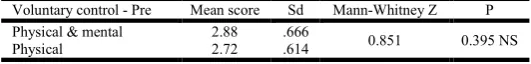
in conventional group and 41.7 years in experimental group 16. Table 1 indicates that, there was no significant difference
in voluntary control scores of both the groups. (p>0.05). Graph 3 indicates comparison of voluntary control scores
before and after intervention indicates comparison of voluntary control before and after intervention. There was statically significant difference in the voluntary control score before and after intervention between the groups. (p<0.05, significant and p<0.001, highly significant). Table 2 indicates comparison of voluntary control of both the groups after the intervention. There was statistically significant difference in the score between the groups. (p value < 0.001 highly significant). Table 3 indicates that, there was no significant difference in time duration of 9-hole peg test of both the groups. (p>0.05) 18. Graph 4 indicates comparison of 9-hole peg test of both the groups indicates comparison of time duration of 9 hole peg test before and after intervention. It showed that there was statically significant difference in both the groups before after intervention (p < 0.001 highly significant). Table 4 indicates comparison of time duration of 9-hole peg test of both the groups after the intervention, there was statistically significant difference in the time duration after treatment between the groups (p value < 0.001 highly significant). Table 5 indicates that, there was no significant difference in motor assessment scores of both the groups (p>0.05). Graph 5 indicates comparison of motor assessment score of both the groups indicates comparison of motor assessment score before and after intervention. There was statistically significant difference in the motor assessment score before and after intervention between both the groups. (p< 0.001, highly significant). Table 6 indicates comparison of motor assessment score of both the groups after the intervention, there was no statistically significant in improvement in the scores after treatment in both groups (p value > 0.05 not significant)
Table 1. Comparison of pre voluntary control in both the groups
Voluntary control - Pre Mean score Sd Mann-Whitney Z P
Physical & mental 2.88 .666
0.851 0.395 NS
Physical 2.72 .614
Table 2. Comparison of post voluntary control in both the groups
Voluntary control - Post Mean score Sd Mann-Whitney Z P
Physical & mental 3.80 .645 4.098 <0.001 HS
Physical 2.88 .666
Table 3. Comparison of pre 9 hole peg test in both the groups
9HPT - Pre Mean in secs SD Unpaired t P
Physical & mental 478.24 7.710 0.465 0.644 NS
Physical 477.20 8.088
Table 4. Comparison of post 9hole peg test in both the groups
9HPT- Post Mean in secs Sd Unpaired t P
Physical & mental 395.28 10.557 12.463 <0.001 HS
Physical 432.48 10.548
Table 5. Comparison of pre motor assessment score in both the groups
MAS - Pre Mean score Sd Mann-Whitney Z P
Physical & mental 18.16 1.519 1.00 0.317 NS
Physical 17.76 1.451
Table 6. Comparison of post motor assessment score in both the groups
MAS - Post Mean score Sd Mann-Whitney Z P
Physical & mental 24.72 2.072 1.797 0.072 NS
Graph 1. Gender distribution
Graph 2. Age distribution
Graph 3. Comparison of pre and post voluntary control in both the groups
Graph 4. Comparison of pre and post 9hole peg test in both the groups
Graph 5. Comparison of pre and post motor assessment score in both the groups
DISCUSSION
The main focus of the study was to compare the effect of physical and combination of physical & mental practice in improving hand function in stroke patients. The hand function was evaluated using voluntary control of hand, 9-hole peg test, and motor control. Improvement in voluntary control was observed in both the groups but was more in experimental group. The similar results were observed in 9-hole peg test Voluntary control is defined as pattern utilizing functionally linked muscles that are constrained by the CNS to act
cooperatively to produce an action (Tareq, 2012). During
intervention the exercise in the form of picking up beads, peg board activity, active exercise helped in opposition of the thumb and movements of fingers. Improvement of voluntary control would be due to motor learning. Motor learning is defined as “acquisition/modification of movement.” (Shumway -Cook and Woollacott, 2001; Carr and Shepherd, 1998) Along with motor processes involves learning of various different strategies for sensing as well as moving. This motor learning makes behavioural changes by acquiring the skilled activity. Learning maximally takes place due to practice of the skilful action (O'Sullivan and Schmitz, 2007). Rehearsing the task used for evaluation helps in better results in the form of outcome. Repetition of the same task improves the strength of the muscles but also helps in learning of the task. This reacquisition of movement skills lost through injury is coined as “motor recovery.” Hence the ability to perform the task is
due to motor recovery (Tunney et al., 2006). Along with this
there are some neuronal changes taking place at the cellular level. Pyramidal neurons (PMN) in layers II/III and V have enlarged dendritic fields.
This enlargement of dendritic surface is accompanied by an increase in the number of synapses per neuron in layer V PMNs suggesting that learning promotes synaptogenesis (Hosp
and Luft, 2011).With learning, adult neurogenesis takes place
resulting in activation of hippocampus. Learning accelerates the maturation of the dendritic trees of new-born neurons and promotes their integration into functional hippocampus neural
networks (Wu et al, 2006). Specific training to induce motor
Another mechanism that could improve the values in the form
of neuroplasticity is reorganization (Shumway-Cook and
Woollacott, 2001). This reorganization will be in the form of perilesional extensions of representations via enlargement of hand area in the brain by recruitment of the dormant area of the brain, shifting from primary to secondary processing systems, and recruitment of homologous areas of the unaffected hemisphere. At greater distances from the infarct, in the area surrounding the scar, inhibitory perineuronal networks degrade due to inflammatory processes and free radical formation, thereby facilitating axonal sprouting. In this area, growth-promoting genes are unregulated, and inhibitory genes are down regulated It is known that sensory experience in the absence of movement results in a selective expansion of the specific regions of somatosensory cortex that are associated with the sensory exposure, leading to change in sensory receptive field size that reflect the characteristics of the
adaptation (Von Lewinski et al., 2009).
Mentamove is an EMG based biofeedback modality. In mentamove there mental practice involves cognitive rehearsal of a task without overt physical movement; it has a display showing the amplitude of potentials attained through mental activity, and it is used as a visual feedback. In addition, electrical stimulation linked with mental activity acted as a somatosensory cue to support the execution of the training. At the same time the therapist can view the mental threshold on the instrument of Mentamove can sense weather the subject is truly performing the movement. Patient is the active participant in the treatment. Mentamove also helped in facilitating the thumb and fingers movements. Thereby improving overall, the voluntary control of hand. Rehearsing by mentally imagining the physical activity in the mind may assist in focussing attention on the action to be performed. Mental imagery is performed for a person who is unable to practice a task physically as it improves the ability to sustain attention and plan task performance (Casey, 2003). Appropriate motor connections may be activated during mental practice helping to establish and reinforce an appropriate coordination pattern (Rajesh, 2015). Mental practice enhances dependent brain reorganization, in which new cortical areas are
recruited to assist in movement of the affected hand (Wu et al.,
2005).The combination of physical practice along with mental
practice helps in recruitment of supplementary motor area, occurring due to continuous bombardment to the cortex via mental stimulus, in form of Mentamove and physical stimulus in the form of exercise. Leading to expansion of the area in the
brain (Meilink et al., 2008).
The shrinking of dendritic trees distant to the lesion may be a consequence of a lesion-related reduction in afferent signals. This phenomenon resembles the model of diaschisis (Casey, 2003), describing (dysfunctional) effects of focal brain injuries on remote areas, for example, caused by neuronal
deafferentation or redistribution of blood perfusion(Wu et al.,
2005). Though motor assessment score showed improvements before and after intervention, there was no difference in the mean of both the groups. Since motor assessment score was not constrained to only hand activities evaluation. It included evaluation of transitions from different positions, sitting balance, trunk and lower limb, upper arm movements, walking etc. Treatment in the form of mental practice was restricted only to hand, therefore giving treatment to the hand will not have any generalise effects on the body as a whole. Since the treatment was given to hand, there wasn’t much of
improvement in the overall score between the groups. This questions its effect on mental practice and physical practice. This might support that mental practice doesn’t have any generalize effect. It emphasises on the localizing effect of the hand. Hence null hypothesis was rejected and alternate hypothesis was accepted.
Conclusion
The results of this study show that physical practice and the combination of physical and mental practice facilitates improvement of hand functions in stroke patients. Combination of mental practice and physical practice is proved to be more effective in improving hand function in stroke patients. Along with intervention in the form of physical exercise, mental training can be used as an adjunct to facilitate improvement in hand function.
REFERENCES
An S, Park C. 2015. The Reliability and Validity of Short Form of the Fugl-Meyer Assessment Scale in Patients with Stroke. Jsers, 54(1):159.
Braun S, Beurskens A, van Kroonenburgh S, Demarteau J, Schols J, Wade D. 2007. Effects of mental practice embedded in daily therapy compared to therapy as usual in adult stroke patients in Dutch nursing homes: design of a
randomized controlled trial. BMC Neurology, 7(1):34.
Braun S, Kleynen M, van Heel T, Kruithof N, Wade D, Beurskens A. 2013. The effects of mental practice in neurological rehabilitation; a systematic review and
meta-analysis. Frontiers in Human Neuroscience, 7.
Brewer L, Horgan F, Hickey A, Williams D. 2012. Stroke
rehabilitation: recent advances and future therapies. QJM.,
106(1):11-25.
Carr J, Shepherd R. 1998. Neurological rehabilitation. Oxford: Butterworth-Heinemann.
Casey B. 2003. Brain plasticity, learning, and developmental
disabilities. Mental Retardation and Developmental
Disabilities Research Reviews, 9(3):133-134.
Dickstein R, Deutsch J. 2007. Motor Imagery in Physical
Therapist Practice. Physical Therapy, 87(7):942-953.
Duncan P, Zorowitz R, Bates B, Choi J, Glasberg J, Graham G et al. 2005. Management of Adult Stroke Rehabilitation Care: A Clinical Practice Guideline. Stroke, 36(9):e100-e143.
Edwards DF, Hahn MG, Baum CM, Perlmutter MS, Sheedy C, Dromerick AW. 2006. Screening patients with stroke for rehabilitation needs: Validation of the post-stroke
rehabilitation guidelines. Neurorehabilitation and Neural
Repair, 20(1): 42-48
Ghodge S, Joshi S. 2014. Effect of Mental Practice on Non-Dominant Side Hand Function. Ind Jour of Physioth and
Occupat Therapy - An Inter Jour., 8(2):9
Hosp J, Luft A. 2011. Cortical Plasticity during Motor
Learning and Recovery after Ischemic Stroke. Neural
Plasticity., 2011:1-9.
Hutchinson E. 2010. Neuroplasticity: Functional recovery after
stroke. Nature Reviews Neuroscience, 12(1):4-4.
Langhammer B, Stanghelle J. 2010. Can Physiotherapy after Stroke Based on the Bobath Concept Result in Improved Quality of Movement Compared to the Motor Relearning
Programme. Physiotherapy Research International, 16(2):
69-80.
System in Recovery from Strokes and Other Forms of
Brain Trauma. JNSK, 1(3).
Malouin F, Richards C, Doyon J, Desrosiers J, Belleville S. 2004. Training Mobility Tasks after Stroke with Combined Mental and Physical Practice: A Feasibility Study. neurorehabil neural repair, 18(2):66-75.
Meilink A, Hemmen B, Seelen HA, Kwakkel G. 2008. Impact of EMG triggered neuromuscular stimulation of the wrist and finger extensorsof the paretic hand after stroke: a
systematic review of the literature. Clin Rehabil.,
22:291-305.
Mentamove.com. Mentamove [Internet]. 2015 [cited 25 November 2015]. Available from: http://www.mentamove. com/en/therapy/
Mulder T. 2007. Motor imagery and action observation:
cognitive tools for rehabilitation. Journal of Neural
Transmission, 114(10):1265-1278.
O'Sullivan S, Schmitz T. 2007. Physical rehabilitation. Philadelphia: F.A. Davis.
Page S, Levine P, Hill V. Mental Practiceâ€, 2004 “Triggered Electrical Stimulation in Chronic, Moderate,
Upper-Extremity Hemiparesis After Stroke. Am J Occup Ther.,
69(1):6901290050p1.
Park J, Choi J. 2013. Effects of Mental Imagery Training Combined with Electromyogram-Triggered Electrical Stimulation on Upper Extremity Motor Function and Activities of Daily Living in Chronic Stroke Patients: A
Randomized Controlled Trial. The Journal of Korean
Society of Occupational Therapy, 21(4):91-106.
Rajesh T. 2015. Effects of Motor Imagery on Upper Extremity Functional Task Performance and Quality of Life among
Stroke Survivors. Disability, CBR & Inclusive
Development, 26(1):109.
Sharma N, Pomeroy V, Baron J. 2006. Motor Imagery: A Backdoor to the Motor System After Stroke? Stroke, 37(7):1941-1952.
Shumway-Cook A, Woollacott M. 2001. Motor control. Philadelphia: Lippincott Williams & Wilkins.
Sirigu A, Duhamel J. 2001. Motor and Visual Imagery as Two
Complementary but Neurally Dissociable Mental
Processes. Journal of Cognitive Neuroscience,
13(7):910-919.
Tareq, 2012. Functional Recovery of Upper Limb Post-Stroke: Mental Practice with Motor and Non-Motor Imagery.
American Medical Journal, 3(1):50-55.
Tareq, 2012. Functional Recovery of Upper Limb Post-Stroke: Mental Practice with Motor and Non-Motor Imagery.
American Medical Journal, 3(1):50-55.
Teasell R, Foley N, Salter K. 2008. Article 12: The Stroke Rehabilitation Evidence-Based Review: 10th Edition. Archives of Physical Medicine and Rehabilitation, 89(10): e4.
Teong Han D. 2009. Motor learning and neuroplasticity in humans [PhD]. Institute of Neurology University College London.
Teresa J. Kimberley et al. 2003. Electrical stimulation driving functional improvements and cortical changes in subjects with stroke. "Experimental Brain Research", 15, University of Minnesota.
Tunney N, Billings K, Blakely B, Burch D, Hill M, Jackson K. 2006. Mental Practice and Motor Learning of a Functional
Motor Task in Older Adults: A Pilot Study. Physical &
Occupational Therapy in Geriatrics, 24(3):63-80.
Von Lewinski F, Hofer S, Klaus J, Merboldt KD, Rothkegel H, Schweizer R, et al. 2009. Efficacy of EMG-triggered electrical arm stimulation in chronichemiparetic stroke
patients. Restor Neurol Neurosci., 27:189-97.
Wu CW, Seo H-J, Cohen LG. 2006. Influence of electric somatosensorystimulation on paretic-hand function in
chronic stroke. Arch Phys Med Rehabil., 87:351-7
Wu CW, Van Gelderen P, Hanakawa T, Yaseen Z, Cohen LG.
2005. Enduring representational plasticity after
somatosensory stimulation. Neuroimage, 27:872-84.