ABSTRACT
SRIPERUMBUDUR, RAJAGOPAL. The Role of TGF-β in Luteinization in the Pig. (Under the direction of Dr. John E. Gadsby).
TGF-β2 to theca cells (TC) in the pre-ovulatory follicle, TGF-β1 and TGF-β2 expression in developing luteal cells (LC) in the post-ovulatory follicle, and TβRI and TβRII expression in GC, TC and LC of the pre- and post-ovulatory follicle. Since hCG, whose actions simulate those of naturally-occurring LH, upregulates some components of the TGF-β signaling pathway and induces luteinization, we suggest that TGF-β signaling may play a role in mediating luteinization that occurs during the normal cycle.
THE ROLE OF TGF-β IN LUTEINIZATION IN THE PIG
By
RAJAGOPAL SRIPERUMBUDUR
A dissertation submitted to the Graduate Faculty of North Carolina State University
in partial fulfillment of the requirements for the Degree of
Doctor of Philosophy
COMPARATIVE BIOMEDICAL SCIENCES
Raleigh, North Carolina 2006
APPROVED BY:
_________________________ _________________________ Dr. Charlotte E. Farin Dr. William L. Flowers
DEDICATION
BIOGRAPHY
ACKNOWLEDGMENTS
The author would like to express his heartfelt gratitude to the members of his advisory committee. Dr. John Gadsby, his major advisor, was instrumental in providing guidance and support during the entire course of his study at North Carolina State University. Without his constant guidance and encouragement, this work would not have been possible. The other members of the committee, Drs. Char Farin, William Flowers and Jorge Piedrahita have also been extremely helpful throughout, and have always been available to answer questions or address concerns. The author wishes to thank Drs. Peter Farin, William Flowers, Maria Correa and Mr. Derek Coombs for their help with statistical analysis. The author is also thankful to Ms. Leah Zorrilla, Vickie Hedgepeth, and all faculty members and friends at the NCSU CVM for their friendship and technical support.
The author is grateful to Dr. Neil Olson, Research Dean, for providing the CVM stipend during the first four years in his program, and to Dr. Prema Arasu, Director of Graduate Programs, for providing encouragement and advice whenever needed.
TABLE OF CONTENTS
page
LIST OF TABLES... viii
LIST OF FIGURES ...x
LITERATURE REVIEW ...1
Follicular development ...1
Ovulation...5
Luteinization and Corpus luteum...6
Progesterone production and the CL...9
Regulation of the CL formation and function...10
Members of the TGF-β superfamily in the ovary ...13
TGF-β members in primordial and primary follicles ...14
TGF-β members in primary to antral stage follicles...15
TGF-β members in ovulation and luteinization...17
TGF-β superfamily ...20
TGF-β structure and activation ...21
Overview of TGF-β signaling...22
TGF-β receptors...22
Mechanisms of transforming growth factor-β receptor activation ...23
Structural requirements for transforming growth factor-b receptor activation...24
Smads...25
Structure of Smads...25
The function of the Smad anchor for receptor activation ...27
Summary of literature review ...27
References...30
TGF-β ligands, receptors and Smad expression in peri-ovulatory porcine follicles ...46
Abstract ...47
Introduction...48
Methods and materials ...51
Reagents...51
Animals and tissue collection ...51
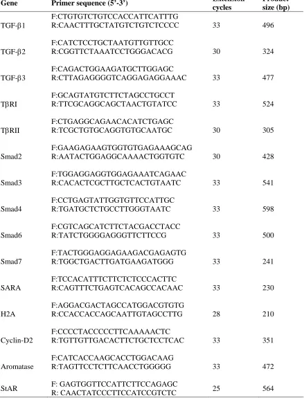
Semi-quantitative analysis of mRNA expression patterns of TGF-β ligands, receptors and Smad proteins...52
Western Blotting ...54
Immunohistochemistry ...55
Statistical analysis...56
Results...57
Discussion ...60
References...78
Expression of TGF-β and components of its signaling pathway in cultured porcine granulosa cells: a possible role of TGF-β in luteinization?...81
Summary ...81
Methods and materials ...84
Granulosa cell isolation and culture...85
Progesterone RIA...86
Semi-quantitative RT-PCR ...87
Results...89
Discussion ...92
References...108
General conclusions ...110
References...115
LIST OF TABLES
TGF-β ligands, receptors and Smad expression in peri-ovulatory porcine follicles
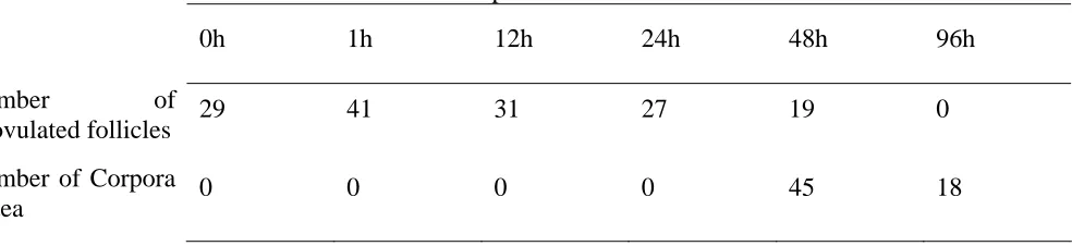
1. Primers used for semi-quantitative RT-PCR ...66 2. Summary table of ovarian characteristics at ovariectomy across all animals...67
Expression of TGF-β and components of its signaling pathway in cultured porcine granulosa cells: a possible role of TGF-β in luteinization?
Appendix
LIST OF FIGURES LITERATURE REVIEW
1. An overview of TGF-β signaling...29
TGF-β ligands, receptors and Smad expression in peri-ovulatory porcine follicles
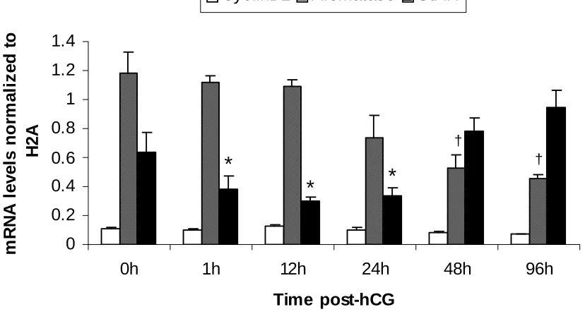
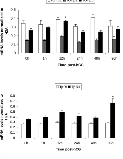
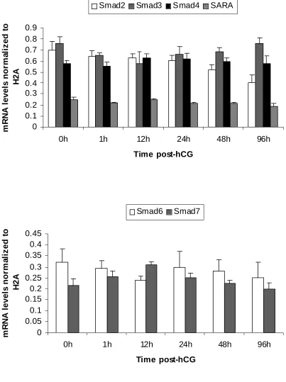
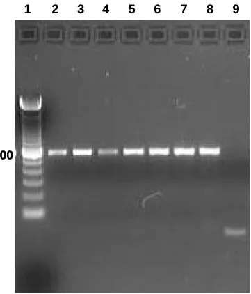
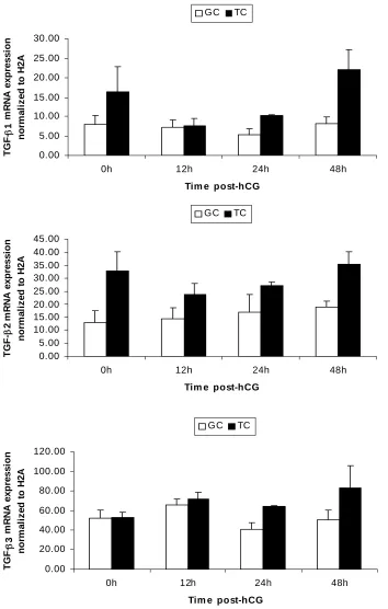
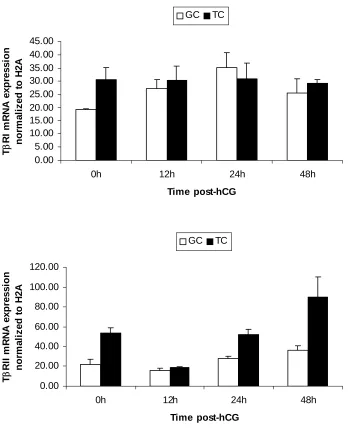
1. mRNA expression patterns of some LH-responsive genes in hCG-treated follicles...68 2. mRNA expression patterns of TGF-β ligands and receptors in hCG-treated follicles ..69 3. mRNA expression patterns of Stimulatory Smads, SARA and inhibitory Smads in hCG-treated follicles...70 4. Example of an ethidium bromide-stained agarose gel of Smad3 amplification products from hCG-treated follicles ...71 5. mRNA expression patterns of TGF-β ligands in granulosa and theca compartments of hCG-treated follicles...72 6. mRNA expression patterns of TGF-β receptors in granulosa and theca compartments of hCG-treated follicles...73 7. Protein expression patterns of TGF-β ligands and receptors in hCG-treated follicles ..74 8. Protein expression patterns of Smad3 and pSmad3 in hCG-treated follicles ...75 9. Representative Western blots of TGF-β ligands, receptors, Smad3 and pSmad3 ...76 10. Immunohistochemical localization of TGF-β ligands and receptors in hCG-treated follicles...77
1. Effect of treatment on progesterone levels in granulosa cell cultures ...101 2. mRNA expression patterns of LH-responsive genes in luteinizing granulosa cells in vitro ...105 3. mRNA expression patterns of some TGF-β pathway components and LH-responsive genes in luteinizing granulosa cells in vitro...106
General conclusions
LITERATURE REVIEW
The first part of this section takes one through all the stages of folliculogenesis, from the primordial germ cell stage to ovulation and luteinization. The second part discusses the
role of TGF-β superfamily members in folliculogenesis and luteinization.
Follicular development
Development of the follicle begins soon after conception in the female fetus. After the the primordial germ cells migrate to the gonadal ridge, they are called oogonia [1].A layer of mesenchymal cells then surround the oogonia to form oogonial clusters or syncytia [2]. Depending on the species, the oogonia undergo a predetermined number of mitotic divisions until they enter meiosis to give rise to oocytes [1]. This occurs usually around mid to late gestation, depending on the species [1]. Follicle formation starts when the mesenchymal cells surrounding the oogonia transform into flattened granulosa cells [3], to give rise to primordial follicles. In rodents, follicular assembly begins immediately after birth, while in primates and domestic animals, it occurs around mid to late gestation [4]. Meiosis marks the end of replication of female germ cells; thus, all the oocytes that a female will have in her lifetime are set at this stage [2]. After initiation of meiosis, the primary oocyte progresses through leptotene, zygotene and pachytene stages of the first prophase, before arresting in the diplotene/dictyate stage until it receives further signals to resume growth [1]. In the pig, there is evidence that the oogonial to oocyte transformation continues after birth [5]
in which they were formed [207]. It is evident from the vast literature available on follicle development that there is a myriad of endocrine and paracrine factors that can signal the follicle to resume development, although how they act singly or together to induce the resumption of follicular growth remains to be worked out. Among these signals are growth factors like insulin and insulin-like growth factors (IGFs), transforming growth factor
(TGF)-α, members of the transforming growth factor-beta superfamily such as TGF-β, bone
morphogenic proteins (BMPs) inhibin, activin, anti Müllerian hormone (AMH), and growth differentiation factors (GDFs), epidermal growth factor (EGF), vascular endothelial growth
factor (VEGF), cytokines like tumor necrosis factor-α (TNF-α), and intra-ovarian steroid
hormones like estrogen (E2) and progesterone (P4) [3, 6-10]. These signals initiate a multitude of signaling cascades to bring about changes in gene expression, which in turn cause follicle selection, and growth and differentiation of the follicular components.
and secondary follicles [15]. The accumulation of mitochondria, and smooth and rough endoplasmic reticulum in the bovine oocyte at the primary follicle stage provides a clue to the increasing energy and synthetic requirements of the oocyte for growth [16]
When the follicle develops two or more layers of granulosa cells, it is called a secondary or preantral follicle [17]; at this stage, the granulosa cells proliferate, and the theca cells which arise from interstitial stromal cells, form a layer around the granulosa layer. Also at this stage, oocyte RNA synthesis is first detectable [18]. Also during this stage, the oocyte secretes a glycoprotein structure around it, called the zona pellucida. The granulosa cells form gap junctions with each other and with the oocyte via connexin proteins [19, 20]. These gap junctions enable critical communication between the oocyte and the granulosa cells. Thus, the oocyte can promote the growth and differentiation of the granulosa cells surrounding it, while the granulosa cells can signal the growth and differentiation of the oocyte [9, 21]. The cells surrounding the oocyte continue to proliferate and differentiate into two layers of theca cells, theca interna and externa, which surround the granulosa cells [22]. At this stage, the follicle may become receptive to FSH [23, 24] and LH [23, 24], as indicated by the appearance of their receptors on granulosa and theca cells [25]. Estrogen is also thought to play a role at this stage of follicle development; in the rat, estrogen receptors
ERα and ERβ have been found in theca and granulosa cells respectively of primary follicles
stage, when theca interna cells appear [29], the granulosa cells with their FSH and estradiol receptors also proliferate. Under the influence of FSH and estradiol, theca interna cells acquire LH receptors [30]. Based on rodent models, it is clear that theca cells, under LH influence, convert cholesterol to androstenedione, which then diffuses across the basement membrane to granulosa cells [31]. The granulosa cells possess P450aromatase, used in converting the androstenedione to estradiol [30]. The difference between the rodent and the pig is that the porcine theca interna cells also possess aromatase activity and hence are capable of producing estradiol [32].
The rapid increase in steroid biosynthesis is thought to result in the increase and accumulation of follicular fluid, forming the antral cavity, and consequently, the follicle is now called an antral follicle. Antral fluid production increases with increasing follicular vascularization and permeability of blood vessels [17]. The factors responsible for the formation of the antrum are not clear, but in vitro studies in rodent, porcine and bovine follicles suggest that FSH [33, 34], LH [35], activin [36], Kit ligand [37], EGF [38] may all be involved. The antral follicle has a layer of granulosa cells closely associated with the oocyte (together called the cumulus-oocyte complex or COC), with the antral fluid separating the COC from the other granulosa cells (called mural granulosa cells) along the inside of the follicular wall [22]. The oocyte acquires the ability to resume nuclear maturation, and the competency to undergo fertilization and cell cleavage [17].
which, via a negative feedback on the hypothalamo-pituitary axis, control FSH secretion (FSH levels decline to levels below what is necessary for further follicular selection) and thus inhibit the growth of subordinate follicles [40, 41]. At the same time, granulosa cells acquire LH receptors that are necessary for continued development [42]. The dominant follicle(s) with LH receptors continue their growth and development, while the subordinate follicles undergo atresia [42, 43]. Whereas FSH promotes rapid growth of GCs in the preantral follicles by the regulation and/or induction of genes involved in cell cycle control, LH promotes the exit from cell cycle and terminal differentiation of the GCs in the mature follicle, and induces genes related to ovulation and luteinization [25]. A very interesting fact is that both of these hormones act via seemingly identical cAMP signaling pathways, and through structurally related receptors, but yet produce completely different outcomes.
Ovulation
macrophages to invade the follicle [51]; (ii) increased expression of proteolytic enzymes such as ADAMTS-1 and cathepsin L [44], plasminogen activator [52], the matrix metalloproteinases MMP2, MMP9 and MMP 13 [53], all of which digest the extracellular matrix of the follicle, thus weakening the follicular wall and facilitating rupture [54, 55]; and (iii) activation of adenylyl cyclase/protein kinase A pathways, which in turn cause the expression of genes involved in ovulation, like progesterone receptor, cyclooxygenase (COX)-2, the CAAT enhancer binding protein, prostaglandin endoperoxidase synthase-2 etc. [44, 53]; the targets of progesterone receptor, in turn, include plasminogen activators, matrix metalloproteinases (MMPs) and their inhibitors (tissue inhibitors of MMPs; TIMPs) [53, 56-60]), all of which help to degrade the extracellular matrix around the follicle, while the TIMPs are thought to protect other growing follicles from the degrading actions of the MMPs [52]. With the help of all these factors, the COC is released from the follicle.
Luteinization and the corpus luteum
The corpus luteum (CL), an endocrine gland, develops from the follicle tissue remnants after ovulation, by a process called luteinization. The CL is the source of progesterone, the steroid hormone responsible for creating a uterine environment conducive to maintaining pregnancy. If pregnancy does not occur, the CL undergoes a process of regression, at which time progesterone production declines (functional luteolysis), and ultimately the CL itself degenerates (structural luteolysis), a process mediated by prostaglandin F2α [61]. Luteolysis brings an end to the lifespan of the CL, and initiates the
entire duration of pregnancy to ensure progesterone availability and pregnancy maintenance. The role of progesterone for maintenance of pregnancy is underlined by the fact that the hormone, by itself, can support gestation in ovariectomized gilts [62]. In the pig, ovulation occurs 36-48 hours after the LH surge [63]. In sows, this time of ovulation after the LH surge is extremely variable, reported anywhere from 26 to 34 hours [64]. After ovulation, the follicle undergoes hyperemic and hypertrophic changes [62, 65]; the granulosa cell layer is thrown into extensive folds around the follicular antrum; there is breakdown of the basement membrane and the theca cells are carried by the vascular and connective tissue into the core of the developing CL along these folds. The development of vascular supply proceeds in centripetal as well as lateral directions [66]. The hypertrophic changes in the granulosa cells and the proliferation of endothelial cells in the CL is phenomenally rapid, even exceeding similar changes in some of the most malignant tumors [65]. All these changes transform the fluid-filled follicle into a solid mass of cells that comprise the CL, within a few days (4-6) days after ovulation. Analyses of the cell types in the CL in the guinea pig [67], sheep [68, 69] and the cow [70] have yielded information about the composition of the CL during various stages of its development. Based on the classification described by others [61, 69, 70], it is accepted that the cell types of the CL can be broadly divided into steroidogenic and non-steroidogenic components. The steroidogenic cells can be further subdivided into small luteal cells (SLC) and large luteal cells (LLC). Endothelial cells, fibroblasts, pericytes and macrophages form the non steroidogenic component [68, 70].
The LLCs are characterized by their larger size (>26-31 μm in sheep) and round
shape, and by the presence of abundant rough and smooth endoplasmic reticulum, numerous mitochondria, lipid droplets and secretory granules in the cytoplasm, all characteristics of a steroidogenically active cell type [70]. Although the LLCs make up about a third of the CL volume, they account for over 80 % of progesterone secretion at peak production (mid luteal phase) [68, 70]. The SLC constitute around 20 % of the CL volume; together, the LLCs and the SLCs roughly make up about 35 % of the total cell number in the CL [68, 72]. During CL development, the LLCs increase in diameter by 3 to 4 fold, though the total cell number of
LLCs remains fairly constant [68]. The LLCs, but not the SLCs, also possess PGF2α
receptors, and hence appear to mediate the luteolytic actions of PGF2α [73]
The SLCs are about a third of the size of the LLCs in domestic ruminants, and are irregularly shaped, and have a lot of smooth endoplasmic reticulum, lipid droplets, a lot less rough endoplasmic reticulum and secretory granules compared to the LLCs [68, 70]. The SLCs maintain a constant size throughout the luteal phase, but increase in cell number by up to five fold [68]. The SLCs, but not the LLCs, possess LH receptors; therefore, they are able to respond to LH and increase their otherwise low basal P4 production [74].
Luteinization is also generally associated with the loss of cytochrome P450aromatase expression in most species, and with the onset of synthesis of progesterone in significant quantities [62]. In the pig, however, aromatase activity is never completely lost and the porcine CL continues to synthesize small quantities of estradiol [76-78]. Steroid production switches from predominantly estradiol production by the follicle, to predominantly progesterone production by the CL. There is also a concomitant upregulation in the amounts of other critical components involved in progesterone synthetic pathway – low density
lipoprotein (LDL) receptor, the cholesterol side chain cleavage enzyme P450scc, and 3β -HSD, another enzyme involved in progesterone synthesis, as well as the cholesterol transport protein StAR (Steroidogenic Acute Regulatory protein) [79, 80].
Progesterone production by the CL
Cholesterol, in the form of low-density and high-density lipoproteins (LDL and HDL), is used by the CL as the substrate for steroid production [81]. Of the two kinds of lipoproteins, LDL is the major substrate used by the porcine CL [80].
inner mitochondrial membrane, cleaves the cholesterol side chain to yield pregnenolone, which is then transported to the smooth endoplasmic reticulum; here, it is converted to
progesterone by the enzyme 3β-HSD [61].
In the pig, as the CL continues to develop, progesterone production increases exponentially, peaking between days 8-12 after ovulation [63]. Progesterone levels decrease thereafter, and by day 16, they are reduced to approximately one twelfth of that measured on day 8 as CL undergoes regression [63].
Regulation of CL formation and function
The initial stimulus for luteinization is believed to be the LH surge, and LH initiates a number of downstream events that result in successful ovulation and the formation and maintenance of the CL. There have been a number of models employed to study the process of luteinization, both in-vivo and in-vitro [85]. Although these models have increased our understanding of the process of luteinization, the regulatory mechanisms inducing luteinization still remain unclear. It is clear, however, that the process begins before ovulation, at the cellular level [86]. At the genetic level, a number of genes have been identified, mostly from rodent studies [44, 45, 47, 48, 86-91], and a recent study in primates [92], as being involved in luteinization. Based on these studies, the genes can be classified into major classes such as
(i) genes involved in steroidogenesis – P450scc, StAR, LDLr, 3β-HSD,
P450aromatase, 17β-HSD etc. The genes involved in progesterone synthesis such
as P450scc, StAR, LDLr, and 3β-HSD and estrogen degradation such as 17β
(ii) genes involved in extra cellular matrix modeling – ADAMTS-1, Metallothionein (MT)-1, Tissue Inhibitor of Metallo Proteinase (TIMP-1) etc. ADAMTS-1, a metalloproteinase, is upregulated around ovulation and early luteinization, possibly contributing to the rupture of the follicle and tissue remodeling that transforms the follicle into the CL; TIMP-1, an inhibitor of metalloproteinases like ADAMTS-1, and MT-1, are both upregulated after ovulation and possibly help to moderate the degradative actions of metalloproteinases, mitigate the local inflammatory reaction caused by ovulation, help in healing and promote angiogenesis [45, 90]
(iii) genes involved in inflammatory processes and angiogenesis: for example, there is evidence that the expression of the enzyme cycloxygenase-2 (COX-2) is induced by the LH surge [95]. Along with lipoxygenase enzymes, COX-2, catalyzes the arachidonic acid pathway, the interruption of which has been shown to reduce the rate of ovulation and luteinization [96, 97]. Message levels of VEGF, a potent angiogenic factor, has been shown to remain constant during the luteal phase and decline only during luteal regression in the mare [98]. In the porcine CL, VEGF receptor 1 (VEGFR-1) mRNA levels increase from low levels after ovulation, to highest levels in mid to late luteal phase [99].
IGF-1, correlates with the early stages of CL development (days 4-10), strongly suggesting a role of IGF-1 in this process in the pig [100-104].
(v) genes involved in cell cycle regulation: luteinization marks the exit of granulosa and theca cells from the cell cycle [62]. In this respect, luteinization can be defined as the terminal differentiation of the follicular cells and marks the transition from a proliferative to a differentiated state. The differentiation of the follicle into CL is also marked by the phenomenal rate of tissue growth and cellular proliferation [61]. The proliferation is evident from mitotic markers such as the proliferating cell nuclear antigen (PCNA) [62] during early CL development, but the labeling index gradually decreases and eventually is undetectable in the LLC over the lifespan of the CL [105]. Thus, it is thought that terminal differentiation occurs in the porcine CL, with increasing restriction in the proliferating capacity over time. Recent evidence [53, 106] shows that in the early CL in rodents, mitosis is initiated by members of the retinoblastoma gene family such as pRb, p107 and p130 [107] but in the late CL, these proteins are inactivated, thereby inactivating positive cell cycle regulators such as cyclins D1, D2 and E . Another factor known to be involved in regulation of the cell cycle is p27Kip1, a member of the family of cyclin-dependent kinases. It is an inhibitor of the cell cycle progression and in knockout studies in mice, it has been shown that p27 levels decrease after the ovulatory surge of LH, thereby preventing granulosa cells from proliferating and facilitate their differentiation into luteal cells [108] (vi) genes that regulate the transcription of other genes: gene transcription plays an
modification, coactivator recruitment, and synthesis and activation of transcription factors. It is well known that the LH surge increases cAMP levels, which in turn activate the protein kinase A pathway and phosphorylation of the cAMP response binding protein (CREB) [106]. Among other genes, the ones for P450scc and StAR transcription, are activated by CREB. The transcription of several genes is also stimulated by VEGF, including its own receptors [109].
Members of the TGF-β superfamily in the ovary
Members of the TGF-β superfamily are involved in every stage of follicular
development, ovulation and luteinization [110, 111]. Because of their pleiotropic roles in these processes, they are described in this section separately. These members include bone morphogenic proteins (BMPs), TGF-βs, growth differentiation factors (GDFs),
anti-Mullerian hormone (AMH), activins, inhibin, etc. Most data on the role of TGF-β family
members in the ovary come from studies in rodent species, but recently, a number of studies point to their role as intraovarian factors in other species such as the sheep, cat, cow, primates etc. A review of the existing literature shows that TGF-β superfamily ligands and
luteinization (as reviewed in [111]). The following few sections illustrate the patterns of stage-dependent expression of these proteins during folliculogenesis.
TGF-β members in primordial and primary follicles
In rats, it has been shown that intra bursal administration of BMP-7 decreases the number of primordial follicles, while increasing the number of follicles from later stages such as primary, secondary and antral follicles [112]. In another rat study [113], BMP-4 produced similar results in a whole ovary-organ culture system; primary follicles increased while primordial follicles decreased. In the same study, it was also found that when ovaries were exposed to a neutralizing antibody to BMP-4, they were much smaller than controls, with accompanying changes such as progressive loss of oocytes and primordial follicles and also increased cellular apoptosis, eventually resulting in loss of ovarian tissue morphology.
Other TGF-β superfamily members have been shown to be expressed specifically in
oocytes in the primordial and primary stage follicles. In rodents and ruminants, GDF-9, BMP-15 and BMP-6 have been identified in oocytes [114-118]. The receptors for these ligands have been identified in the pre-granulosa and granulosa cells, indicating that these cells are targets of signaling by these ligands. Mice null for gdf-9 gene are infertile and have follicles arrested at the primary stage [119, 120]. In another study [121], GDF-9 treatment of rat follicles in vitro caused progression of follicle growth from early to late primary stage. Sheep with homozygous mutations in the bmp-15 or gdf-9 genes are infertile [122, 123].
secreted by growing primary stage follicles and all the way up to pre-ovulatory stage follicles suppress the premature recruitment of primordial follicles.
TGF-β members in primary to antral stage follicles
Several members of the TGF-β superfamily are thought to be involved in the growth
of primary follicles to secondary and antral follicles. In vitro studies in rodents [121, 126-128] have shown that exposing ovaries to GDF-9 promotes primary follicle progression. In other studies, in mice null for GDF-9 [119], and in sheep with homozygous mutations in the
suppressed FSH-stimulated progesterone production and caused granulosa cells to proliferate. In the pig, BMP-2 reduced FSH-stimulated progesterone production by granulosa cells [137]. In the cow, BMP-4, BMP-6 and BMP-7 also suppressed basal as well as LH-induced androgen production by theca cells [138]
Activin βA and βB subunits have also been detected in primary stage follicles onward in many species including human [139, 140]. Though the exact role of activin in follicle development is still unclear, there is some evidence that it may promote granulosa cell proliferation and differentiation (as reviewed in [141]). In the antral follicle however, activin A has been shown to accelerate oocyte maturation and improve oocyte developmental competence [142, 143]. On the other hand, AMH is thought to have a negative effect on primordial follicle recruitment, as seen earlier. It is also known that AMH inhibits FSH-dependent growth of late preantral follicles in cultured mouse ovaries [144].
Message and protein from TGF-β ligands (TGF-β1, TGF-β2, TGF-β3) and receptors
(type-I and type-II) have been detected first at the preantral follicle stage of several species, including the rat [145], cow [146], sheep [147] and human [148], but there is species to species as well as spatio-temporal expression variation and the role of these proteins is still
unclear. In the antral follicle of the rat [149], TGF-β and in the human [150], TGF-β1 and 2 have been shown to be produced by both theca and granulosa cells. In sheep, cows and pigs
[11], TGF-β1 is mainly produced by thecal cells of antral follicles. In the rat study, TGF-β
(the specific ligand unknown) was shown to be expressed in theca cells and exhibited proliferative and cytodifferentiative actions on granulosa cells [151]. In another rat study,
TGF-β was shown to increase FSH receptor mRNA and protein [152]. In pig studies, it was
granulosa cells, in contrast to the rat study [154, 155]. The actions of TGF-β on granulosa
cells has been further substantiated by the discovery of TGF-β receptors on porcine
granulosa cells [156]. More evidence that pig granulosa cells may not produce TGF-β comes
the studies of Mulheron and coworkers [157], who showed that TGF-β2 mRNA was not
expressed by these cells.
TGF-β members in ovulation and luteinization
In most species, inhibin/activin subunit expression is down-regulated after ovulation and during CL formation [111]. Follicular expression of BMP-2, BMP-3b, BMP-4, BMP-6, BMP-7, BMP-15 and GDF-9 is also downregulated in the rat after ovulation [131, 135]. It is thought that these factors inhibit luteinization in the follicle until ovulation has occurred.
Mouse luteal cells express both TGF-β1 and TGF-β2 [158], and so does the rat luteal
macrophage [159]. In another rat study, prolactin, a progesterone production enhancing
peptide, and TGF-β had similar effects on cultured luteal cells; they both suppressed 20α
-HSD, whose activity decreases progesterone [160]. In this study, it was shown that prolactin also stimulates TGF-β2 expression in luteal macrophages. In another study, human
granulosa-lutein cells treated with TGF-β1 showed decreased apoptosis [161]. Taken
together, it appears that TGF-β, in concert with prolactin, supports CL function by increasing progesterone and decreasing apoptosis.
Although there is evidence of expression of TGF-β ligands and its signaling pathway
components during follicular development, there is very little information on the exact role of these proteins on ovulation and luteinization. The following studies suggest the possible role
(i) When immature mice were sequentially treated with FSH for 3 consecutive days and hCG on the fourth day[162], it was shown that the receptors TβRI and TβRII were
immunolocalized to both theca and granulosa cells, the oocyte stained for TβRI only while
Smads were present in all cell types, with Smad 4 staining the strongest. Protein and mRNA
expression analysis showed that TGF-β2, TβRI, Smad 2 and Smad 4 were upregulated,
whereas that of Smad 6 was downregulated, in response to the gonadotropins (FSH and LH). This study thus shows that components of the TGF-β pathway are upregulated in the
peri-ovulatory follicle in response to LH and FSH. We used a similar model for our study with pre-pubertal pigs treated sequentially with gonadotropins (P.G.600, a combination of hCG and eCG, followed by hCG 3 days later). Our findings appear in the next chapter.
(ii) In the marmoset, TGF-β1 and TβRII were co-immunolocalized with the
luteinization marker 3β-HSD in granulosa and theca cells of periovulatory follicles; on the
other hand, the expression of TGF-β1 and TβRII was absent in atretic follicles [163]. In
another marmoset study, when large, non-luteinized follicles were cultured and induced to luteinize with treatments known to induce luteinization, such as LH, TGF-β and cAMP,
increased expression of TβRII was observed, along with other expected changes such as
breakdown of basement membrane of the follicle, increased expression of 3β-HSD, and decreased expression of connexin-43 (an ovarian gap junction protein that decreases during
luteinization) and TβRII [164]. The consistent, increased expression of TβRII in luteinizing
cells only prompted the authors to start using TβRII as a marker of luteinization in
subsequent studies.
(iii) In a follow up study to the studies described in (ii) above, [165], the authors
inhibitory effect on luteinization), on porcine granulosa cells in vitro and monitored the cells for luteinization. TNF-α downregulated TβRII along with other luteinization-inhibition
parameters such as decreased progesterone receptor expression, and influenced the balance between proliferation and apoptosis.
(iv) In the chicken, TGF-β1, on its own or in combination with FSH, increased the
expression of FSH receptor and LH receptor in undifferentiated granulosa cells; the increase in LH receptor sensitizes the granulosa cells to LH action by increasing P450scc and StAR expression, thus increasing the cells’ ability to produce more progesterone [166].
(v) The presence of TGF-β1 and 2 in murine luteal cells [158] and rat luteal macrophages [159] has been reported. A few studies have also indicated the possible role of
TGF-β in the CL: Prolactin, a luteotropic hormone, and TGF-β increased progesterone
production in cultured rat luteal cells by suppressing 20α-HSD expression, and in rat
macrophage cells, prolactin stimulated TGF-β expression [160]. Also, in the same study,
when the luteal cells were treated with an anti-TGF-β antibody, the suppressive action of
prolactin on 20α-HSD was decreased, thus indicating that the luteotropic actions of prolactin
may partly be mediated by TGF-β.
(vi) More evidence about the role of TGF-β in CL formation and maintenance comes from two studies in the rhesus monkey. In the first study, when CLs at various stages of
pregnancy were examined for the presence of StAR, TGF-β1 and TβRII mRNA and protein,
it was found that their expression progressively increased during early pregnancy, when the CL was being formed and was functional, while in the regressing CL, their expression
decreased [167]. Also, in this study, Interferon (IFN)-γ, a luteolytic cytokine decreased the
development was induced in the rhesus monkey using eCG/hCG treatment, and the expression of TGF-β1, TβRI, TβRII and StAR was examined in CLs at various stages of
development using immunohistochemistry and in-situ hybridization [168]. It was found that their expression increased to reach peak levels when the CLs were functional, and decreased
when the CL regressed, and IFN-γ reduced their expression. These studies strongly suggest
that TGF-β and its receptors may play a role in CL formation and maintenance of its
function.
All of the above data support the view that TGF-β working via TβRI and TβRII plays
a role in maintaining luteal function in these species. In the sheep however, a study shows that TGF-β1 and TGF-β2 inhibit progesterone production in cultured granulosa cells [147],
suggesting that TGF-β may be suppressing luteinization in this species.
Before describing our studies to investigate the role of TGF-β in luteinization in the
pig, a review of TGF-β and its signaling pathways is provided below.
TGF-β superfamily
Members of the transforming growth factor (TGF)-β superfamily are important
regulators of cellular processes in many cell types [169]. The members of this superfamily are structurally and functionally similar, and influence such crucial processes as cell growth and differentiation, early embryonic development, extracellular matrix deposition, immunoregulation, cell motility and apoptosis [170]. Some of the prominent members of the
For the purposes of describing the signal transduction mechanisms of members of the TGF-β superfamily, the prototype member, TGF-β, will be considered here, especially as it
is the focus of the studies described in this thesis.
TGF-β structure and activation
TGF-β has three structurally similar isoforms, TGF-β1-3, which are encoded by three
distinct genes in mammals [171]. They all have similar bioactivity in vitro [169], with
TGF-β1 being the most prevalent isoform [172]. When initially synthesized, the precursor TGF-β
protein is large, consisting of around 390-412 amino acids [169]. The precursor protein is comprised of three domains: 1) an N-terminal signal domain called the latency associated peptide (LAP, 249 amino acids), 2) a signal peptide (SP, 29 amino acids) and 3) a C-terminal domain (112 amino acids) [173]. The C-terminal fragment is small and forms the mature portion of the protein, which is involved in triggering intracellular signal transduction. The signal domain functions to target the precursor to the secretory pathways, while the pro-domain is thought to help in folding, dimerization and regulation of the biological activity of the C-terminal fragment. The mature TGF-β molecule usually has 7-9 cysteine residues, and
exists as a homodimer formed by disulfide bonds [174].
The TGF-β precursor is thought to be activated through different mechanisms [173,
Overview of TGF-β signaling
Members of the TGF-β superfamily use common mechanisms to signal to the nucleus
[170]. They bind to membrane receptors possessing a cytoplasmic serine/threonine kinase domain. Once the ligand binds to its receptor, the ligand-receptor complex phosphorylates members of the Smad (the vertebrate homologue of Drosophila Mad (Mad =Mothers against decapentaplegic ) and the related Caenorhabditis elegans gene Sma) or MAPK (Mitogen Activate Protein Kinase) protein families. These protein families can in turn translocate to the nucleus themselves, or phosphorylate downstream targets which can move to the nucleus and control target gene expression [169, 170]. There is now evidence to suggest that the Smad
and the MAPK pathways intersect [169]. A simplified schematic of TGF-β signaling is given
in figure 1. The components of the TGF-β signaling pathway are discussed in detail in the
following paragraphs.
TGF-β receptors
Signaling by TGF−β is initiated by its binding to its receptor complex. All members
of the TGF-β superfamily transduce their signals through two serine/threonine transmembrane receptors, called Type I and Type II receptors and their downstream effectors, the Smad proteins [173, 176].
To date, five type II receptors and seven type I receptors have been discovered in
mammals, each binding to different ligands [177]. The type II receptor TGF−β RII (TβRII)
binds only to TGF−β isoforms, with a higher affinity for TGF−β1 and TGF−β3, and a lower
affinity for TGF−β2. The kinase domain of the type II receptor is the most conserved domain
[177]. Of the type I receptors, ALK5, ALK1 and ALK2 can transmit TGF-β signaling, but
transmembrane serine/threonine kinase [177]. The receptors TβRI and TβRII receptors are
structurally similar, although TβRI does have a shorter extracellular domain, a much shorter
C-terminal tail, and a highly conserved glycine and serine (GS)-rich region immediately preceding the kinase domain. The SGSGLP amino acid sequence is considered to be a
signature of TβRI. The phosphorylation of serines and threonines in the GS domain by TβRII
is required for the activation of TβRI and TGF−β signaling [177].
There are also type III or accessory receptors for TGF-β signaling, and include
betaglycan and endoglin [177]. The biological functions of both betaglycan and endoglin are
not clear. It was thought that betaglycan may function to present the TGF−βs, especially
TGF−β2, to TβRII [178]. More recently, it has been shown that betaglycan is responsible for endocardial cell transformation (Brown et al., 1999). In addition, some cell lines can secrete soluble betaglycan, corresponding to the extracellular domain of betaglycan, which can
inhibit the binding of TGF−β to the TβRI-TβRII complex [179]. Supporting evidence comes from studies showing that this soluble betaglycan can inhibit tumorigenesis and metastasis of
MDA-MB-231 human breast cancer cells by reducing the activity of TGF−β1 and TGF−β2
secreted by these cells [180].
Mechanisms of transforming growth factor-β receptor activation
All Type I and Type II TGF−β receptors exist as homodimers in the absence of ligand
[177, 181]. For TGF−β receptors, TGF−β binds poorly TβRI to alone [182]; TGF−β must
first bind to the TβRII homodimer, which then recruits TβRI to form a heterotetrameric
complex [183, 184]. TβRII is constitutively hyperphosphorylated at several serine residues
phosphorylation of serine and threonine residues in the TβRI GS domain by TβRII [183].
The phosphorylated TβRI then becomes activated, and signals are transduced to cytoplasmic
targets [186, 187]. Since TGF−β2 has only low affinity for TβRII, the binding of TGF−β2 to
Type III receptors can facilitate the binding of TGF−β2 to TβRII, which, in turn, can recruit
TβRI to form an active receptor complex [188, 189]. However, TGF−β2 can also directly
bind to TGF−β receptor complexes in the absence of RIII in some cell types [190-192].
Structural requirements for transforming growth factor-β receptor activation
Specific domains in TβRI and TβRII are important for TGF−β signaling [170]. The extracellular domains of both receptors are required for ligand binding [193, 194]. The
cytoplasmic domains of both TβRI and TβRII are required for the dimerization of the
receptors, which is essential for TGF−β signaling [187, 194, 195]. In addition, the kinase
domain of TβRII is required for TβRII to phosphorylate TβRI, while the kinase domain of
TβRI is required for TβRI to phosphorylate downstream targets [183, 194, 196, 197]. It is
also possible that TβRII can directly phosphorylate other downstream targets. The glycine
and serine-rich GS domain is essential for the activation of TβRI by TβRII [183, 196]
Some of the serine and threonine residues in the Type I and Type II receptors are
important for TGF−β receptor signaling as well [177]. TβRII primarily autophosphorylates
threonines in vitro, but serines in vivo [183]. TβRII autophosphorylates at least three serine
residues: Ser409 and Ser416 in the protein kinase domain and Ser213 in the juxtamembrane
domain [187]. Phosphorylation of Ser213 and Ser409 is essential for TβRII kinase signaling,
Phosphorylation of TβRII has also been observed on Ser551 and Ser553 of the
terminus and Ser225, Ser228, and Ser229 in the juxtamembrane domain [183, 198]. Since C-terminal tail deletion did not affect TβRII actions [194, 197], the phosphorylation of the
C-terminal tail is not thought to play a critical role in TβRII signaling. However, it is still not known whether the phosphorylation of the serine residues in the juxtamembrane domain
plays a role in TβRII signaling. In vitro, TβRII can autophosphorylate on Tyr259 in the
kinase domain, Tyr336 in subdomain V, and Try424 in subdomain VIII [199]. The role of
TβRII tyrosine autophosphorylation in TGF−β signaling is not clear. TβRII also
phosphorylates TβRI at four sites in the GS domain (Thr186, Ser187, Ser189, and Ser191) and at Ser165 in the juxtamembrane domain. The phosphorylation of Ser165 was thought to
be the direct effect of TβRII, and may modulate the signaling specificity of TGF−β receptors
[198].
Smads
Smads can be divided into three groups based upon their structure and function. The first group, referred to as receptor-activated Smads (RSmads), includes Smads 1-3, 5, and 8.
The RSmads specific for TGF-β signaling are Smad2 and Smad3. The second group consists
of the single member Smad 4, and is referred to as common-partner Smad (Co-Smad). The last group is referred to as the inhibitory Smads, including Smads 6 and 7 [170].
Structure of Smads
Co-Smad, also have a characteristic serine-rich SXS motif in their MH2 domain. The phosphorylation of the two serine residues in the SXS motif of the MH2 domain activates the RSmad [200, 201].
Smad signaling
The general mechanisms mediating TGF−β receptor activation of Smads are as
follows: binding of the ligand to the TGF−β receptor complex induces the phosphorylation of
RSmads. Generally, Smads 1, 5, and 8 can be phosphorylated by BMPs, while TGF−β and
activin can phosphorylate Smad2 and Smad3. It is known that TGF−β can also phosphorylate
Smad1 in Hs578T human breast cancer cells and IEC 4-1 cells [169]. In addition, it has been shown that TGF−β can phosphorylate Smad5 in human hematopoietic cells [202].
The active TβRI phosphorylates the RSmad at the serine-rich sequence SXS in the
C-terminal tail of the MH2 domain, thus activating it. Smad4 does not have this SXS motif, and
hence is not phosphorylated by TβRI [169]. Structural analysis suggests that the inability of
Smad4 to bind to ΤβRI is due to a lack of a basic, positively-charged surface patch at the
MH2 domain of Smad4 [203]. The phosphorylation of RSmad causes conformational
linker region of RSmads, there are several consensus MAPK phosphorylation sites that are responsible for crosstalk between the Smad and MAPK pathways [169, 205].The inhibitory
Smads, 6 and 7, appear to block the activation of RSmads by TGF−β receptors [183].
The function of the Smad anchor for receptor activation
After ligand binding, RSmads can transiently associate with Type I receptors with the help of the auxiliary protein, Smad anchor for receptor activation (SARA) [196]. The SARA protein contains a FYVE (a double zinc finger) domain, which can bind to phosphatidylinositol-3-phosphate in the lipid layer endosomal membrane. There is also a
peptide sequence of SARA that can interact with the RSmads [206]. TβRI-mediated
phosphorylation of SARA-bound RSmad is more efficient in SARA-rich endosomes ([201]. The role of SARA as a regulator of TGF-β signaling has been demonstrated in studies where
a mutation in SARA in Mv1Lu or HepG2 cell cultures caused disruption of TGF-β signaling
[206]
Summary of literature review
Members of the TGF-β superfamily have been shown to be involved at all stages of
follicle development. There is evidence that the prototypical member of this superfamily, TGF-β, is involved in pre-antral to antral follicle development and ovulation. But there are
only a few studies that have examined the role of this cytokine in the post-ovulatory follicle. Even among these studies, data for the pig is limited. Preliminary studies from our laboratory indicated that TGF-β2 precursor as being one of the genes that was upregulated very early in
response to the pre-ovulatory surge of LH. This observation led us to believe that TGF-β may
be involved in the process of luteinization.
To test our hypothesis, the following research was performed. In the first study, the message and protein expression of TGF-β and components of its signaling pathway was
examined in peri-ovulatory porcine follicles at various time points after hCG administration.
In the second study, the expression of TGF-β and components of its signaling pathway were
examined in granulosa cells induced to luteinize in vitro with LH/IGF-1. The effects of
TGF-β alone or in combination with LH/IGF-1 on luteinization were also examined in by
REFERENCES
1. Picton HM. Activation of follicle development: the primordial follicle. Theriogenology 2001; 55:1193-1210.
2. Smitz JE, Cortvrindt RG. The earliest stages of folliculogenesis in vitro. Reproduction 2002; 123:185-202.
3. Kezele P, Skinner MK. Regulation of ovarian primordial follicle assembly and development by estrogen and progesterone: endocrine model of follicle assembly. Endocrinology 2003; 144:3329-3337.
4. Fortune JE. The early stages of follicular development: activation of primordial follicles and growth of preantral follicles. Anim Reprod Sci 2003; 78:135-163.
5. Fulka J, Kopecny V, Trebichavsky I. Studies on oogenesis in the early postnatal pig ovary. Biol Reprod 1972; 6:46-50.
6. Fair T. Follicular oocyte growth and acquisition of developmental competence. Anim Reprod Sci 2003; 78:203-216.
7. Filicori M, Cognigni GE, Samara A, Melappioni S, Perri T, Cantelli B, Parmegiani L, Pelusi G, DeAloysio D. The use of LH activity to drive folliculogenesis: exploring uncharted territories in ovulation induction. Hum Reprod Update 2002; 8:543-557.
8. Fortune JE, Rivera GM, Yang MY. Follicular development: the role of the follicular microenvironment in selection of the dominant follicle. Anim Reprod Sci 2004; 82-83:109-126.
9. Gilchrist RB, Ritter LJ, Armstrong DT. Oocyte-somatic cell interactions during follicle development in mammals. Anim Reprod Sci 2004; 82-83:431-446.
10. Zeleznik AJ. The physiology of follicle selection. Reprod Biol Endocrinol 2004; 2:31. 11. McNatty KP, Heath DA, Lundy T, Fidler AE, Quirke L, O'Connell A, Smith P, Groome N, Tisdall DJ. Control of early ovarian follicular development. J Reprod Fertil Suppl 1999; 54:3-16.
12. Fortune JE, Cushman RA, Wahl CM, Kito S. The primordial to primary follicle transition. Mol Cell Endocrinol 2000; 163:53-60.
14. Oktay K, Briggs D, Gosden RG. Ontogeny of follicle-stimulating hormone receptor gene expression in isolated human ovarian follicles. J Clin Endocrinol Metab 1997; 82:3748-3751.
15. Roy SK, Wang SC, Greenwald GS. Radioreceptor and autoradiographic analysis of FSH, hCG and prolactin binding sites in primary to antral hamster follicles during the periovulatory period. J Reprod Fertil 1987; 79:307-313.
16. Fair T, Hulshof SC, Hyttel P, Greve T, Boland M. Oocyte ultrastructure in bovine primordial to early tertiary follicles. Anat Embryol (Berl) 1997; 195:327-336.
17. van den Hurk R, Zhao J. Formation of mammalian oocytes and their growth, differentiation and maturation within ovarian follicles. Theriogenology 2005; 63:1717-1751. 18. Fair T, Hulshof SC, Hyttel P, Greve T, Boland M. Nucleus ultrastructure and transcriptional activity of bovine oocytes in preantral and early antral follicles. Mol Reprod Dev 1997; 46:208-215.
19. McGee EA, Hsueh AJ. Initial and cyclic recruitment of ovarian follicles. Endocr Rev 2000; 21:200-214.
20. Amleh A, Dean J. Mouse genetics provides insight into folliculogenesis, fertilization and early embryonic development. Hum Reprod Update 2002; 8:395-403.
21. Hunter MG, Brankin V, Quinn RL, Ferguson EM, Edwards SA, Ashworth CJ. Oocyte-somatic cell-endocrine interactions in pigs. Domest Anim Endocrinol 2005; 29:371-384.
22. Driancourt MA. Follicular dynamics in sheep and cattle. Theriogenology 1991; 35:55-68.
23. van den Hurk R, Bevers MM, Dieleman SJ. Comparative endocrinology and reproduction. 1999; p.296-312.
24. Van den Hurk R, Abir R, Telfer EE, Bevers MM. Primate and bovine immature oocytes and follicles as sources of fertilizable oocytes. Hum Reprod Update 2000; 6:457-474. 25. Conti M. Specificity of the cyclic adenosine 3',5'-monophosphate signal in granulosa cell function. Biol Reprod 2002; 67:1653-1661.
26. Palter SF, Tavares AB, Hourvitz A, Veldhuis JD, Adashi EY. Are estrogens of import to primate/human ovarian folliculogenesis? Endocr Rev 2001; 22:389-424.
27. Robker RL, Richards JS. Hormonal control of the cell cycle in ovarian cells: proliferation versus differentiation. Biol Reprod 1998; 59:476-482.
29. Foxcroft GR, Hunter MG, Grant SA. The physiology of follicular maturation in the pig. Acta Physiol Pol 1989; 40:53-63.
30. Ainsworth L, Tsang BK, Downey BR, Marcus GJ. The synthesis and actions of steroids and prostaglandins during follicular maturation in the pig. J Reprod Fertil Suppl 1990; 40:137-150.
31. Hillier SG, Whitelaw PF, Smyth CD. Follicular oestrogen synthesis: the 'two-cell, two-gonadotrophin' model revisited. Mol Cell Endocrinol 1994; 100:51-54.
32. Tsang BK, Ainsworth L, Downey BR, Marcus GJ. Differential production of steroids by dispersed granulosa and theca interna cells from developing preovulatory follicles of pigs. J Reprod Fertil 1985; 74:459-471.
33. Hartshorne GM. In vitro culture of ovarian follicles. Rev Reprod 1997; 2:94-104. 34. Mao J, Wu G, Smith MF, McCauley TC, Cantley TC, Prather RS, Didion BA, Day BN. Effects of culture medium, serum type, and various concentrations of follicle-stimulating hormone on porcine preantral follicular development and antrum formation in vitro. Biol Reprod 2002; 67:1197-1203.
35. Cortvrindt R, Hu Y, Smitz J. Recombinant luteinizing hormone as a survival and differentiation factor increases oocyte maturation in recombinant follicle stimulating hormone-supplemented mouse preantral follicle culture. Hum Reprod 1998; 13:1292-1302. 36. Zhao J, Taverne MA, van der Weijden GC, Bevers MM, van den Hurk R. Effect of activin A on in vitro development of rat preantral follicles and localization of activin A and activin receptor II. Biol Reprod 2001; 65:967-977.
37. Driancourt MA, Reynaud K, Cortvrindt R, Smitz J. Roles of KIT and KIT LIGAND in ovarian function. Rev Reprod 2000; 5:143-152.
38. Gutierrez CG, Ralph JH, Telfer EE, Wilmut I, Webb R. Growth and antrum formation of bovine preantral follicles in long-term culture in vitro. Biol Reprod 2000; 62:1322-1328.
39. Fortune JE, Rivera GM, Evans AC, Turzillo AM. Differentiation of dominant versus subordinate follicles in cattle. Biol Reprod 2001; 65:648-654.
40. Ireland JJ. Control of follicular growth and development. J Reprod Fertil Suppl 1987; 34:39-54.
42. Webb R, Nicholas B, Gong JG, Campbell BK, Gutierrez CG, Garverick HA, Armstrong DG. Mechanisms regulating follicular development and selection of the dominant follicle. Reprod Suppl 2003; 61:71-90.
43. Campbell BK, Scaramuzzi RJ, Webb R. Control of antral follicle development and selection in sheep and cattle. J Reprod Fertil Suppl 1995; 49:335-350.
44. Robker RL, Russell DL, Yoshioka S, Sharma SC, Lydon JP, O'Malley BW, Espey LL, Richards JS. Ovulation: a multi-gene, multi-step process. Steroids 2000; 65:559-570. 45. Espey LL, Ujioka T, Okamura H, Richards JS. Metallothionein-1 messenger RNA transcription in steroid-secreting cells of the rat ovary during the periovulatory period. Biol Reprod 2003; 68:1895-1902.
46. Russell DL, Doyle KM, Ochsner SA, Sandy JD, Richards JS. Processing and localization of ADAMTS-1 and proteolytic cleavage of versican during cumulus matrix expansion and ovulation. J Biol Chem 2003; 278:42330-42339.
47. Richards JS, Hernandez-Gonzalez I, Gonzalez-Robayna I, Teuling E, Lo Y, Boerboom D, Falender AE, Doyle KH, LeBaron RG, Thompson V, Sandy JD. Regulated expression of ADAMTS family members in follicles and cumulus oocyte complexes: evidence for specific and redundant patterns during ovulation. Biol Reprod 2005; 72:1241-1255.
48. Hernandez-Gonzalez I, Gonzalez-Robayna I, Shimada M, Wayne CM, Ochsner SA, White L, Richards JS. Gene expression profiles of cumulus cell oocyte complexes during ovulation reveal cumulus cells express neuronal and immune-related genes: does this expand their role in the ovulation process? Mol Endocrinol 2006; 20:1300-1321.
49. Shimada M, Hernandez-Gonzalez I, Gonzalez-Robayna I, Richards JS. Paracrine and autocrine regulation of epidermal growth factor-like factors in cumulus oocyte complexes and granulosa cells: key roles for prostaglandin synthase 2 and progesterone receptor. Mol Endocrinol 2006; 20:1352-1365.
50. Sriraman V, Rudd MD, Lohmann SM, Mulders SM, Richards JS. Cyclic guanosine 5'-monophosphate-dependent protein kinase II is induced by luteinizing hormone and progesterone receptor-dependent mechanisms in granulosa cells and cumulus oocyte complexes of ovulating follicles. Mol Endocrinol 2006; 20:348-361.
51. Norman RJ, Brannstrom M. White cells and the ovary--incidental invaders or essential effectors? J Endocrinol 1994; 140:333-336.
52. Tsafriri A, Reich R. Molecular aspects of mammalian ovulation. Exp Clin Endocrinol Diabetes 1999; 107:1-11.
54. Espey LL. Ovulation as an inflammatory reaction--a hypothesis. Biol Reprod 1980; 22:73-106.
55. Espey LL. Current status of the hypothesis that mammalian ovulation is comparable to an inflammatory reaction. Biol Reprod 1994; 50:233-238.
56. Curry TE,Jr, Osteen KG. The matrix metalloproteinase system: changes, regulation, and impact throughout the ovarian and uterine reproductive cycle. Endocr Rev 2003; 24:428-465.
57. Simpson KS, Komar CM, Curry TE,Jr. Localization and expression of tissue inhibitor of metalloproteinase-4 in the immature gonadotropin-stimulated and adult rat ovary. Biol Reprod 2003; 68:214-221.
58. Jo M, Thomas LE, Wheeler SE, Curry TE,Jr. Membrane type 1-matrix metalloproteinase (MMP)-associated MMP-2 activation increases in the rat ovary in response to an ovulatory dose of human chorionic gonadotropin. Biol Reprod 2004; 70:1024-1032. 59. Jo M, Curry TE,Jr. Regulation of matrix metalloproteinase-19 messenger RNA expression in the rat ovary. Biol Reprod 2004; 71:1796-1806.
60. Smedts AM, Curry TE,Jr. Expression of basigin, an inducer of matrix metalloproteinases, in the rat ovary. Biol Reprod 2005; 73:80-87.
61. Niswender GD, Juengel JL, Silva PJ, Rollyson MK, McIntush EW. Mechanisms controlling the function and life span of the corpus luteum. Physiol Rev 2000; 80:1-29.
62. Murphy BD, Gevry N, Ruiz-Cortes T, Cote F, Downey BR, Sirois J. Formation and early development of the corpus luteum in pigs. Reprod Suppl 2001; 58:47-63.
63. Anderson LL, Dyck GW, Mori H, Henricks DM, Melampy RM. Ovarian function in pigs following hypophysial stalk transection or hypophysectomy. Am J Physiol 1967; 212:1188-1194.
64. Soede NM, Helmond FA, Kemp B. Periovulatory profiles of oestradiol, LH and progesterone in relation to oestrus and embryo mortality in multiparous sows using transrectal ultrasonography to detect ovulation. J Reprod Fertil 1994; 101:633-641.
65. Tamanini C, De Ambrogi M. Angiogenesis in developing follicle and corpus luteum. Reprod Domest Anim 2004; 39:206-216.
66. Corner GW. On the origin of the corpus luteum of the sow from both granulosa and theca interna. American Journal of Anatomy 1919; 27:117-183.
68. Farin CE, Moeller CL, Sawyer HR, Gamboni F, Niswender GD. Morphometric analysis of cell types in the ovine corpus luteum throughout the estrous cycle. Biol Reprod 1986; 35:1299-1308.
69. Farin CE, Sawyer HR, Niswender GD. Analysis of cell types in the corpus luteum of the sheep. J Reprod Fertil Suppl 1989; 37:181-187.
70. Wiltbank MC. Cell types and hormonal mechanisms associated with mid-cycle corpus luteum function. J Anim Sci 1994; 72:1873-1883.
71. Alila HW, Hansel W. Origin of different cell types in the bovine corpus luteum as characterized by specific monoclonal antibodies. Biol Reprod 1984; 31:1015-1025.
72. Rodgers RJ, O'Shea JD, Bruce NW. Morphometric analysis of the cellular composition of the ovine corpus luteum. J Anat 1984; 138 ( Pt 4):757-770.
73. Pate JL, Condon WA. Effects of prostaglandin F2 alpha on agonist-induced progesterone production in cultured bovine luteal cells. Biol Reprod 1984; 31:427-435.
74. Niswender GD, Juengel JL, McGuire WJ, Belfiore CJ, Wiltbank MC. Luteal function: the estrous cycle and early pregnancy. Biol Reprod 1994; 50:239-247.
75. Barboni B, Turriani M, Galeati G, Spinaci M, Bacci ML, Forni M, Mattioli M. Vascular endothelial growth factor production in growing pig antral follicles. Biol Reprod 2000; 63:858-864.
76. Lemon M, Loir M. Steroid release in vitro by two luteal cell types in the corpus luteum of the pregnant sow. J Endocrinol 1977; 72:351-359.
77. Conley AJ, Howard HJ, Slanger WD, Ford JJ. Steroidogenesis in the preovulatory porcine follicle. Biol Reprod 1994; 51:655-661.
78. Slomczynska M, Duda M, Sl zak K. The expression of androgen receptor, cytochrome P450 aromatase and FSH receptor mRNA in the porcine ovary. Folia Histochem Cytobiol 2001; 39:9-13.
79. Juengel JL, Guy MK, Tandeski TR, McGuire WJ, Niswender GD. Steady-state concentrations of messenger ribonucleic acid encoding cytochrome P450 side-chain cleavage and 3 beta-hydroxysteroid dehydrogenase/delta 5,delta 4 isomerase in ovine corpora lutea during the estrous cycle. Biol Reprod 1994; 51:380-384.
80. LaVoie HA, Benoit AM, Garmey JC, Dailey RA, Wright DJ, Veldhuis JD. Coordinate developmental expression of genes regulating sterol economy and cholesterol side-chain cleavage in the porcine ovary. Biol Reprod 1997; 57:402-407.
82. Brown MS, Goldstein JL. A receptor-mediated pathway for cholesterol homeostasis. Science 1986; 232:34-47.
83. Johnson WJ, Phillips MC, Rothblat GH. Lipoproteins and cellular cholesterol homeostasis. Subcell Biochem 1997; 28:235-276.
84. Stocco DM. A StAR search: implications in controlling steroidgenesis. Biol Reprod 1997; 56:328-336.
85. Murphy BD. Models of luteinization. Biol Reprod 2000; 63:2-11.
86. McRae RS, Johnston HM, Mihm M, O'Shaughnessy PJ. Changes in mouse granulosa cell gene expression during early luteinization. Endocrinology 2005; 146:309-317.
87. Richards JS. Hormonal control of gene expression in the ovary. Endocr Rev 1994; 15:725-751.
88. Fitzpatrick SL, Carlone DL, Robker RL, Richards JS. Expression of aromatase in the ovary: down-regulation of mRNA by the ovulatory luteinizing hormone surge. Steroids 1997; 62:197-206.
89. Robker RL, Richards JS. Hormone-induced proliferation and differentiation of granulosa cells: a coordinated balance of the cell cycle regulators cyclin D2 and p27Kip1. Mol Endocrinol 1998; 12:924-940.
90. Espey LL, Yoshioka S, Russell DL, Robker RL, Fujii S, Richards JS. Ovarian expression of a disintegrin and metalloproteinase with thrombospondin motifs during ovulation in the gonadotropin-primed immature rat. Biol Reprod 2000; 62:1090-1095.
91. Espey LL, Richards JS. Temporal and spatial patterns of ovarian gene transcription following an ovulatory dose of gonadotropin in the rat. Biol Reprod 2002; 67:1662-1670. 92. Stouffer RL, Xu F, Duffy DM. Molecular control of ovulation and luteinization in the primate follicle. Front Biosci 2007; 12:297-307.
93. Meduri G, Vu Hai MT, Jolivet A, Takemori S, Kominami S, Driancourt MA, Milgrom E. Comparison of cellular distribution of LH receptors and steroidogenic enzymes in the porcine ovary. J Endocrinol 1996; 148:435-446.
94. Pescador N, Houde A, Stocco DM, Murphy BD. Follicle-stimulating hormone and intracellular second messengers regulate steroidogenic acute regulatory protein messenger ribonucleic acid in luteinized porcine granulosa cells. Biol Reprod 1997; 57:660-668.
95. Sirois J, Sayasith K, Brown KA, Stock AE, Bouchard N, Dore M. Cyclooxygenase-2 and its role in ovulation: a 2004 account. Hum Reprod Update 2004; 10:373-385.
97. Richards JS, Russell DL, Ochsner S, Hsieh M, Doyle KH, Falender AE, Lo YK, Sharma SC. Novel signaling pathways that control ovarian follicular development, ovulation, and luteinization. Recent Prog Horm Res 2002; 57:195-220.
98. Al-zi'abi MO, Watson ED, Fraser HM. Angiogenesis and vascular endothelial growth factor expression in the equine corpus luteum. Reproduction 2003; 125:259-270.
99. Boonyaprakob U, Gadsby JE, Hedgpeth V, Routh P, Almond GW. Expression and localization of vascular endothelial growth factor and its receptors in pig corpora lutea during the oestrous cycle. Reproduction 2003; 126:393-405.
100. Gadsby JE, Lovdal JA, Samaras S, Barber JS, Hammond JM. Expression of the messenger ribonucleic acids for insulin-like growth factor-I and insulin-like growth factor binding proteins in porcine corpora lutea. Biol Reprod 1996; 54:339-346.
101. Ge Z, Nicholson WE, Plotner DM, Farin CE, Gadsby JE. Insulin-like growth factor I receptor mRNA and protein expression in pig corpora lutea. J Reprod Fertil 2000; 120:109-114.
102. Ge Z, Miller E, Nicholson WE, Hedgpeth V, Gadsby JE. Insulin-like growth factor (IGF)-1 and IGF binding proteins -2, -3, -4, -5 in porcine corpora lutea during the estrus cycle; evidence for inhibitory actions of IGFBP-3. Domestic Animal Endocrinology 2003; 25:183-197.
103. Miller EA, Ge Z, Hedgpeth V, Gadsby JE. Steroidogenic responses of pig corpora lutea to insulin-like growth factor I (IGF-I) throughout the oestrous cycle. Reproduction 2003; 125:241-249.
104. Gadsby JE, Rose L, Sriperumbudur R, Ge Z. The role of intra-luteal factors in the control of the porcine corpus luteum. In: Control of Pig Reproduction VII; 2006; Netherlands.
105. Ricke WA, Redmer DA, Reynolds LP. Growth and cellular proliferation of pig corpora lutea throughout the oestrous cycle. J Reprod Fertil 1999; 117:369-377.
106. Richards JS. Graafian follicle function and luteinization in nonprimates. J Soc Gynecol Investig 2001; 8:S21-3.
107. Classon M, Dyson N. p107 and p130: versatile proteins with interesting pockets. Exp Cell Res 2001; 264:135-147.
110. Knight PG, Glister C. Local roles of TGF-beta superfamily members in the control of ovarian follicle development. Anim Reprod Sci 2003; 78:165-183.
111. Knight PG, Glister C. TGF-beta superfamily members and ovarian follicle development. Reproduction 2006; 132:191-206.
112. Lee WS, Otsuka F, Moore RK, Shimasaki S. Effect of bone morphogenetic protein-7 on folliculogenesis and ovulation in the rat. Biol Reprod 2001; 65:994-999.
113. Nilsson EE, Skinner MK. Bone morphogenetic protein-4 acts as an ovarian follicle survival factor and promotes primordial follicle development. Biol Reprod 2003; 69:1265-1272.
114. McGrath SA, Esquela AF, Lee SJ. Oocyte-specific expression of growth/differentiation factor-9. Mol Endocrinol 1995; 9:131-136.
115. Bodensteiner KJ, Clay CM, Moeller CL, Sawyer HR. Molecular cloning of the ovine Growth/Differentiation factor-9 gene and expression of growth/differentiation factor-9 in ovine and bovine ovaries. Biol Reprod 1999; 60:381-386.
116. Jaatinen R, Laitinen MP, Vuojolainen K, Aaltonen J, Louhio H, Heikinheimo K, Lehtonen E, Ritvos O. Localization of growth differentiation factor-9 (GDF-9) mRNA and protein in rat ovaries and cDNA cloning of rat GDF-9 and its novel homolog GDF-9B. Mol Cell Endocrinol 1999; 156:189-193.
117. Elvin JA, Yan C, Matzuk MM. Oocyte-expressed TGF-beta superfamily members in female fertility. Mol Cell Endocrinol 2000; 159:1-5.
118. McNatty KP, Juengel JL, Wilson T, Galloway SM, Davis GH. Genetic mutations influencing ovulation rate in sheep. Reprod Fertil Dev 2001; 13:549-555.
119. Dong J, Albertini DF, Nishimori K, Kumar TR, Lu N, Matzuk MM. Growth differentiation factor-9 is required during early ovarian folliculogenesis. Nature 1996; 383:531-535.
120. Carabatsos MJ, Elvin J, Matzuk MM, Albertini DF. Characterization of oocyte and follicle development in growth differentiation factor-9-deficient mice. Dev Biol 1998; 204:373-384.
121. Nilsson EE, Skinner MK. Growth and differentiation factor-9 stimulates progression of early primary but not primordial rat ovarian follicle development. Biol Reprod 2002; 67:1018-1024.
123. McNatty KP, Smith P, Moore LG, Reader K, Lun S, Hanrahan JP, Groome NP, Laitinen M, Ritvos O, Juengel JL. Oocyte-expressed genes affecting ovulation rate. Mol Cell Endocrinol 2005; 234:57-66.
124. Durlinger AL, Gruijters MJ, Kramer P, Karels B, Ingraham HA, Nachtigal MW, Uilenbroek JT, Grootegoed JA, Themmen AP. Anti-Mullerian hormone inhibits initiation of primordial follicle growth in the mouse ovary. Endocrinology 2002; 143:1076-1084.
125. Durlinger AL, Kramer P, Karels B, de Jong FH, Uilenbroek JT, Grootegoed JA, Themmen AP. Control of primordial follicle recruitment by anti-Mullerian hormone in the mouse ovary. Endocrinology 1999; 140:5789-5796.
126. Hayashi M, McGee EA, Min G, Klein C, Rose UM, van Duin M, Hsueh AJ. Recombinant growth differentiation factor-9 (GDF-9) enhances growth and differentiation of cultured early ovarian follicles. Endocrinology 1999; 140:1236-1244.
127. Wang XF, Lin HY, Ng-Eaton E, Downward J, Lodish HF, Weinberg RA. Expression cloning and characterization of the TGF-beta type III receptor. Cell 1991; 67:797-805.
128. Wang J, Roy SK. Growth differentiation factor-9 and stem cell factor promote primordial follicle formation in the hamster: modulation by follicle-stimulating hormone. Biol Reprod 2004; 70:577-585.
129. Hanrahan JP, Gregan SM, Mulsant P, Mullen M, Davis GH, Powell R, Galloway SM. Mutations in the genes for oocyte-derived growth factors GDF9 and BMP15 are associated with both increased ovulation rate and sterility in Cambridge and Belclare sheep (Ovis aries). Biol Reprod 2004; 70:900-909.
130. Juengel JL, Hudson NL, Heath DA, Smith P, Reader KL, Lawrence SB, O'Connell AR, Laitinen MP, Cranfield M, Groome NP, Ritvos O, McNatty KP. Growth differentiation factor 9 and bone morphogenetic protein 15 are essential for ovarian follicular development in sheep. Biol Reprod 2002; 67:1777-1789.
131. Erickson GF, Shimasaki S. The spatiotemporal expression pattern of the bone morphogenetic protein family in rat ovary cell types during the estrous cycle. Reprod Biol Endocrinol 2003; 1:9.
132. Shimasaki S, Moore RK, Otsuka F, Erickson GF. The bone morphogenetic protein system in mammalian reproduction. Endocr Rev 2004; 25:72-101.
133. Juengel JL, McNatty KP. The role of proteins of the transforming growth factor-beta superfamily in the intraovarian regulation of follicular development. Hum Reprod Update 2005; 11:143-160.