Western University Western University
Scholarship@Western
Scholarship@Western
Electronic Thesis and Dissertation Repository
9-4-2015 12:00 AM
Degrees of Damage: Quantifying male vs. female
Degrees of Damage: Quantifying male vs. female
exercise-induced muscle damage through magnetization transfer ratios
induced muscle damage through magnetization transfer ratios
Nicholai Michael Clausius Crawford The University of Western Ontario
Supervisor Dr. Greg Marsh
The University of Western Ontario
Graduate Program in Kinesiology
A thesis submitted in partial fulfillment of the requirements for the degree in Master of Science © Nicholai Michael Clausius Crawford 2015
Follow this and additional works at: https://ir.lib.uwo.ca/etd
Part of the Exercise Physiology Commons, Kinesiotherapy Commons, Musculoskeletal System Commons, and the Sports Sciences Commons
Recommended Citation Recommended Citation
Clausius Crawford, Nicholai Michael, "Degrees of Damage: Quantifying male vs. female exercise-induced muscle damage through magnetization transfer ratios" (2015). Electronic Thesis and Dissertation Repository. 3291.
https://ir.lib.uwo.ca/etd/3291
Degrees of Damage: Quantifying male vs. female exercise-induced muscle damage
through magnetization transfer ratios
by
Nicholai M. Clausius Crawford
Graduate Program in Kinesiology
A thesis submitted in partial fulfillment of the requirements for the degree of
Masters of Science
The School of Graduate and Postdoctoral Studies Western University
Abstract:
No direct, quantitative, and non-invasive method presently exists to assess exercise-induced muscle damage (EIMD). However, magnetization transfer ratios
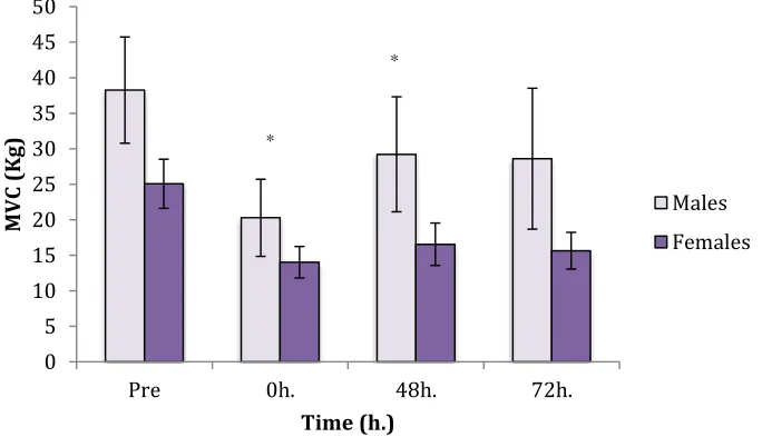
(MTR), quantified via magnetic resonance imagery, may provide a more direct indication of muscle integrity following EIMD. This study compared the temporal pattern of the MTR following EIMD to alternative indirect markers in male vs. female populations, to establish whether there were sex differences in initial damage and recovery. In animals, the antioxidant properties of estrogen have been shown to minimize muscle trauma, maintain membrane stability, and limit swelling resulting in heightened tissue integrity and resilience to EIMD in females. Six men and three women completed a standardized eccentric biceps preacher curl protocol of 10 sets of 10 reps (1 minute rest between sets) with 110% of the subjects MVC for a 3-second eccentric component. Joint angle, rate of perceived pain (RPP), maximum voluntary contraction (MVC), muscle cross-sectional area (CSA), transverse relaxation times (T2), and MTR were established at baseline prior to testing, and again at 0h, 48h, and 72h post EIMD. While joint angle and RPP revealed no significant changes, MVC showed a significant decrease in men but not women (males: 38.25Kg ± 7.46Kg to 20.28Kg ± 5.42Kg; females: 25.06Kg ± 3.44Kg to 14.02Kg ± 2.21Kg). Mean male and female CSA increased without significance; however,
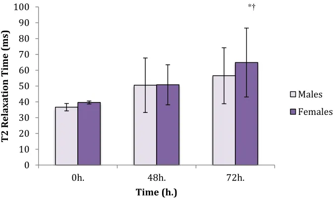
significant differences were observed between the sexes at baseline and 48h Mean T2 relaxation times of the brachialis, short, and long heads of the biceps similarly
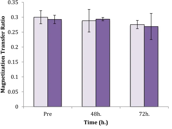
experienced no significant increases within or between sexes (P>0.05). MTR additionally showed no statistical significance within or between males and females. In a
recreationally-active population, our investigation concluded that females do not demonstrate an improved maintenance of MVC post eccentric exercise, a resilience to DOMs as determined by RPP, maintenance of joint angle, suppression of swelling as evaluated by CSA, or a decrease in changes in MTR or T2 times. While the study revealed little significance between participant groups, this may have been due to the small sample size.
Keywords:
Acknowledgments
I would like to sincerely thank the following individuals and organizations for their constant support and encouragement throughout my research
Dr. Greg D. Marsh
Jeff Watson at Kirkley Stadium
Dr. Charles Rice Neuromuscular Lab
Mr. Alex Moull, Mr. Colin Moore, Mr. Eric Kirk, and Mr. Brendan Major
The numerous participants who generously offered their time and effort to the study.
Special thanks to Western University Kinesiology graduate office
Table Of Contents:
Abstract:………..ii
Acknowledgements:………....iii
Table of Contents:………...iv
List of Tables:………...vi
List of Figures:………....vii
List of Abbreviations:……….viii
List of Appendices:………...…..ix
Chapter 1.0 Introduction:………...1
Chapter 2.0 Literature Review:………. 2
2.1 Normal Muscle Function………...3
2.2 Evaluating and Quantifying Exercise-Induced Muscle Damage………..5
2.3 Sex Differences In Exercise-Induced Muscle Damage ………...6
2.4 Mechanisms of Damage………...10
2.5 Electric Contraction Coupling & Cellular Response………12
2.6 Inflammation………...12
2.7 Metabolic Accumulation, Protein Markers, and Endocrine Reaction…...16
2.8 Magnetization Transfer Ratio & Contrast………...24
Chapter 3.0 Methods:……….25
3.1 Participants……….…...25
3.2 Protocol Overview………25
3.3 Joint Angle……….…...26
3.4 Rate of Perceived Pain ……….26
3.5 Evaluating Maximum Voluntary Contraction………..27
3.6 Eccentric Damage Protocol………..27
3.7 MR Sequencing………....28
3.8 Magnetization Transfer………....29
3.9 T2 Relaxation Time……….30
3.10 Cross-Sectional Area………...30
4.1 Participant Characteristics ………..31
4.2 Joint Angle………...32
4.3 Rate of Perceived Pain……….33
4.4 Maximum Voluntary Contraction………34
4.5 Cross-Sectional Area ………..35
4.6 T2 Relaxation Time……….37
4.7 Magnetization Transfer Ratio………..39
Chapter 5.0 Discussion:………...43
5.1 Maximum Voluntary Contraction………...43
5.2 Rate of Perceived Pain ………...46
5.3 Cross-Sectional Area………..48
5.4 T2 Relaxation Time & Magnetization Transfer Ratio………....49
5.5 Effects of Estrogen On Markets of Inflammation………..52
Chapter 6.0 Conclusion:……….55
Chapter 7.0 Study Limitations:………..57
Chapter 8.0 Future Directions:………...58
References:……….60
Appendices:………....68
List of Tables:
Table 1: Timeframe of Procedures………26
Table 2: Subject Characteristics ……….. 32
Table 3: Joint Angle Values………..32
Table 4: Rate of Perceived Pain Values………...….33
Table 5: Maximum Voluntary Contraction Values………...34
Table 6: Cross-Sectional Area Values………...35
Table 7: T2 Relaxation Times ………..37
List of Figures:
Figure 1: Mean Elbow Angle (Males vs. Females)………. ………..33
Figure 2: Maximum Voluntary Contraction Sex Differences ………...35
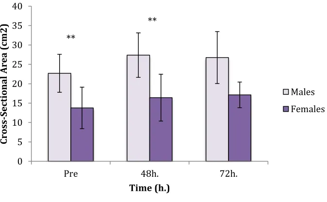
Figure 3:Cross-Sectional Area Sex Differences……….……36
Figure 4: Axial Resonance Images Depicting CSA Increases………....37
Figure 5: T2 Values of the Brachialis in Males and Females………38
Figure 6: Combined T2 Values of the Brachialis and Short / Long (b) Heads of the Biceps in Males & Females………...39
Figure 7: Mean MTR Value for Slice 25 Sex Differences……….40
Figure 8(a) and (b): Mean MTR Values for Slices 20(a) and 10(b) for Males and Females ……41
List of Abbreviations:
1RM: One Rep Max ALD: Aldolase
ANOVA: Univariate Analyses of Variance AMP: Adenosine Monophosphate
AO: Antioxidant
ATP: Adenosine Triphophate CON: Concentric Contraction COX-2: Cyclooxygenase CK: Creatine Kinase CSA: Cross-Sectional Area
DOMs: Delayed Onset Muscle Soreness E1: Estrone
E2: Estradiol E3: Estriol
ECC: Eccentric Contraction
EcSOD: Extracellular Superoxide Dismutase EIMD: Exercise-Induced Muscle Damage ES: Effect Size
EU: Eumenorrheic GH: Growth Factor
HIF-ß1: Hypoxia Inducible Factor HRT: Hormone Replacement Therapy IL: Interleukin
ISO: Isometric Contraction
LCA: Lymphocyte Chemoattractant Activity LDH: Lactate Dehydrogenase
LIF: Leukemia Inhibitory Factor Mb: Myoglobins
NMR: Nuclear Magnetic Resonance
MnSOD: Manganese Superoxide Dismutase MT: Magnetization Transfer
MTR: Magnetization Transfer Ratio MVC: Maximum Voluntary Contraction NOC: Non Contraceptive
OC: Oral Contraceptive OUX: Ovariectomized PCr: Phosphocreatine Pi: Inorganic Phosphate PL: Placebo
ROI: Region of Interest RPP: Rate of Perceived Pain SD: Standard Deviation
T2: Transverse Relaxation Time TGF-ß1: Transforming Growth Factor TNF: Tumour Necrosis Factor
List of Appendices:
Chapter 1.0 Introduction:
The extant literature establishes the potent antioxidant properties of estrogen as a
critical component in heightened muscle tissue integrity characteristic of females. While
much of the research concentrates on the animal model, Amelink et al. (1990), Moordian
(1993), and Hubal et al. (2008), ground the argument with the inclusion of human
subjects. The antioxidant properties of estrogen hypothetically minimize muscle trauma,
maintain membrane stability, and limit creatine kinase (CK) secretion and inflammation
resulting in heightened tissue integrity and resilience to exercise-induced muscle damage
(EIMD) (Amelink et al., 1990; and Hubal et al., 2008). In comparison to females, males
will experience an exaggerated decrease in skeletal muscle tissue integrity following
severe trauma (Moordian 1993). However, this research, focused on the biochemical
interactions of estrogen, fails to establish the functional or practical repercussions of
estrogen on stabilizing and improving tangible markers of performance and recovery,
post EIMD. Likewise, the consistency and correlation of serum CK and certain
inflammatory markers as indicators of acute muscle damage are contested (Friden and
Lieber 2000).
No direct, quantitative, and non-invasive markers presently exist to assess EIMD.
Markers, such as soreness, strength loss, cross-sectional area, T2 relaxation times, protein
markers, and muscle biopsies provide merely inferential indicators of EIMD. However,
magnetization transfer ratios (MTRs), quantified via magnetic resonance imagery, may
provide a more direct indication of muscle integrity following EIMD. By comparing Mo
(signal emitted without a pre-saturation pulse) and Msat (signal emitted with a
and the tightly bound macromolecular protons found in proteins (Henkelman et al.,
2001). A decrease in the MTR thus reflects a reduction in the structural integrity of the
tissue or the extent of decay or damage. Typically applied in a neurological context, MTR
histograms are sensitive to subtle changes in the grey matter of the cerebral cortex and
cerebellum and are subsequently a critical measure of cognitive function alongside
standard MR protocols including T2, volume, and cross-sectional area (CSA) (Pinter et
al., 2015). Potentially capable of qualifying diverse tissue types, MTR should be
applicable in the context of sport medicine and evaluating muscle integrity.
The purpose of this study was to compare the temporal pattern of the MTR to
alternative indirect markers of EIMD in male vs. female populations. Based on previous
work that had shown a protective effect of estrogen, it was hypothesized that males
would sustain greater EIMD than females as indicated by a lower MTR and greater
changes in muscle soreness, strength loss, cross-sectional area, and T2 relaxation times.
Females would also show a more rapid recovery of these variables. The temporal pattern
of the MTR was expected to most closely resemble that of the CSA, an indicator of
swelling, with both peaking at around 72-96h and be consistent with other markers of
EIMD. The elucidation of sex differences in recovery from training will have
implications for both high performance athletes as well as for therapy regimes among
senescing populations.
Chapter 2.0 Literature Review:
Readily adaptive to mechanical contractile activity, skeletal muscle hypertrophy is
thoroughly documented in the extant literature. Skeletal muscle adaption and
the mechanical stress of exercise, the severity of EIMD is dependant on exercise
familiarity, type, duration, and intensity. Controlled EIMD is generally associated with
localized macromolecular and sarcolemmal tearing, as well as with disruption of
contractile elements, connective tissue, and the cytoskeleton (Schoenfeld, 2012).
Exercise-induced trauma is additionally identified by acute and chronic metabolic and
endocrine responses and cellular signaling. Outwardly, EIMD is characterized by an
immediate decrease in muscle function (Bryne et al., 2001), performance (Sargeant &
Dolan, 1987), an increase in intramuscular swelling and localized discomfort (Chapman
et al., 2008), and an increase in circulating muscle proteins, notably creatine kinase (CK),
lactate dehydrogenase (LDH), and myoglobins (Mb) (Nosaka & Clarkson, 1996; Sayer et
al., 2003).
2.1 Normal Muscle Function:
Characterizing EIMD requires a preliminary appreciation of normal
neuromuscular function. Skeletal muscle motor units contain a range of motor neurons
that each innervates a set of muscle fibres demonstrating particular characteristics. Motor
units are selectively recruited by the central nervous system according to the “size
principle”: a motorneuron’s susceptibility to discharge and excitation corresponds
inversely to size (Luscher et al., 1979). Smaller fibre diameter correlates with quicker
depolarization and a denser excitatory current - resulting in hypersensitivity. An increase
in contraction intensity reflects the ordered and selective recruitment of individual muscle
fibres into larger cohorts during excitation by contraction coupling. Excitation
contraction coupling defines the process by which physiological mechanical movement is
cascade at the cell surface that results in muscle contraction.
Muscle contraction is ultimately achieved through the longitudinal activation of
sarcomeres in series. However, multiple contraction types define movement: concentric
contraction (CON), eccentric contraction (ECC), and isometric contraction (ISO). CON
suggests that summation of force produced by contracting sarcomeres exceeds external
impeding forces, such as gravity, antagonist muscles, or load. ISO indicates the
summation of sarcolemmal force is equal to antagonistic influences. Finally, ECC
denotes that external forces exceed the force of contracting sarcomeres, resulting in the
lengthening of the muscle fibres.
Although large muscle mass is popularly associated with the male sex hormone
testosterone, skeletal muscles expresses estrogen receptors. These receptors provide an
equally important facet of healthy neuromuscular and mechanical function. The
estrogen receptor, most prevalent in skeletal muscle, remains responsive to circulating
estrogen as well as cues released by exercise. Receptor expression is significantly
increased post exercise, which suggests that estrogen receptors and associated ligands are
critical for skeletal muscle recovery and function (Spangenburg et al., 2012). The
research of Phillips et al. (1993) examines the preservation of strength in postmenopausal
women who have been administered estrogen hormone therapy. These results are
supported by a meta-analysis of extant literature by Spangenburg et al. (2012) concerning
estrogen as a preservative of muscle strength in postmenopausal women: estrogen
therapy provides a minor benefit to strength (effect size = 0.23) as well as force
development (effect size = 0.66).
contractile proteins, specifically permeabilized fibers (Qaisar et al., 2013). Lacking intact nerve
endings or distinct membranes, permeabilized fibers bypass excitation contraction coupling and
respond directly to protein contractile function. Permeabilized rat soleus fibers demonstrated a
20% decrease in muscle force generation 10-14 weeks following ovariectomy in comparison to
the control (Widrick et al., 2003). Indeed the research of Spangenburg et al. (2012) confirms
decreases in 17 b-estradiol impaired strong-binding myosin and subsequently limited the
muscle’s force generating capacity. Estradiol positively affects muscle force generation
independent of nerve enervation or muscle activity. Deficiencies in ovarian hormones
contributed to a decrease in isometric titanic force (normalized to CSA) as well as a 20%
decrease in soleus contractile protein content in hindlimb suspended and muscle denervation
models respectively (Spangenburg et al., 2012). Normal - optimal - muscle function, regardless
of EIMD, thus necessitates the presence of estrogen receptors as well as 17 b-estradiol.
2.2 Evaluating and Quantifying Exercise-Induced Muscle Damage:
Currently there exists no direct, non-invasive, quantitative marker to assess EIMD
(Paulsen et al., 2012). Indirect markers such as soreness, strength loss (MVC),
cross-sectional area, T2 relaxation times, and changes in muscle torque and optimal length
provide merely inferential indicators of EIMD. While muscle biopsies and blood protein
markers offer more directly quantifiable means of measurement, their invasive and
inherently damaging nature and subsequent effect on muscle performance and tissue
sample integrity make these deterrent. Additionally, the varied pattern of whole-muscle
damage is perhaps only roughly approximated by localized muscle biopsies.
Nuclear Magnetic Resonance (NMR) offers a viable alternative for assessment of
immediately observed following exercise, particularly eccentric exercise, yet dissipates
rapidly. A secondary increase in T2 relaxation times occurring 12h - 24h post exercise
and climaxing at 72h -120h correlates directly with increases in CSA, muscle volume,
protein markers, and delayed onset muscle soreness (DOMs). Such correlation suggests
that increases in T2 reflect cell rupture and edema as well as restorative adaptations
(Takahashi et al., 1994; Mair et al., 1992). The use of magnetic resonance imagery, and
more specifically magnetization transfer ratios, provides a direct visual indication,
qualification, and quantification of whole muscle integrity following EIMD (Jayaraman
et al., 2004). Correlating direct and indirect markers of EIMD to MTR guarantees a
complete and comprehensive appreciation of muscle trauma on the cellular level and its
subsequent effects on indirect performance measures. While these indirect and direct
markers are not specifically associated with sex differences in cellular biochemistry or
indeed estrogen, they might serve to reveal trends suggestive of the hormone’s potential
stabilizing, anti-inflammatory, and restorative properties.
2.3 Sex Differences of Exercise-Induced Muscle Damage:
An 18-carbon corticosteroid protein, estradiol, is secreted predominantly by the
ovaries in females and in the Leydig and Sertoli cells of the testes in males. Estrogen is
also synthesized from its precursors, progesterone via androgen intermediates such as
testosterone, in other tissues such as adrenal, liver, adipose, breast, and neural tissues
(Simpson, 2003; Carreau et al., 2003). The hormone is implicated in sexual and
reproductive function as well as cardiovascular, musculoskeletal, and immunological
systems. In skeletal muscle, estrogen bears a significant effect on muscle contractile
of estrogen exist: estradiol (or E2, which is the most abundant), estriol (or E3), and
estrone (or E1). In the context of musculoskeletal function, estrogens reportedly attenuate
creatine kinase concentrations, intramuscular lysosomal acid hydrolase ß-glucuronidase,
and the infiltration of inflammatory neutrophils and macrophages following
exercise-induced muscle damage (Amelink et al., 1990). Estrogen additionally stimulates
regenerative systems through the localized activation and proliferation of satellite cells –
a result of its intercalation into the phospholipid bilayer that has the effect of stabilizing
cellular membranes, probably by conferring antioxidant protection (Deborah et al., 2010).
There are many other indications of an estrogen-mediated effect upon muscle. For
example, there is a decreased incidence of cardiac disease in pre-menopausal women,
who presumably enjoy higher relative titers of estrogens, as well as a reduction in the
grade of myocardial trauma following ischemia-reperfusion injury (Booth et al., 2008).
Indeed, in rabbit studies inhibition of estrogen-activated cyclooxygenase (COX-2) or of
prostaglandin pathways, particularly PG12, blunted cardioprotective effects of estrogen.
Acute administration with 17ß-estradiol following reperfusion “significantly decreased
infract size as a percent of area at risk” (Booth et al., 2008). Estrogen’s stabilizing effect
upon cardiac muscle is presumably replicated in skeletal muscle.
Estrogen’s influence on regenerative processes via activation and proliferation of
satellite cells is attributed to its structural resemblance to antioxidants, vitamin E in
particular. Indeed, the hormone successfully “scavenges free radicals” while
simultaneously activating antioxidant enzymes responsible for minimizing oxidative
damage (Deborah et al., 2010). Strehlow et al. (2003) illustrates estrogen’s
manganese- and extracellular- superoxide dismutase (MnSOD and ecSOD respectively)
expression in circulating human monocytes (255 ± 52% and 219 ± 15%).
Correspondingly, estrogen deficiencies were characterized by a “downregulation” of
MnSOD and ecSOD mRNA (43 ± 6% and 63 ± 3% of control respectively) in vessel
walls and an increase in circulating free radicals. This suggests a greater propensity
towards membrane instability (Strehlow et al., 2003).
Typically, steroids activate or repress particular target genes. Some of them can
do this by two separate routes. For example, estradiol will bind to Steroid Nuclear
Receptor superclass (estrogen receptors ER and ) or to G-coupled membrane-bound
class receptors. The former will traffic directly to the nucleus where the ligand/receptor
complex interacts with specific DNA sequence motifs. The latter activates a cascade of
signaling steps that transfer the membrane stimulus to a nuclear effect
(Heldring et al., 2007; Prossnitz et al., 2007). Additionally, three potential non-genomic
venues for steroid action exist: membrane bound receptor binding, neurotransmitter
receptor binding, and integration of lipophilic steroids into the membrane.
Lipophilic in nature, estrogen additionally bears structural similarity to
cholesterol. As a consequence, it possesses the capacity to intercalate in the cell plasma
membrane, specifically the phospholipids, altering membrane fluidity and providing a
stabilizing effect, possibly also by scavenging free radicals and thereby preserving
membrane integrity. Variances in lipid fluidity provide a “mechanism of action” for
steroids, specifically estrogen, since membrane fluidity changes membrane function and
thereby modulates the activation and proliferation of membrane-bound enzymes (Whiting
membrane bilayer (Velten & Kempfle, 1993), 17ß-estradiol significantly increases lipid
fluidity of the membrane altering the mobility of enzymes and subsequently increasing
Ca++ ATPase from the sarcoplasmic reticulum membrane (Whiting et al., 2000). The
effects of estrogen on the membrane act jointly with other “potential non-genomic
effects” that stimulate rapid steroid reactions. Whiting et al. (2000) identifies estrogen
intercalation as a contributing factor, not as a substitute, for conventional
receptor-mediated mechanisms. In part, this diversity of modes of action may explain why
estrogen has such a broad spectrum of effects that can exert their influences in such a
cell- and tissue specific manner. More germane to this discussion, steroidal intercalation
could contribute to greater membrane stability, and subsequently, to resistance to
exercise-induced muscle damage.
Moreover, estrogen facilitates recovery processes. Specific hormone variances
during the menstrual cycle are related to the effect of estrogen on muscle’s regenerative
mechanisms following EIMD. During the ovulatory phase, muscle strength recovered and
returned to baseline faster than damage incurred during the follicular phase. 17ß-estradiol
remained significantly higher (P<0.005) in the ovulatory phase versus the follicular phase
(229.6 ± 81.8 pmol/L vs. 119 ± 61.7 pmol/L) (Sipaviciené et al., 2013). Maximum
voluntary contraction as well as changes in torque evoked by 1 second trains of electrical
stimulation at 20 Hz and 100 Hz recovered at 72h post exercise if the challenge was
issued during the ovulatory phase, yet still remained suppressed in the follicular phase.
Alternative markers of muscle damage and recovery (CK, soreness, and fatigue)
remained relatively consistent between the ovulatory and follicular phases suggesting that
– they were unlikely to be acting via a systemic influence. Sipaviciené (2013)
hypothesizes that the protective effects of estrogen correlate to its suppression of
leukocyte infiltration, containment of membrane damage, and the promotion of recovery.
Carter et al. (2001) similarly confirm the protective effects of estrogen. A
comparison of oral contraceptive (OC) and eumenorrheic (EU) subjects in their
mid-luteal and mid-follicular phase, respectively, revealed significant changes in CK secretion
and DOMs following an eccentric exercise protocol of 30 minutes downhill running at
60% VO2 Max. While DOMs increased significantly respective of contraceptive use, EU
subjects demonstrated greater CK at 72h post exercise (P<0.05), which suggests a
reduced degree of muscle damage in the estrogen contraceptive group (Carter et al.,
2001). The elevated estrogen and progesterone characteristic of the mid-luteal phase
confer a greater protective effect in comparison to the low levels of estrogen and
progesterone during the mid-follicular phase of the menstrual cycle. Carter attributes the
stabilizing effect of estrogen to its antioxidant properties and membrane stabilizing
nature. While the research of Sipaviciené (2013) and Carter (2001) differ regarding the
localized vs. systemic nature of estrogen’s protective effects, the authors mutually
attribute the suppression of an inflammatory response to the hormone’s protective
potency.
2.4 Mechanisms of Muscle Damage:
Fridén & Lieber (1992) identify skeletal muscle damage or a disruption of muscle
homeostasis as “primary or secondary” disruption of the sarcolemmal, alteration of
myofibril contractile units, cytoskeletal damage, and finally, disruption of the
sarcomere theory describing the progressive “popping” of sarcomeres as individual fiber
load exceeded maximum tensile strength (Morgan, 1990). By contrast, Fridén & Lieber
(1992) found that neural recruitment patterns during eccentric muscle contraction
irregularly loaded individual sarcomeres, and that this resulted in uneven force
distribution along actin filaments. This in turn caused excessive elongation of sarcomeres
and potential Z-disk streaming. The degree of muscle trauma and Z-disk streaming was
similarly identified as “broadening, smearing,” and “total myofibrillar disruption” (Fridén
& Lieber, 1992). Such a disruption of myofilament substructures is perhaps attributed to
sarcomerogenesis, deteriorative events / enzymes, or to a localized imbalance in
myofibrillar protein homeostasis (Fridén & Lieber, 1992; Morgan, 1990).
Following consecutive strenuous eccentric contractions, overstretched sarcomeres
fail to “re-interdigitate” eventually causing membrane damage and an immediate
reduction in maximum voluntary contraction (Proske & Morgan, 2001). The magnitude
of damage increases in correlation with an increase contraction velocity. Individual fibers
experience greater mechanical stress when contraction velocity increases (Chapman et
al., 2006). Indeed, an eccentric contraction at 1% V max increases tension in excess of
50% Po suggesting cross-sectional tension increases “10 times faster” during the
eccentric in opposition to the concentric when velocity is normalized (Fridén & Lieber,
1992). Such variances in eccentric vs. concentric tensions reflect irregular sarcomere
loading and predispose towards sarcolemmal disruption. Actin-myosin bonds are
mechanically separated in contrast to control ATPase enabled disengagement (Edwards et
al., 1977). Fridén & Lieber (1988) suggest mechanical tearing contributes to the efflux of
Proske & Morgan (2001), it seems that overstretching and mechanical tearing of the
sarcomere occur on the descending leg of the length tension curve.
2.5 Excitation Contraction Coupling and Cellular Response:
The ultimate failure of excitation contraction coupling is associated with the
mechanical tearing of actin and myosin filaments during eccentric contraction and with
excessive Ca++ muscle saturation, rather than controlled ATPase separation (Morgan &
Allen, 1999 and Armstrong, 1990). Trauma to the sarcoplasmic reticulum may heighten
membrane permeability and subsequently increase intracellular Ca++ concentrations
(Armstrong, 1990). The presence of Ca++ in the sarcoplasm activates proteolytic
pathways integral to muscle fiber degradation and repair. Indeed, calcium is directly
responsible for muscle protein turnover via the activation of proteases, phopsolipases,
and lysosomal enzymes (Zang et al., 2008). However, Belcastro identifies the
calcium-activated neutral protease, calpain, as the dominant mediator of muscle damage
post-eccentric contractions, specifically Z-line protein degradation. The introduction of
leupeptin, a calpain inhibitor, significantly stifles excitation-contraction uncoupling and
titin degradation following excessive Ca++ saturation (Belcastro et al., 1998). Verburg et
al. (2005), confirmed titan degradation via endogenous calpains following prolonged
Ca++ exposure. Following moderate intensity eccentric contractions, titin
immunostaining, reflective of titin degradation, increased significantly (P>0.05). Indeed
the research of Zhang et al. (2008) confirms that the removal of extracellular Ca++
improves muscle function post exercise-induced muscle damage.
2.6 Inflammation:
produces inflammation – a result of mechanical tearing, suppressed excitation contraction
coupling / calcium signaling, and stimulation of “calcium-sensitive degradation
pathways” (Peake et al., 2005). Localized inflammation responsive to EIMD is
recognized by leukocyte infiltration, the production of pro-inflammatory cytokines, and
adjustments in leukocyte receptor expression (Peake et al., 2005). Following eccentric
exercise, localized neutrophil circulation dramatically increases as does cytokine activity
and responsiveness. Neutrophils are predominantly responsible for the containment and
disposal of damaged tissue via phagocytosis, respiratory burst, and degranulation
subsequent to EIMD (Butterfield, 2010). Alternatively, cytokines limit muscle fibrosis
and stimulate regenerative processes.
While the prevalence of neutrophils gradually increases (peaking at 175%) within
the initial 24h period of EIMD, macrophage concentrations increase thereafter alongside
pro-inflammatory cytokines including interleukin 1ß, tumor necrosis factor (TNF-a), IL-6
and transforming growth factory (TGF-ß1), and finally, inflammatory leukemia
inhibitory factor (LIF) and hypoxia inducible factor (HIF-1ß). Macrophage
concentrations correlate with the secondary cytoskeletal disruption associated with EIMD
(Tidball, 2005; Pizza et al., 2005). The research of Tidball (1995) indicates that
pro-inflammatory cytokines released by traumatized fibres are responsible for the emergence
and accumulation of neutrophils; this suggests that inflammation remains secondary and
responsive to cellular disruption. However, Pizza et al. (2005) demonstrate the presence
of neutrophils in the extracellular matrix of intact skeletal muscle. Inflammation may
potentially precede or occur exclusive of cytoskeletal damage. The localized
(Peake et al., 2005). However, systemic increases in pro-inflammatory cytokines,
specifically IL- 1ß and TNF-a, are minor. Indeed, a systemic increase in
anti-inflammatory cytokines IL-1ra, IL-4, IL-10, and IL-14 suggests a systemic inhibitory
response to localized inflammation (Peake et al., 2005).
Ovarian hormones are integral to immune system function and subsequently
inflammatory processes. McClung et al. (2006) evaluated the inflammatory response
associated with muscle damage and “ovarian hormone depletion”. Muscle neutrophil
concentration remained 43% greater in ovariectomized (OVX) female rats three days
following exercise in comparison to the control. ED1+ macrophage concentrations
similarly increased (exponentially) by 511% and 593% at one and three days post-EIMD
respectively. However, in OVX muscle ED1+ remained increased (558%) seven days
post-exercise protocol. Localized increases of ED1+ are indicative of skeletal myofiber
necrosis. Ovariectomy therefore increased myofiber necrosis 51% (P=0.008). In OVX
muscle ED2+ macrophages additionally increased 111% immediately and remained
elevated for 3 and 7 days at 64% and 59% respectively. In contrast, in the intact control,
ED2+ macrophage concentration only increased on day 3 of recovery (McClung et al.,
2006). Inflammatory cytokines, indicative of muscle regeneration, multiplied in both
OVX and control groups. IL-6 mRNA increased 138% and 291% 1 and 3 days
post-exercise, returning to baseline within a week. IL-mRNA concentrations additionally
increased 1692% and 77% 1 and 3 days post-exercise (McClung et al., 2006).
The research of Moordian (1993) further confirms the antioxidant properties of
estriol and 17 beta-estradiol in comparison to testosterone. The decay of fluorescence
17 alpha and beta estradiol as well as estriol (84.0 ± 5.42, 74 ± 6.3, and 64.2 ± 2.53
respectively as percentage of AAPH slope). Testosterone, in comparison, conferred no
such benefit with regards to suppressing decay. Indeed the percentage of AAPH slope for
testosterone was 108.6 ± 8.74, well above (P<0.05) those percentages of 17 alpha and
beta estradiol as well as estriol. Moordian (1993) concludes that estrogens impart
significant antioxidant “potency.”
Markers of inflammation, specifically muscle neutrophil (myeloperoxidase) and
macrophage (SOD1 and 2) concentrations, were significantly altered in men following
17-estradiol (E2) supplementation. A 250% increase in E2 concentration effectively
blunted neutrophil infiltration at 3h and 48h post-exercise (P<0.001) as well as
macrophage SOD1 content at 48h (MacNeil et al., 2009). SOD2 concentration increased
230% regardless of E2 treatment. Plasma total antioxidant capacity as well as expression
of membrane homeostasis genes (forkhead box O1 or FOXO1, caveolin, and sterol
regulatory element binding protein-2) were similarly unaffected by E2 supplementation
(MacNeil et al., 2009). While E2 successfully hampers neutrophil infiltration, the
hormone’s anti-inflammatory properties are not anti-oxidant related.
Savage et al. (2000) parallels the research of McClung et al. (2006) regarding the
effects of estrogen suppression and increased inflammation. In a comparison of oral
contraceptive (OC) and non-contraceptive (NOC) use following eccentric muscle damage
of the elbow flexors, estrogen-impaired subjects demonstrated quantifiably greater
inflammation. Inflammation was evaluated via arm circumferences 4cm above (MID)
and 8 cm below (DIS) the elbow. Inflammation was significant 3-5 days post-exercise in
inflammation merely reached a maximum of 24.0 cm, 0.9 cm above baseline, while
estrogen-mediated inflammation reached 26.1 cm, 1.6 cm above baseline. DIS
measurements revealed a similar tendency. NOC inflammation measurements
demonstrated no significance while OC inflammation increased to 24.5 cm, 0.9 cm above
baseline (Savage et al., 2000). A greater propensity towards inflammation in estrogen
deficient subjects following traumatic exercise is confirmed by elevated concentrations of
neutrophils, macrophages ED1+ and ED2+, as well as IL-6 and IL-mRNA, and finally
a tangible increase in muscle circumference.
2.7 Metabolite Accumulation, Protein Markers, & Endocrine Reaction:
In skeletal muscle, specific metabolic and protein markers identify severe cell
disruption. Metabolic stress provoked by resistance exercise is defined by a depletion of
adenosine triphosphate (ATP) and phosphocreatine (PCr), in conjunction with a decrease
in intramuscular pH and an increase in inorganic phosphate (Pi), adenosine
monophosphate (AMP) and lactate. The hypoxic and ischemic intramuscular
environment initiated by resistance exercise remains an essential precursor of skeletal
muscle damage and subsequently physical adaptation (Goto et al., 2005). Intramuscular
metabolites and pH as well as the Pi surges, indicative of fast twitch fiber recruitment and
subsequently increased muscle fatigue and damage, were significantly higher (P<0.001)
following conventional high intensity exercise (Suga et al., 1985).
The metabolic stress model identifies metabolic accumulation as a dominant
factor provoking EIMD and subsequent symptoms: metabolite accumulation increases
the vulnerability of muscle fibers to mechanical stress (Tee et al., 2007). The decreased
removal of Ca++ thus provoking an increase in cytosolic Ca++ and an influx of
metabolites that cause muscle fiber degeneration (Armstrong et a., 1991). Alterations in
oxidative metabolism following exercise additionally allude to the potential influence of
metabolic variances on muscle integrity.
An increase in the ratio of phosphocreatine to inorganic phosphate (PCr/Pi), as
well as increased titers of free ADP, and blood lactate indicate an increased metabolic
rate and an increased reliance on non-oxidative metabolism (Tee et al., 2007). Walsh et
al. (2001) identify the increase in resting muscle oxygen consumption, the decrease in
oxygen availability due to restricted diffusion, and finally the decrease in maximal
mitochondrial respiration and ADP sensitivity, as mechanisms potentially altering
oxidative metabolism following exercise.
Interrupted ovarian function or low estrogen secretion - exerts a negative effect on
metabolism in other ways too. Jackson et al. (2012) identified a disposition towards
metabolic disorder, specifically dysfunction of skeletal muscle lipid metabolism and
mitochondrial function that result from a disruption of peripheral glucose homeostasis in
skeletal muscle. Ovariectomized mice demonstrated significantly increased (P<0.05)
skeletal muscle intramyocellular lipids and an equivalent increase in fatty acid
transporters CD36/Fat and FABPpm (160% and 115% increase respectively) (Jackson et
al., 2012).
While intramyocellular lipids are not traditionally associated with hindering
skeletal muscle function, excessive accumulation promotes nonesterfied free fatty acid to
circulate and effectively sabotage (increase) insulin resistance via stimulation of the
significant decrease in acyl-carnitine species and critical amino acids; this suggests a
fundamental impairment of mitochondrial oxygen consumption and subsequently of lipid
and amino acid metabolism (Jackson et al., 2012). A comparative 33% decrease in
circulating sort-chain acyl-carnitines (P<0.05) in ovariectomized mice indicates a
decrease in -oxidation pathways and a subsequent impairment of skeletal muscle lipid
metabolism at a basal level following exercise (Jackson et al., 2012). However, groups
demonstrated no variance in CPT-1 mRNA or malonyl CoA content. CPT-1 determines
fatty acid transportation through -oxidation while CPT-1 remains negatively regulated
by malonyl CoA (Rasmussen, 2002).
Intense eccentric contractions inevitably correlate with increases in markers of
acute muscle damage, namely creatine kinase (CK), cytokines, interleukin-6 (IL-6),
myoglobins, and tumor necrosis factor alpha (TNF ). Kanda et al. (2014) monitored the
effects of eccentric exercise on “serum leaking enzymes” and “new biomarkers” of
EIMD. Following an intensive (10 sets of 40 reps) calf lifting protocol, protein markers
of muscle trauma increased significantly. Creatine kinase (CK) and aldolase (ALD)
increased at 72h (P<0.05), remaining elevated until 96h (P <0.01) post-exercise bout.
Indeed, ALD activity corresponded directly to “tenderness” of the middle gastrocnemius.
Lactate dehydrogenase (LDH) demonstrated similar temporal increases (P<0.05) (Kanda
et al., 2014). However, following an identical eccentric exercise protocol, the authors
established no significance in variations of cytokine concentrations. Plasma and urine
samples demonstrated increased neutrophil migratory activity (P<0.05) 4h post eccentric
exercise bout. However, no significant changes in alternate plasma and urinary
and IL-12p70 (Kanada et al., 2013). Isolated eccentric exercise likely provokes limited
localized inflammation and regional neutrophil mobilization associated with preliminary
muscle damage and inflammation.
Plasma CK concentrations, indicative of muscle membrane trauma, are
significantly higher in male and ovariectomized female rats. Amelink et al. (1990)
similarly indicate an inverse relationship between estrogen supplementation and CK
concentrations, thus further solidifying the membrane-stabilizing effects of estrogen post
EIMD. Indeed, the efflux of CK remained 172% higher in the estrogen suppressed (male)
model (2.25 vs. 1.31 U, P=0.035). Sipaviciené et al. (2013) additionally promote the
protective or mitigating effects of estrogen either post exercise or post intensive
stretch-shortening contractions. During the follicular phase of the menstrual cycle, when
estrogen remains relatively suppressed (in comparison to the ovulation phase), CK
concentrations remained elevated post exercise (11 IU/L vs. 8 IU/L 24h post exercise and
7 IU/L vs. 4 IU/L 48h post exercise) (Sipaviciené et al., 2013). Decreased sensitivity and
increased membrane stability during peak ovulation and subsequently peak estrogen
further solidify the mitigating nature of estrogen.
Wolf et al. (2012) additionally documented an increase in CK concentrations from
baseline at 24h post exercise in men vs. women following an acute heavy resistance
exercise protocol of six sets of five squats at 90% of the subjects’ one repetition
maximum. However, estradiol receptor expression on granulocytes, indicative of
neutrophil granulocyte expression (inflammatory response), demonstrated no significant
variances between the sexes, suggesting disparity in CK concentrations and tissue quality
2012). While CK levels may demonstrate alarming variance and variability, CK
concentrations provide (at the very least) indirect means of quantifying and qualifying
exercise-induced membrane damage. Increased permeability of the sarcolemma, a result
of membrane trauma, facilitates the diffusion of CK from muscle into the bloodstream
(Sipaviciené et al., 2013).
The accumulation of metabolites increases intramuscular reperfusion and
intracellular swelling – effectively sabotaging the structural integrity of cell membranes.
The anabolic effects of intracellular swelling on protein synthesis, myocellular hydration,
proteolysis, and subsequent skeletal muscle damage and hypertrophy are readily
confirmed (Lang et al., 1988). Acute increases in muscle effect size (ES) are attributed to
the transfer of fluid from vascular plasma to the stimulated muscle. The decrease in
plasma volume is measurable and correlates to an acute 4% increase in muscle CSA (Abe
et al., 2010). Acute muscle swelling is thought to prompt G-protein-mediated activation
of tyrosine kinase and subsequently the mTOR and MAPK pathways, indicative of
muscle damage leading to adaptive muscle hypertrophy (Loenneke et al., 2012).
Responsive to acidosis and metabolite / proton accumulation, endocrinal
hormones, specifically growth hormone (GH) and testosterone, are certainly affected by
resistance exercise. Indeed following an intense bout of exercise (6 sets of 10 repetition
of squats), GH and testosterone concentrations increased 9.5 ± 7.3µg·L(-1) and 31.4 ±
10.3nmol·L(-1) respectively, and remained elevated at 15 (P15) and 30 (P30) minutes
post bout (Shanner et al., 2014). The increase in GH is attributed to metabolite
accumulation and the ensuing stimulation of sympathetic nerve activity that is
and the mTOR pathways via janus kinase 2 and phosphatidulinositol-3 kinase signaling;
however, the extent to which acidosis elicits hypertrophic adaptation via this GH pathway
remains unclear. Nevertheless, Reeves et al. (2006) determined “no change” in resting
total testosterone (T; mean 15.7 ± 1.6 nmol/L), free testosterone (FT; 54.1 ± 4.5 pmol/L),
and cortisol (267.6 ± 11 nmol/L) pre-exercise, immediately post-exercise, and 15min post
moderate intensity exercise. Static testosterone concentrations are perhaps reflective of
low (moderate) exercise intensity. In the context of an acute systemic hormonal response
to resistance exercise, higher intensity and total muscle mass stimulation are likely
necessary to provoke alterations in testosterone secretion.
Nunes et al. (2011) confirm an increase in testosterone, cortisol, and
immunoglobulin post exercise in elite female athletes. Following endurance, strength, or
power resistance exercise regimes, salivary samples revealed a significant increase in
testosterone, cortisol, and immunoglobulin (P<0.05) associated with an increase in
metabolic demand and subsequently “training strain” (Nunes et al., 2011).
While testosterone remains a critical stimulant of recovery and adaptation post
exercise, estrogen, particularly 17-estradiol (E2), exhibits similar recuperative
properties. Evaluating salivary , interlukin-6 production, and metabolic substrate use,
Ives et al. (2010) compared the hormone profile of 10 female subjects during the
midfollicular and midluteal menstrual phases following 1 hour of treadmill exercise at
65% of peak VO2. E2 demonstrated an acute increase immediately post exercise yet
decreased most notably upon cessation of exercise between 60 and 90 minutes post
exercise. Concentrations rapidly decreased to baseline values between 60 to 90 minutes
the midfollicular and midluteal menstrual phase groups respectively. The greatest
estradiol concentrations were observed 30 minutes post exercise at 6.75pg/ml (1.16pg/ml
above baseline P<0.05) (Ives et al., 2010). The data suggests estradiol remains hyper
sensitive and dependent on exercise intensity and duration.
Willoughby and Wilborn (2006) indicate a similar increase in 17-estradiol (E2)
following eccentric contractions of the knee extensors (7 sets of 10 at 150% of 1RM).
Blood and muscle samples obtained during the mid-luteal phase prior to exercise and
again 6, 24, 48 and 72h post exercise demonstrate an increase in serum E2. Elevated E2
correlated to myostatin mRNA at 6h (r=0.739, P=0.036) and 24h (r=0.813, P=0.014) post
exercise as well as myostatin LAP propeptides at 6h (r=0.713, P=0.047) and 24h
(r=0.735, P=0.038). Myostatin mRNA and LAP propeptides did not increase significantly
following eccentric exercise indicating a dampening of myostatin signaling and
expression by E2 and subsequently an inhibition of muscle damage (Willoughby &
Wilborn, 2006).
However, following an exhaustive swimming protocol ranging from low to
medium intensity, female adolescents demonstrated no significant increase in E2 plasma
concentration (46.4 ± 12.1 pg/ml at baseline to 58.6 ± 13.3 1h post exercise) despite a
significantly higher baseline (125%) in comparison to male counterparts (Tauler et al.,
2008). The extensive research of Wojtys et al. (2014) additionally suggests a moderate
level of exercise does not influence systematic (urinary) concentrations of estrogen,
progesterone, or luteinizing hormones. The profiling of estradiol and progesterone in 106
adolescent girls, who experienced a minimum of 3 menstrual cycles, revealed no
and creatine demonstrating a minor increase in the non-athletic control (Wojtys et al.,
2014). The authors associated the subjects’ merely “moderate” level of activity with the
lack of hormone variance between participants. A “high” intensity or level of activity
might provoke a greater systemic hormonal response.
Complementing the anabolic properties of testosterone, estrogen undoubtedly
confers protective benefits regarding muscle membrane stability and recovery post
exercise-induced damage. While a systematic increase of estrogen post exercise yet
remains unconfirmed, the hormone’s absence certainly contributes to a greater
susceptibility to muscle damage. The literature verifies estrogen’s influence on strength
through the reinforcement of strong binding myosin (Lowe et al., 2011). Its affect as a
potent radical scavenging antioxidant as well as a mimic of cholesterol capable of
intercalating in the membrane corresponds to heightened tissue resilience. The extant
literature additionally supports estrogen’s recuperative ability via the suppression of CK
plasma concentration as well as leukocyte and neutrophil infiltration (Stupka et al.,
2000). A comparison of MTR as a direct marker of EIMD to alternative indirect markers
of EIMD in male vs. female populations would effectively quantify and qualify sex
differences in biochemical activity during recovery particular to estrogen.
2.8 Magnetization Transfer Ratio or Contrast:
While muscle research pertaining to magnetization transfer ratio or contrast
imaging remains relatively scarce in comparison to T2 imaging, specifically with regards
to exercise-induced muscle damage and eccentric exercise, Yoshioka et al. (1994)
successfully quantified the effects of acute exercise-induced effects with MT techniques.
minutes post exercise in comparison to immediately post exercise. Yoshioka
characterized the increase in signal intensity as indicative of a change in
water-macromolecule interaction in skeletal muscle – in this case hyperemia post exercise.
Similarly, McDaniel et al. (1999) differentiated tissue characteristics of patients suffering
from muscular dystrophy. MT contrast was suppressed (11%-38%) in diseased muscle in
contrast to healthy tissue (38%-41%), a difference demonstrating an “inverse relationship
between symptom duration and suppression ratios” (McDaniel et al., 1999). MT
suppression rates were significantly limited with gross fatty infiltration – symptomatic of
limb girdle muscular dystrophy. Providing a quantitative as well as a qualitative measure
of disease progression, MT imaging can serve as a useful and noninvasive alternative or
complement to traditional methods of evaluating muscle tissue integrity.
While Fulford et al. (2015) observed no significant changes in MTR values
following an exhaustive ECC squat protocol of 10 sets of 10 repetitions with 70% of the
subjects’ body weight, Fulford nonetheless notes the potential of the MR technique and
suggests a more stringent restraining of the muscle during scans to minimize image noise.
Sinclair et al. (2010) additionally quantified magnetization transfer imaging in healthy
human skeletal muscle in vivo. Tracking the T(1) and B(1) excitation radiofrequency
currents utilized in the MT of seven muscles, the mean T2 relaxation times of the
restricted bound pool was 5.9 ± 0.2 1H-NMR.
The objective of the present study is to compare MTR with alternative indirect
markers of muscle damage as a means of evaluating sex differences in response to EIMD.
Joint angle, rate of perceived pain, pain algometer, MVC, MTR, T2, and CSA will be
stability, reducing EIMD, and subsequently shorting the period of recovery following
eccentric exercise.
Chapter 3.0 Methodology:
3.1 Participants:
Nine healthy, recreationally-active students, six males and three females aged
21-31, were recruited from the Western University population. Eligibility was determined by
a training frequency of, at minimum, one upper body specific resistance exercise training
session every 2 weeks consistently for 3 months. Varsity and “intensely” active
individuals as well as those suffering any injuries were excluded. The study protocol was
approved by Western University’s Ethics Board for Health Science Research Involving
Human Subjects (See Appendix A) and conformed to the Declaration of Helsinki.
Written and oral consent of the subjects were obtained following full disclosure and
explanation of the research and protocol prior to any testing.
3.2 Protocol Overview:
Table 1 shows the timeline for the various tests and measures in the study.
Following the acquisition of the baseline measures the nine subjects performed the
eccentric damage protocol (see section 3.6). Data was then acquired at 48 and 72 hr.
Table 1: Timeframe of test protocols
3.3 Joint Angle:
Prior to measurements of pain, strength, or muscle quality, participants’ resting
(hanging) elbow extension angle was measured with a goniometer. The goniometer
descended distally from the shoulder, was centered at the lateral epicondyle, and
paralleled the ulna. Joint angle, and subsequently tensile elasticity of the biceps brachii
tendon and muscle, ultimately illustrates the flexibility and tension of the muscle.
Following traumatic damage of the membrane, muscle remains stiff and partially
contracted. A reduction in joint angle thus indirectly indicates the degree of membrane
damage.
3.4 Rate of Perceived Pain:
Muscle soreness or rate of perceived pain (RPP) was determined using a visual
analog scale (VAS). Participants were instructed to qualify the pain in their elbow flexors
prior to completion of the MVC follow-up protocols. Zero centimeters suggested no
discomfort while ten centimeters represented unbearable pain. Individual RPPs were
measured after completion of each of the MVC attempts (pre and 48 / 72h post). The
standard visual analogue ensured participants’ RPP bore reference to previous MVC
Baseline
• Arm Angle
• Magnetic Transfer Ratio
• Transverse Relaxation • Cross Sectional Area • Maximum Voluntary
Contraction
48h Post
• Arm Angle
• Rate of Percieved Pain • Maximum Voluntary
Contraction • Magnetic Transfer
Ratio
• Transverse Relaxation • Cross Sectional Area
72h Post
• Arm Angle
• Rate of Percieved Pain • Maximum Voluntary
Contraction • Magnetic Transfer
Ratio
attempts and discomfort ratings.
3.5 Evaluating Maximum Voluntary Contraction:
Following baseline MRI screening and prior to any eccentric muscle damage,
participants’ MVCs were evaluated using CSMI Medical Solutions Cybex Humac Norm
Dynamometer (Stoughton MA 1994, Research Toolkit Software). The integrated
brushless motor possesses a wide performance range from 1/10 ft/lb and 1/16 deg/sec to
500 ft/lbs and 500 deg/sec. The isokinetic extremity system boasts 22 exercise patterns
including isokinetic elbow flexion (biceps contraction). The point of axis for the elbow
flexion remains distal to the lateral epicondyle and shifts slightly shortly to accommodate
for the increase in flexion. Subjects were instructed to create a peak contraction and
sustain maximum effort indicative of near maximum motor unit recruitment. The peak
mean was evaluated and 110% of the subject’s MVC applied to the Hammer Strength
preach curl by converting Nm into Kg.
3.6 Eccentric Damage Protocol:
The eccentric damage protocol, consisting of 10 sets of 10 reps (1 minute rest
between sets), was executed on the same Hammer Strength preacher curl machine. 10%
was added to the participant’s MVC. Two experimenters lifted the weight stack until the
participant’s biceps remained fully contracted at an angle ranging from 140º-160º of
elbow flexion. Following release of the load the subject lowered the weight, lengthening
the muscle to a count of 3 seconds. The subjects were verbally encouraged and instructed
to provide maximal effort and maintain the desired 3-second eccentric component. The
weight was returned to the fully contracted start position by the experimenters upon
with the implications and details of the protocol and granted the opportunity to practice
until thoroughly confident with the study’s requirements. This familiarization protocol
effectively minimized potential learning effects while additionally ensuring accurate data
trends. All participants regardless of gender exceeded 58 total reps: combined group
mean was 87.5, the lowest being 58 and highest being 100 (male mean: 86.1 reps; female
mean: 90 reps). While standard deviation remained at 18.09 reps for men and 17.32 reps
for women, ultimately achieving complete muscle failure remained the equalizing factor
regardless of total reps performed.
3.7 MR Sequencing:
Magnetic resonance imaging was gathered using a 3.0 Tesla magnet (Siemens
Magnetom Verio, Siemans AG, Munich, Germany) at St. Joseph’s Hospital, London,
Ontario. MTR calculations incorporated 3-4 specific regions of interest (ROI) within the
bicep brachii to provide a complete appreciation of the muscle. Proximity to the humerus
(central region) and brachialis (peripheral region) permitted effective assessment of both
fast twitch glycolytic fibers as well as slow twitch oxidative fibers within the brachii. T2
relation times, obtained using a gradient-recall 10-echo, similarly included 3-4 ROI
within the brachii. CSA was determined via proton density 2D flash images. Subjects
were inserted into the magnet head first in a supported angled prone position with their
non-dominant arm set firmly above their head. To ensure minimal movement and
subsequently image clarity, participants’ arms were supported by a flex coil. Thirty-two
transverse T2 weighted images were acquired using the following parameters: acquisition
time 6 minutes and 26 seconds, 2000ms repetition time, ten echos of 14.0ms duration,
5.0 mm and slices thickness was 5mm. A series of 32 contiguous slices were obtained.
3.8 Magnetization Transfer:
Magnetization transfer alludes to the transfer of angular or magnetic momentum
from elementary particles. Skeletal muscle tissue (protein) contains two categories of
water molecule associations: free or bulk and bound or hydration molecules.
Magnetization transfer manipulates the relationship of the proton resonance frequencies
(energy levels) that differentiate these two categories of water molecule through an
energy exchange compelled and measured via nuclear magnetic resonance. Free water
molecules, via their protons, demonstrate a greater range of mechanical freedom and
subsequently possess relatively homogenous resonance frequencies and a delayed
transverse magnetization dephasing. In contrast, bound molecules appear mechanically
confined resulting in broad resonance and rapid dephasing. Energy applied to bound
molecules is transferred to free molecules via dipole-dipole interactions, and this provides
a mechanism for establishing signal detection and contrast.
More specifically, application of an MR excitation pulse at a frequency specific to
bound pools leaves free protons unaffected. However, the transfer of magnetization from
bound to free protons results in a decrease in signal intensity when a secondary excitation
pulse is emitted at frequencies matching the free proton pool. The degree of this MT
effect is quantifiable by digitally subtracting images with and without an MT saturation
pulse (or bound proton excitation pulse). Any discrepancy in signal intensity represents
the scale of bound to free coupling. Representative of macromolecules, any change in the
bound pool reflects tissue quality. MTR is represented or expressed by the equation:
3.9 Transverse Relaxation:
T2 relaxation times were calculated via semi-automated and manual techniques
with open source OsiriX image processing software (version 3.7, Geneva, Switzerland).
Images demonstrating the greatest cross-sectional area were used for the evaluation of
CSA as well as T2 relaxation time. The ROI, 2.4 cm squared in area, was manually
outlined in three precise sites within the biceps brachii and brachialis. Connective tissue
and adipose tissue were deliberately excluded. The ROI were outlined on the initial
image and subsequently automatically propagated by OsisriX over the remaining images
– each corresponding directly to the placement of the initial ROI. The software
subsequently generated a T2 Fit Map of each ROI. This process was repeated across all
timelines: pre-damage protocol, 48h and 72h post eccentric damage. Final T2 times were
calculated by subtracting the T2 time at a specific time point from its corresponding
(location) T2 baseline value.
3.10 Cross-Sectional Area:
Muscle cross-sectional area was calculated via manual and semi-automatic
techniques feature on open-source OsiriX image processing software (version 3.7,
Geneva, Switzerland). CSA of the biceps brachii was calculated at slice 25, 20, and 10 of
the MR series at each time point (pre, 48h and 72h) to emphasize the proximal (to the
elbow) portion of the muscle that exhibited the greatest signs of EIMD. Image analysis
began proximal to the elbow at the insertion of the biceps brachii. The biceps brachii was
manually outlined and subsequently automatically quantified. Inter-muscular space and
unavoidable connective and adipose tissue were additionally erased to ensure exclusively
baseline total CSA of the muscle from the 48h and 72 h values respectively. With a
baseline value of 33.34cm2 at slice 25 and a value of 40.09cm2 at 48h post EIMD,
participant ECC001 experienced 6.75cm2 of inflammation (40.09-33.34=6.75). This
calculation was repeated at all slice and time points for all participants.
3.11 Statistics:
GraphPad Prism (version 6.01) was used for data analyses. One-way analyses of
variance (ANOVA) determined differences in MVC, CSA, elbow angle, T2 relaxation
time, and MTR over time. Paired-samples t tests determined differences in rate of
perceived pain. Unpaired-sample t tests determined differences between sex in age,
height, body mass, MVC, CSA, elbow angle, RPP, and T2 relaxation time. Results are
reported as group means ± standard deviation (SD) while the level of significance
remained P<0.05.
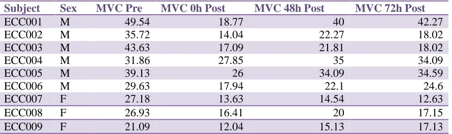
Chapter 4.0 Results:
4.1 Participant Characteristics:
Table 2 displays the means and standard deviations of the two groups (males vs.
females) for age, height, and mass. Student participants reflected a recreationally-active
population – having completed a minimum of one upper body resistance exercise session
every two weeks for one month Mean age for male participants was 24.7 ± 3.6 years old
while mean height and weight were 1.79 ± 0.08 m and 81.1 ± 15.2 kg respectively.
Females, mean age 24.7 ± 2.1, measured 1.64 ± 0.08 m and weighed 66 ± 3.5 kg. No
significant differences were observed in age (P=0.099), height (P=0.054), or weight
Subject Characteristic
Group Age (y) Height (m) Weight (Kg)
Male (n = 6) 24.7 ± 3.6 1.79 ± 0.08 81.1 ± 15.2
Female (n = 3) 24.7 ± 2.1 1.64 ± 0.08 66.0 ± 3.5
Table 2: Age, height (M), and weight (KG) of the nine participants, Values are mean ± standard deviation; n number of subjects.
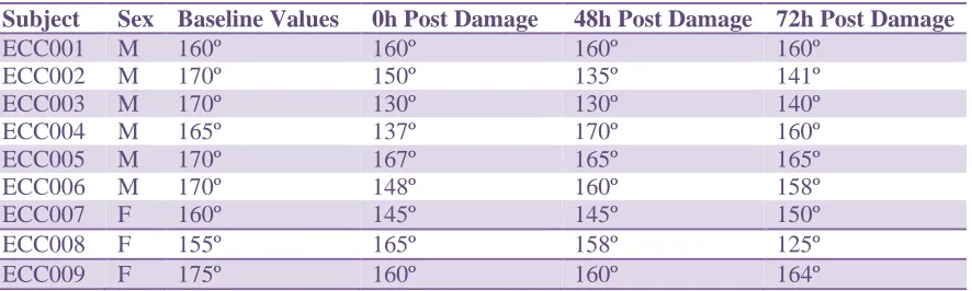
4.2 Joint Angle (Elbow Extension):
Subject Sex Baseline Values 0h Post Damage 48h Post Damage 72h Post Damage
ECC001 M 160º 160º 160º 160º
ECC002 M 170º 150º 135º 141º
ECC003 M 170º 130º 130º 140º
ECC004 M 165º 137º 170º 160º
ECC005 M 170º 167º 165º 165º
ECC006 M 170º 148º 160º 158º
ECC007 F 160º 145º 145º 150º
ECC008 F 155º 165º 158º 125º
ECC009 F 175º 160º 160º 164º
Table 3: Changes in joint (elbow) angle at baseline, immediately post damage, and 48h and 72h post EIMD
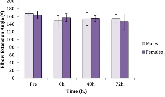
Mean elbow extension angle for all subjects was 166.1º ± 11.1º pre MVC; 151.3º
± 21.3º immediately post eccentric muscle damage; 153.6º º ± 23.6º following 48h; and
finally 151.4º ± 26.4º at 72h post EIMD. Male mean angle immediately decreased post
damage to 148.7º ± 13.8º from 167.5 º ± 4.18º prior to EIMD and steadily returned
toward baseline values at 153.3º ± 16.6º and 154º ± 10.7º at 48h and 72h respectively.
While female means immediately declined to 156.6 º ± 10.4º following the lengthening
protocol from a baseline of 163.3º ± 10.4º, mean continued to decline from 154.7º ± 8.1º
at 48h to 146.3º ± 19.8º at 72h Neither males nor females demonstrated any significant
difference in elbow angle between time points (males, P=0.058; females, P=0.402).
Additionally, there were no differences between sexes at baseline (P=0.401), immediately
Figure 1: Mean values for elbow angle (males and females) with standard deviation at baseline, immediately post EIMD, and at 48h and 72h Within group differences from baseline are signified by (*). (†) denotes significance from previous time point, while (**) signifies between group differences at the same time point. Significance determined at P<0.05 following post hoc testing.
4.3 Rate of Perceived Pain:
Subject Sex RPP 48h Post (cm) RPP 72h Post (cm)
ECC001 M 4.5 5.5
ECC002 M 5 6
ECC003 M 5 4
ECC004 M 5 5
ECC005 M 6 6.5
ECC006 M 5 2
ECC007 F 6 9
ECC008 F 2 6
ECC009 F 3 3
Table 4: Visual analogue scale rate of perceived pain (1-10) for male and female subjects at 48h and 72h post ECC.
Rate of perceived pain, as evaluated through a standardized numeric visual scale,
increased from 4.61cm ± 2.6cm units at 48h post EIMD to 5.22cm ± 3.8cm 72h post
damage (P<0.05). Pain reported by males remained relatively static declining slightly
from 5.08cm ± 0.92cm to 4.83cm ± 2.53cm over the 24h period (P=0.727). Similar trends
were reported by females where no statistical significance (P=0.330) was observed
0 20 40 60 80 100 120 140 160 180 200
Pre 0h. 48h. 72h.