2016 Joint International Conference on Artificial Intelligence and Computer Engineering (AICE 2016) and International Conference on Network and Communication Security (NCS 2016)
ISBN: 978-1-60595-362-5
A Robust Watermarking Algorithm of Encrypted Medical Volume Data
Based on 3D DWT and 3D DCT
Lian XU
a, Jing-Bing LI
b*, Wei-Bin FU
cHainan University, Haikou, Hainan, P.R. China
a[email protected], b[email protected], c[email protected]
*Corresponding author
Keywords: Medical Volume Data, Feature Vector, Logistic Map, Zero Watermarking, Robustness.
Abstract. This work proposed a novel scheme for robust watermarking in the encrypted medical volume data, which protects the original medical volume data from the third party embedders. The original medical volume data is encrypted using a selective partial encryption method based on 3D DCT and Logistic Map. In the watermark embedding phase, the watermark is associated with the feature vector of the encrypted medical volume data, generating a key which will be used in watermark extraction. The feature vector of the encrypted medical volume data is extracted in a way combining 3D DWT and 3D DCT. The watermark extraction is performed on the encrypted domain. The proposed watermarking algorithm realizes zero-watermarking technology in the encrypted domain, which has no effects on the encrypted medical volume data. The experimental results show that the proposed watermarking scheme has good robustness to some normal and geometric attacks. Therefore, it has a good practical value.
Introduction
In modern medical systems, digital information system plays a more and more significant role. On the one hand, digital medical images are convenient to be stored and transmitted, which makes some applications based on network develop rapidly, like telemedicine, long-distance operation, etc. On the other hand, there is a threaten of leakage of patients’ information when transmitting medical images containing lots of privacy information, which creates possibilities that medical images can be easily tempered or forged. Therefore, the authentication of medical images becomes extremely important.[1] utilizes encryption algorithms to encrypt medical images, which protects confidentiality of the content of medical images. [2] uses information hiding technology to embed validation information in medical images, which guarantees the reality of medical images.
more robust than those in the spatial domain. Bianchi[7] proposed the implementation of DFT and FFT in the encrypted domain. Zheng[8] proposed the implementation of DWT in the encrypted domain and the method to reduce the data expansion after encryption. Huang[9] proposed a robust watermarking scheme in the encrypted domain, while the computation cost is expensive and the robustness to geometric attacks should be improved.
There is no research on robust watermarking algorithm for 3D images in the encrypted domain, while 3D images are universal in medical images, such as CT, MRI images. This paper proposes a novel robust watermarking scheme for encrypted medical volume data, which utilizes selective partial encryption in transform domain and zero-watermarking technology. The proposed algorithm is considerably concise and efficient. The experimental results show it is robust against some normal and geometric attacks.
Fundamental Theory
3D Discrete Cosine Transform(3D-DCT)
The formula of 3D(three-dimensional) DCT is as follows:
1 1 1
0 0 0
(2 1) (2 1) (2 1) ( , , ) c( )c( )c( )[ ( , , ) cos cos cos ]
2 2 2
M N P
x y p
x u y v z w
F u v w u v w f x y z
M N P
(1)0,1, , 1; 0,1, , 1; 0,1, , 1;
u M v N w P
Where
1/ 0 1/ 0 1/ 0
( ) , ( ) , ( )
1/ 1,2, , 1 2 / 1,2, , 1 2 / 1,2, , 1
M u N v P w
c u c v c w
M u M N v N P w P
The formula of 3D IDCT is as follows:
1 1 1
0 0 0
(2 1) (2 1) (2 1)
( , , ) [ ( ) ( ) ( ) ( , , ) cos cos cos ]
2 2 2
M N P
x y p
x u y v z w
f x y z c u c v c w F u v w
M N P
(2)0,1, , 1; 0,1, , 1; 0,1, , 1;
x M y N z P
where ( , , )x y z is the sampling value in the spatial domain and ( , , )u v w is the sampling value in the frequency domain.
3D Discrete Wavelet Transform
In 1988, S. Mallat proposed the Wavelet Transform algorithm, which was a method of Time-Frequency analysis. Two-dimensional multi-resolution decomposition theory has been widely applied in the image processing field. In order to realize the multi-resolution decomposition of 3D images, the algorithm of 2D DWT has been promoted to 3D DWT.
The one-level decomposition process of 3D DWT is show as Fig.1, where L and H are low-frequency and high-frequency parts after low-pass and high-pass filters, respectively. Same as the DWT of 2D images, after 3D DWT, medical volume data is decomposed to approximation coefficients and detail coefficients. The subscript “1” represents the first decomposition level of 3D DWT.
Logistic Map
Logistic Map is one of the most widely used Chaotic maps for briefness of expression and good performance. Logistic Map is defined as
( ) (1 ), [0,1]
f x x x x (3)
Where is a constant. Consider only a case when x[0,1], ( ) [0,1]f x , soshould meet the
Assume3.569945672, when 4, the Logistic Mapping sequence
xn has threecharacteristics as follows, a. initial value sensitivity, b. aperiodicity, c. having strange attractors, the first two are exactly basic properties of and encryption key or a stream cipher.
The Algorithm Process
The algorithm process is divided into three parts: the encryption of medical volume data, watermark extraction, watermark extraction and the decryption of medical volume data, the detailed process is expressed as Fig.2:
Figure 1. 1-level Decomposition Figure 2. The Process of the Algorithm. Process of 3D DWT.
The Encryption of the Medical Volume Data
In this phase, a selective partial encryption method based on 3D DCT is used to encrypted medical volume data. In this way, the real content of the medical volume data is hidden, while the feature of the medical volume data in the transform domain is still retained. The encryption process of the medical volume data is described in detail as follows,
a. Apply 3D-DCT on the medical volume data I and obtain a matrix of 3D-DCT coefficients F.
b. Logistic Map is used to generate a sequence of real numbers. A threshold
is used to transform the sequence to a symbol matrix S which contains only ±1.1, ( )
1, n n x S n
x
(4)
( ,[ , , ])
Sreshape S M N P (5)
c. Each coefficient of F is multiplied by the corresponding coefficient of S in the in the same
coordinate respectively.
d. Apply 3D-IDCT on the output of the last step and obtain the encrypted medical volume data
I’.
Watermark Embedding
a. Apply 2-level 3D DWT on the encrypted medical volume data I’ and then 3D DCT is
performed on the LLL2 sub-band, a feature vector v of the encrypted medical volume data is generated by comparing the value of the first L DCT coefficients F’ on the upper left corner of the
coefficient matrix with 0. 1 '( ) 0
( ) 1, 2, , 0 '( ) 0
F n
v n n L
F n
(6)
b. In order to improve the watermark security, the original watermark w is scrambled by Arnold transformation and the scrambled watermark is represented as s.
c. The exclusive OR result of each row of the scrambled watermark s and the feature vector v of
the encrypted medical volume data is calculated as follows, and the output k is saved as a key which
will be used in the watermark extraction phase.
k s v (7)
Watermark Extraction & Image Decryption
In this phase, the third party extracts the watermark in the received encrypted medical volume dataIr. We consider the third party has the watermark embedding key k and the watermark
scrambling key. The detailed procedure is as follows.
a. A feature vector v’ of the received encrypted medical volume data Ir is generated in the
same way as mentioned in the watermark embedding phase.
b. The exclusive OR is applied on the feature vector v’ and the watermark embedding key k and
the output is the extracted scrambled watermark s’.
c. Perform the inverse Arnold transformation on the extracted scrambled watermark s’ and
obtain the extracted watermark w’.
d. The correlation coefficient value is calculated between the extracted watermark and the original one.
e. The encrypted medical volume data is decrypted in the same way mentioned in the 2.1 section.
The Experimental Results
The test medical volume data sized 128 128 27 shown in Fig.3(a) was used as the original image in the experiment. We let 4, the initial value of Logistic Mapx00.135. After image
encryption, the 3D DCT coefficients were encrypted to generate an encrypted medical volume data shown in Fig.3(b). We let the length of the feature vector L= 64, the original watermark is chosen as a binary image sized 64 64 , which is shown in Fig.3(c), and the scrambled watermark is shown in Fig.3(d). The watermarked encrypted medical volume data is shown in Fig.3(e). We could extract the scrambled watermark embedded in the encrypted medical volume data using the watermark embedding key. The extracted scrambled watermark and its decryption are shown in Fig.3(g)-3(h). With the encryption key, the original medical volume data could recover from the watermarked encrypted medical volume data, shown in Fig.3(f).
In order to evaluate the relevance, we compute the Normalized Cross Correlation (NC) of the original watermark and the decryption of the extracted watermark, the expression of NC is given as
2 ( , ) '( , )
( , )
i j
i j W i j W i j NC
W i j
(8)
(a) (b) (c) (d)
[image:5.612.186.438.66.198.2](e) (f) (g) (h)
Figure 3. Experimental Results: (a)The Original Medical Volume Data; (b) The Encrypted Medical Volume Data; (c) The Watermark; (d)The Scrambled Watermark; (e)The Watermarked
Encrypted medical Volume Data; (f)The Decryption of the Watermarked Encrypted Medical
Volume Data; (g)The Extracted Watermark from the Encrypted Medical Volume Data; (g)The Decryption of the Extracted Watermark.
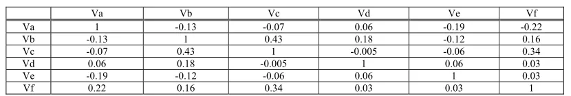
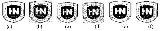
In order to prove that the feature vectors of different encrypted medical volume data in the proposed method are different, experiments were performed on six 3D images, i.e. Head, Liver1, Liver2, Engine, Teddy Bear and Tooth, shown in Fig.4(a)-4(f). They were encrypted using the proposed image encryption scheme. The results are shown in Fig.5(a)-5(f). We calculated the NC values between different encrypted 3D images. The results displayed in the Table 1 show that the relevance of different 3D images after encryption is close to zero, except that NC between liver1 and liver2 after encryption is relatively higher as the original images of liver1 and liver 2 are similar.
[image:5.612.131.491.377.446.2](a) (b) (c) (d) (e) (f)
Figure 4. Different 3D Images: (a)Head; (b)Liver1; (c)Liver2; (d)Engine; (e)Teddy Bear; (f)Tooth.
(a) (b) (c) (d) (e) (f)
Figure 5. The Corresponding Encrypted Images of Different Volume Data: (a)the Encrypted Head;
(b)the Encrypted Liver1; (c)the Encrypted Liver2; (d)the Encrypted Engine; (e)the Encrypted Teddy Bear; (f)the Encrypted Tooth.
Table 1. NC Between Different Encrypted 3D Images.
Va Vb Vc Vd Ve Vf
Va 1 -0.13 -0.07 0.06 -0.19 -0.22
Vb -0.13 1 0.43 0.18 -0.12 0.16
Vc -0.07 0.43 1 -0.005 -0.06 0.34
Vd 0.06 0.18 -0.005 1 0.06 0.03
Ve -0.19 -0.12 -0.06 0.06 1 0.03
Vf 0.22 0.16 0.34 0.03 0.03 1
[image:5.612.127.493.487.561.2] [image:5.612.99.514.629.699.2]performance of the proposed watermarking scheme against different attacks with varying degrees respectively as follows.
a) Salt & Pepper Noise
We use the function imnoise() to add salt & pepper noise to the watermarked encrypted medical volume data. When the variance of noise is 0.04, the decryption of the extracted watermark is shown in Fig.6(b). From Table 2, we can conclude that the proposed watermarking scheme has good robustness against salt & pepper noise.
Table 2. The Experimental Data of Adding Salt & Pepper Noise.
Variance (%) 1 4 8 16 20 30 NC 0.91 0.88 0.84 0.84 0.84 0.75
b)JPEG compression. JPEG compression is applied on the watermarked encrypted medical volume data. When the compression quality is 15%, the decryption of the extracted watermark is shown in Fig.6(c). From Table 3, we can conclude that the proposed watermarking scheme has strong robustness against JPEG compression.
Table 3. The Experimental Data of JPEG Compression.
Compression Quality(%) 4 10 15 20 25 30 NC 0.84 0.78 0.94 0.97 0.94 0.94
c) Rotation
When the watermarked encrypted medical volume data is rotated 5°clockwise, the decryption of the extracted watermark is shown in Fig. 6(d). From Table 4, we can conclude that the proposed algorithm can resist rotation attacks within a small range.
Table 4. The Experimental Data of Rotation.
Rotational Degree -5 -3 -1 1 3 5 NC 0.84 0.88 0.94 0.81 0.62 0.62
d) Resizing
When the resizing factor is 0.5, namely resizing the watermarked encrypted medical volume data from size 128 128 27 to 256 256 27 , the decryption of the extracted watermark is shown in Fig.6(e). From Table 5, we can conclude that the proposed algorithm has strong robustness against resizing attacks.
Table 5. The Experimental Data of Resizing.
Resizing Factor 0.2 0.5 0.8 1.2 2 2.4 NC 0.69 1 0.91 0.91 1 0.97
e) Cropping
When cropping 1/32 of the volume of the watermarked encrypted medical volume data along the YZ plane, the decryption of the extracted watermark is shown in Fig.6 (f). From Table 6, we can conclude that the proposed algorithm has good robustness against random cropping attacks.
Table 6. The Experimental Data of Cropping.
Cropping Percentage(%) 1/2 1/4 1/8 1/16 1/32 1/64
(a) (b) (c) (d) (e) (f)
Figure 6. The Decryption of the Extracted Watermark: (a)under no Attacks(NC=1); (b)under Salt & Pepper Noise(Noise Variance=0.04, NC=0.88; (c)under JPEG Compression(Compression
Quality=15%, NC=0.94; (d) under Rotation of 5° Clockwise(NC=0.84); (e) under
Resizing(from128 128 27 to256 256 27 , NC=1); (f)under Cropping
(1/32 of the Volume Cropped, NC=0.94).
Summary
In this paper, we propose a robust watermarking scheme of medical volume data in the encrypted domain, which consists of image encryption, watermark embedding, watermark extraction & image decryption phases. The medical volume data is encrypted using a selective partial encryption method based on 3D DCT and Logistic Map. A feature vector of the encrypted medical volume data is extracted combining 3D DWT and 3D DCT. The watermark is not actually embedded in the encrypted medical volume data, but associated with the feature vector of that. Therefore, there is no restriction on the number of the associated watermarks and the watermark embedding doesn’t affect the encrypted medical volume data as well. The experimental results show that the proposed watermarking scheme has good robustness to some normal and geometric attacks.
Acknowledgement
This work is partly supported by the National Natural Science Foundation of China (NO:61263033), and by the International Science and Technology Cooperation Project of Hainan (NO: KJHZ2015-04) and the Institutions of Higher Learning Scientific Research Special Project of Hainan(Hnkyzx2014-2).
References
[1]M. Dzwonkowski, M. Papaj, R. Rykaczewski. “A New Quaternion-Based Encryption Method for DICOM Images”, IEEE Transactions on Image Processing, vol. 24, no. 11, pp. 4614-4622, November 2015.
[2]G. Coatrieux, C.L. Guillou, J.M. Cauvin, et al. “Reversible Watermarking for Knowledge Digest Embedding and Reliability Control in Medical Images”, IEEE Transactions on Information Technology in Biomedicine, vol. 13, no. 2, pp. 158-165, March 2009.
[3]X. Zhang. “Reversible Data Hiding in Encrypted Image”, IEEE Signal Processing Letters, vol. 18, no. 4, pp. 255-258, April 2011.
[4]W. Hong. “An Improved Reversible Data Hiding in Encrypted Images Using Side Match”, IEEE Signal Processing Letters, vol. 19, no. 4, pp. 199-202, April 2012.
[5]X. Zhang. “Separable Reversible Data Hiding in Encrypted Image”, IEEE Transactions Information Forensics and Security, vol. 7, no. 2, pp. 826-832, April 2012.
[8]P. Zheng, J. Huang, “Discrete Wavelet Transform and Data Expansion Reduction in Homomorphic Encrypted Domain”, IEEE Transactions on Image Processing, vol. 22, no. 6, pp. 2455-2468, June 2013.
[9]J. Guo, P. Zheng, J. Huang, “Secure Watermarking Scheme Against Watermark Attacks in the Encrypted Domain”, Journal of Visual Communication and Image Representation, Elsevier, no. 30, pp. 125-125, 2015.