Functional selectin ligands mediating human
CD34
+
cell interactions with bone marrow
endothelium are enhanced postnatally
Andrés Hidalgo, … , Linnea A. Weiss, Paul S. Frenette
J Clin Invest.
2002;110(4):559-569. https://doi.org/10.1172/JCI14047.
Hematopoietic progenitor cells (HPCs) can home to the bone marrow (BM) after a simple
intravenous injection, but the adhesive mechanisms mediating the initial interactions of
human HPCs with the BM endothelium have not been evaluated in vivo. Using
fluorescence intravital microscopy and homing assays in NOD/SCID mice, we show that
endothelial selectins are necessary for human adult CD34
+cell homing, since rolling on BM
endothelium and retention in the BM compartment are drastically reduced (>90%) in
endothelial selectin–deficient NOD/SCID mice. Comparative analyses of CD34
+cells
collected from adults and from cord blood (CB) reveal that neonatal cells display reduced
rolling fractions compared with adult CD34
+cells obtained from peripheral blood or BM,
suggesting abnormal selectin ligand function on neonatal progenitors. Flow cytometric and
intravital microscopy studies suggest that this defect results from nonfunctional P-selectin
ligand on a subset (
~
30%) of neonatal CD34
+cells. Further analyses indicate that
P-selectin glycoprotein ligand-1 (PSGL-1) is expressed in a nonfunctional form among
neonatal CD34
+cells that do not bind P-selectin and that this subset is enriched in primitive
CD34
+CD38
lo/–progenitors. These results underscore the potential to improve homing of
CB CD34
+cells to the BM by manipulation of selectins and their ligands.
Article
Find the latest version:
Introduction
Hematopoietic progenitor cells (HPCs) can be trans-planted to the bone marrow (BM) of another individ-ual following a simple intravenous infusion. The abil-ity of HPCs to home to the BM was first demonstrated a few decades ago when Jacobson and colleagues showed that shielding of the spleen allowed mice to recover from a lethal dose of radiation (1). This study, along with others (2, 3), established that pluripotent stem cells present in the spleen or infused exogenously could repopulate distant hematopoietic organs. This feature is the basis for the clinical use of BM transplantation in treatment of hematologic malignancies (4). However, despite an advanced knowledge of the mechanisms that allow the migration of mature leukocytes into inflamed tis-sues, the mechanisms of human HPC homing to the BM are still poorly understood.
The migration of leukocytes to sites of inflammation is initiated by labile but critical adhesive interactions (attachment and rolling) that are largely mediated by the selectin family and its glycoconjugated ligands. Two members of the selectin family, P- and E-selectins, are expressed on endothelial cells (5, 6). P-selectin is stored in granules of endothelial cells and platelets and is rapidly translocated to the cell surface after stimulation with various secretagogues. E-selectin expression is induced by endotoxin or inflammatory cytokines. Although the E-selectin gene is silent in cul-tured endothelial cells, low levels of E-selectin are expressed in most tissues in vivo and regulate leuko-cyte homeostasis together with endothelial P-selectin (7). Mice lacking both P- and E-selectins display severe leukocytosis and can develop spontaneous skin infec-tions (7, 8). The main ligand for P-selectin, P-selectin glycoprotein ligand-1 (PSGL-1), is a disulfide-bonded homodimeric mucin-like glycoprotein expressed on leukocytes (9), platelets (10), and CD34+cells (11, 12).
PSGL-1 requires specific posttranslational modifica-tions in order to be functional. These include sialyla-tion and fucosylasialyla-tion of O-linked sugars, as well as sul-fation of tyrosine residues present in the N-terminus of the protein (13). While P- and E-selectin are critical for myeloid cell rolling in venules of the systemic cir-culation, the leukocyte selectin (L-selectin) mediates lymphocyte rolling in specialized venules of secondary lymphoid organs. The close interaction between leuko-cytes and endothelial cells during the rolling step allows the activation of leukocyte integrins triggered
Functional selectin ligands mediating human
CD34
+cell interactions with bone marrow
endothelium are enhanced postnatally
Andrés Hidalgo, Linnea A. Weiss, and Paul S. Frenette
Department of Medicine, Mount Sinai School of Medicine, New York, New York, USA
Hematopoietic progenitor cells (HPCs) can home to the bone marrow (BM) after a simple intra-venous injection, but the adhesive mechanisms mediating the initial interactions of human HPCs with the BM endothelium have not been evaluated in vivo. Using fluorescence intravital microscopy and homing assays in NOD/SCID mice, we show that endothelial selectins are necessary for human adult CD34+cell homing, since rolling on BM endothelium and retention in the BM compartment are drastically reduced (>90%) in endothelial selectin–deficient NOD/SCID mice. Comparative analyses of CD34+cells collected from adults and from cord blood (CB) reveal that neonatal cells display reduced rolling fractions compared with adult CD34+cells obtained from peripheral blood or BM, suggesting abnormal selectin ligand function on neonatal progenitors. Flow cytometric and intravital microscopy studies suggest that this defect results from nonfunctional P-selectin ligand on a subset (∼30%) of neonatal CD34+cells. Further analyses indicate that P-selectin glycoprotein ligand-1 (PSGL-1) is expressed in a nonfunctional form among neonatal CD34+cells that do not bind P-selectin and that this subset is enriched in primitive CD34+CD38lo/–progenitors. These results underscore the potential to improve homing of CB CD34+cells to the BM by manipulation of selectins and their ligands.
J. Clin. Invest.110:559–569 (2002). doi:10.1172/JCI200214047.
Received for publication August 23, 2001, and accepted in revised form June 24, 2002.
Address correspondence to:Paul S. Frenette, Mount Sinai School of Medicine, One Gustave L. Levy Place, Box 1079, New York, New York 10029, USA. Phone: (212) 659-9693; Fax: (212) 849-2574. E-mail: [email protected].
Conflict of interest:No conflict of interest has been declared.
by chemokines present on the surface of the endothe-lium. This leads to the firm adhesion and migration of leukocytes through the vessel wall into the extravas-cular space (14). Whether a similar paradigm is appli-cable for immature hematopoietic precursors is still unclear. However, studies in mice suggest that the recruitment of HPCs into the BM requires multiple adhesion pathways, including endothelial selectins and VCAM-1 (15–18).
The study of primitive human hematopoiesis in vivo has been facilitated by the generation of NOD/LtSz-scid/scid mice (hereafter referred to as NOD/SCID mice) that have multiple defects in innate and adaptive immunologic functions (19). The BM of NOD/SCID mice can be repopulated by human HPCs (CD34+cells)
after sublethal doses of radiation (20). Clinically, human HPCs are obtained from three different sources: BM, mobilized peripheral blood (mPB), and cord blood (CB). CB-derived progenitors represent an especially promising source of human hematopoietic stem cells because they are widely available and their use is associated with reduced graft-versus-host disease (21). However, transplantation using CB cells has been restricted mostly to children due to the limited num-ber of cells available in the placenta (22).
Here, we describe the molecular mechanisms medi-ating the initial adhesive interactions of human HPCs in the BM microcirculation of NOD/SCID mice using fluorescence intravital microscopy. We show that human CD34+cell rolling on BM endothelium and
extravasation into the BM compartment are complete-ly dependent upon endothelial selectins. In addition, we demonstrate that the initial interactions of CB CD34+cells are reduced compared with those derived
from adult sources. We show that a large subset (∼30%) of neonatal CD34+ cells, enriched in primitive
CD34+CD38lo/–cells, does not bind P-selectin, and that
this inability to bind P-selectin originates from incom-plete posttranslational modifications of PSGL-1. These results may have implications for therapies using CB progenitor/stem cells.
Methods
Ab’s and selectin chimeras. For in vivo studies, rat mAb’s against mouse VCAM-1 (MK 2.7) and P-selectin (clone RB40.34) were purified from hybridoma supernatants (American Type Culture Collection, Rockville, Mary-land, USA), and control rat IgG was obtained from Sigma-Aldrich (St. Louis, Missouri, USA). Potential endotoxin contamination was removed using a polymyxin B column (Detoxi-Gel; Pierce Biotechnolo-gy Inc., Rockford, Illinois, USA). The rat anti–mouse E-selectin Ab (clone 9A9) was a generous gift of B. Wolitzky (MitoKor, San Diego, California, USA). Anti–human PSGL-1 (KPL1) and FITC-conjugated anti–human CD38 were purchased from Pharmingen (San Diego, California, USA), and phycoerythrin-con-jugated (PE-conphycoerythrin-con-jugated) anti–human CD34 (clone AC136) was from Miltenyi Biotec (Bergisch Gladbach,
Germany). The mouse anti–human PSGL-1 Ab (PSL-275) and human P-selectin–IgG chimera were generously provided by R. Schaub (Genetics Institute, Cambridge, Massachusetts, USA). Murine E- and P-selectin–IgM chimeras were produced by transfection of COS-7 cells with selectin-IgM DNA vector (a kind gift of John Lowe, University of Michigan, Ann Arbor, Michigan, USA) using the DEAE-dextran method (23). Human samples and CD34+cell isolation. Fresh human
hematopoietic cell samples were obtained from unused portions of three different clinical sources: steady-state BM, mPB, and CB. BM samples were collected from normal donors at the time of harvest for allogeneic transplantation. mPB cells were collected by leuka-pheresis from either healthy donors (n= 19) or patients with hematopoietic malignancies in remission (n= 15). Both BM and mPB samples were obtained from the Mount Sinai Bone Marrow Transplantation Program. Umbilical CB samples were obtained from normal full-term deliveries (≥38 weeks). All human samples were obtained in accordance with protocols approved by the Internal Review Board of Mount Sinai.
For the enrichment of CD34+ cells, low-density
mononuclear cells (MNCs) were collected after cen-trifugation at 250 gover Ficoll-Hypaque (d= 1.077 g/ml). After two washes in PBS containing 2 mM EDTA and 0.5% BSA, contaminating red blood cells (RBCs) were lysed in a 0.8% NH4Cl solution. CD34+
cells were purified from the MNC fraction using the CD34-isolation mini-MACS kit (Miltenyi Biotec) fol-lowing the manufacturer’s instructions. The purity of the isolated CD34+cells ranged from 70% to 99%
(average ∼89% for CB and ∼95% for mPB). For intrav-ital microscopy experiments, purified human CD34+
cells were fluorescently labeled by incubation with 33
µM carboxyfluorescein succinimidyl ester (CFSE; Molecular Probes Inc., Eugene, Oregon, USA) for 30 minutes at room temperature and washed three times in RPMI before injection into mice via the carotid artery catheter.
Mice. A NOD/SCID mouse colony was established from breeding pairs obtained from The Jackson Labo-ratory (Bar Harbor, Maine, USA). Mice with P- and E-selectin double null mutations (P/E–/–) or a single
E-selectin null mutation (E–/–) (7) were backcrossed
into the NOD/SCID background. After each genera-tion, blood counts were monitored to select for the SCID mutation. Third generation littermates, which display a lymphopenia similar to that in pure NOD/SCID animals, were used to set up colonies of selectin knockouts and control wild-type NOD/SCID mice. Wild-type and selectin-deficient NOD/SCID mice had numbers of T (CD3+) cells, B (B220+) cells,
and NK (NK1.1+) cells similar to the parental
in the barrier facility at Mount Sinai School of Medi-cine. Mice were fed with sterilely irradiated chow and autoclaved water. Experimental procedures on animals were approved by the Animal Care and Use Committee at the Mount Sinai School of Medicine.
Intravital microscopy of the BM. Mice 6–9 weeks old were used for BM intravital microscopy (BM-IVM). To avoid the retention of injected human cells in the spleen, mice were splenectomized upon weaning as described (16) and allowed to recover for at least 2 weeks before BM-IVM experiments. Mice were anesthetized by intraperitoneal injection (6 ml/kg) of 100 mg/ml ure-thane and 20 mg/ml chloralose (both from Sigma-Aldrich) in PBS. This anesthetic combination was cho-sen because it does not significantly alter blood flow in the BM microcirculation. The hair in the sub-mandibular area of the neck and on the skullcap was removed using a hair removal lotion (Nair; Carter Prod-ucts, New York, New York, USA). The trachea was can-nulated with PE-160 polyethylene tubing to facilitate spontaneous respiration, and a PE-10 catheter (Becton, Dickinson and Co., Franklin Lakes, New Jersey, USA) was inserted into the left common carotid artery for injection of fluorescent cells and Ab’s. The scalp was incised in the midline to expose the frontoparietal skull, and the conjunctive tissue covering the skull was carefully removed. A plastic ring was inserted in the incision area to allow application of endotoxin-free PBS. The mouse’s cranium was kept in place using a stereotactic holder (David Kopf Instruments, Tujunga, California, USA). A mouse thus prepared was posi-tioned under a fixed-stage, custom-designed intravital microscope (MM-40) equipped with a mercury fluo-rescent lamp and water immersion objectives (Nikon Corp., Tokyo, Japan). To block potential sites of reten-tion of human cells, 107unlabeled human MNCs from
each sample were injected immediately before the injec-tion of labeled CD34+cells. Human CD34+cells are
rapidly cleared (within minutes) from the circulation of NOD/SCID mice after injection. Approximately 1 ×10⁶to 2 ×10⁶CD34+cells were injected per
experi-ment to visualize 50–500 cells in the left parietal BM using a 10×water-immersion objective (0.3 NA; Nikon Corp.). For determination of the hemodynamic param-eters, 107CFSE-labeled human MNCs or RBCs were
injected into the carotid artery at the end of each exper-iment. In the experiments designed to determine the role of VCAM-1 or P-selectin, mice were preinjected with control rat IgG (or no IgG in two experiments), followed by labeled human CD34+cells. When cells
from the first injection were no longer detected in the circulation (10–15 minutes), the same mouse was injected with anti–VCAM-1 (80 µg) or anti–P-selectin (60 µg) at least 15 minutes before a second injection of labeled human CD34+cells. In the experiments aimed
at evaluating the role of PSGL-1, fluorescently labeled CD34+cells were incubated with 2 µg KPL1 or control
IgG per 106cells prior to intracarotid injection. The
images were captured using an SIT camera with a
C2400 camera controller (Hamamatsu, Hamamatsu City, Japan) and were recorded using a VHS video recorder (SV0-9500MD; Sony Corp., Tokyo, Japan).
Image analysis. Vessel diameter and cell velocities were measured using a video caliper and sequential single-frame analysis. The maximal velocity (Vmax), which
rep-resents the average velocity of free-flowing CFSE-labeled CD34–cells or RBCs, was determined for each
BM vessel. The mean blood flow velocity (Vmean) was
thus calculated as Vmean= Vmax/(2 – ε2), where εis the
ratio of the CD34+cell diameter (∼7 µm) to the vessel
diameter (Dv). The wall shear rate and critical velocity
(Vcrit) were obtained from the following formulas: wall
shear rate = 8(Vmean/Dv), and Vcrit= Vmean× ε(2 – ε). Any
cell traveling below Vcritwas considered to be rolling on
the vessel wall. Cells that remained stationary for 5 sec-onds or more were considered arrested.
Flow cytometry and selectin chimera binding assay. For dou-ble staining of CD34 and P-selectin ligands, 106total CB
or mPB MNCs depleted of RBCs were washed and resus-pended in staining buffer (RPMI, 5% FCS, and 0.02% NaN3). Fc receptors were blocked with 1 µg human IgG
(Sigma-Aldrich), and cells were incubated with PE-con-jugated anti–human CD34. After one wash, cells were incubated with 4 µg of the HUMAN P-selectin–IgG chimera that had been preconjugated with 0.3 µg biotinylated protein A. Preliminary experiments showed that this concentration saturates P-selectin ligands on CD34+cells. Cells were subsequently incubated with an
excess of streptavidin-FITC (1.5 µg; Jackson ImmunoRe-search Laboratories Inc., West Grove, Pennsylvania, USA) and washed once before analysis using a FACSCalibur flow cytometer (Becton, Dickinson and Co.). All incuba-tions were performed for 20 minutes at 6°C. For triple-color labeling with anti-CD34–PE, P-selectin–IgG–Cy5, and anti-CD38–FITC, the procedure was the same except that an anti-CD38–FITC Ab (Pharmingen) was coincubated with an excess of streptavidin-Cy5 (Jackson ImmunoResearch Laboratories Inc.) before the final wash. In control samples, staining was carried out in the presence of 5 mM EDTA. For double staining of CD34 and PSGL-1, CB or mPB MNCs prepared as described above were incubated with an anti–PSGL-1 Ab (PSL-275 or KPL1), washed once, and incubated with an FITC-conjugated anti-mouse Ab (Pharmingen). After washing, cells were incubated with 1 µg mouse IgG (Sigma-Aldrich) and anti-CD34–PE Ab, washed, and analyzed by flow cytometry. To evaluate E-selectin ligand expression, 105CB- or mPB-derived CD34+cells were incubated with
supernatants from COS-7 cells transfected with the murine E-selectin–IgM construct, followed by PE-labeled anti-human IgM goat Ab (Sigma-Aldrich). A similar pro-cedure was followed for the detection of P-selectin lig-ands using the murine P-selectin–IgM chimera, using supernatants from COS-7 cells transfected with the murine P-selectin–IgM construct.
cells (106cells) from F1 generation EGFP-Tg NOD/SCID
mice were injected into sublethally irradiated (300 cGy) NOD/SCID recipients. Fourteen days after transplanta-tion, recipient mice were prepared for fluorescence BM-IVM of the parietal bone as described above.
For homing assays, MNCs were isolated from mPB samples as indicated above and resuspended in RPMI. Irradiated (375 cGy) wild-type and P/E–/–NOD/SCID
mice were injected intravenously with 2 × 10⁷MNCs
immediately after irradiation. Two hours after injec-tion, mice were sacrificed and femoral BM cells were harvested. After lysis of RBCs and blocking of Fc receptors with mouse and human IgG, 106total BM
nucleated cells were stained with both PE-conjugated anti-CD34 and FITC-conjugated anti-CD45 Ab’s (Immunotech, Marseilles, France). Flow cytometry was gated on the lymphocyte population since more than 90% of CD34+cells fall in this region. Analyses were
performed on 5 ×10⁵events per transplanted animal.
Engraftment of human CD34+cells into NOD/SCID mice.
CD34+cells (105) purified from CB or G-CSF mPB
were injected intravenously into sublethally irradiat-ed (350–375 cGy) NOD/SCID or β2 microglobu-lin–deficient NOD/SCID mice (The Jackson Labora-tory). Mice were sacrificed 4 weeks after injection. After lysis of RBCs and blocking of Fc receptors with mouse and human IgG, 106BM nucleated cells were
stained with an FITC-conjugated anti–human CD45 Ab (Immunotech). Depending on the level of engraft-ment found by FACS analysis, 0.25 × 10⁵to 2.5 × 10⁵
BM nucleated cells were plated into methylcellulose media (MethoCult H4433; Stem Cell Technologies, Vancouver, Canada) and incubated at 37°C in a humidified atmosphere containing 5% CO2. Human
myeloid colonies, erythroid colonies, and colonies containing both myeloid and erythroid cells were counted on day 14.
Statistical analysis. All values are reported as mean ± SEM. Statistical significance for two paired or unpaired groups was assessed by the Student ttest. The Mann-Whitney test was used to determine statistical significance in engraftment studies.
Results
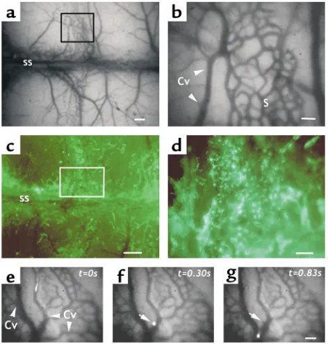
Human CD34+cells roll and arrest in NOD/SCID BM
[image:5.576.58.294.54.301.2]microvessels. The thin cortex of murine cranial bones allows one to observe the behavior of fluorescently labeled cells in the BM of living animals using epifluo-rescence intravital microscopy (17). Because of the pro-tection afforded by the surrounding bone, the BM microvasculature can be observed without direct injury from the surgical preparation. The bulk of cranial BM is concentrated around the bone sutures of the calvaria. However, individual BM microvessels are not clearly visible in most areas by fluorescence intravital microscopy except for a section in the middle of the parietal bone (Figure 1a, box), which displays a well-defined vascular network composed of sinusoids (Fig-ure 1b, “S”, honeycomb appearance) draining into at least two collecting venules that run perpendicular or parallel to the sagittal sinus (Figure 1b, Cv, and Figure 1, e–g). The blood flow in the midparietal area of the NOD/SCID BM is centripetal, draining into the sagit-tal sinus. To evaluate the distribution of active hematopoiesis in the calvaria, BM nucleated cells from EGFP-Tg mice were transplanted into sublethally irra-diated (300 cGy) NOD/SCID recipient mice. Fourteen days after transplantation, EGFP-expressing hematopoietic cells had heavily repopulated the parasagittal areas of the parietal bone (including the aforementioned midparietal section; Figure 1, c and d), but also areas in the frontal and occipital bones near
Figure 1
the coronal and lambdoidal sutures, respectively (not shown). These results indicate that the midparietal area carries active hematopoiesis and that the distribution of hematopoietic sites in the cranium of NOD/SCID animals is similar to that described in C57BL/6 animal studies using rhodamine 6G uptake (17).
Having established that the midparietal vascular net-work in NOD/SCID mice encompasses BM tissue, we next set out to evaluate the behavior of human HPCs in the BM microcirculation. CD34+cells from G-CSF–
mobilized healthy donors or from patients with hema-tologic malignancies in remission were isolated. Puri-fied CD34+cells were fluorescently labeled and
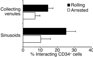
inject-ed via the carotid artery of NOD/SCID mice preparinject-ed for intravital microscopy. Labeled cells can be individ-ually tracked in BM microvessels (Figure 1, e–g) and the percentages of rolling and arrested cells determined by careful analysis of video recordings. To comparatively evaluate CD34+cell interactions in sinusoids and
col-lecting venules, we initially studied the numbers of rolling and arrested HPCs in each vessel type separate-ly. Although there was a trend toward a larger CD34+
cell rolling fraction in sinusoids compared with
collecting venules, the difference was not significant (Figure 2; P= 0.14). Therefore, the numbers of rolling and arrested cells in both types of microvessels were pooled in subsequent analyses. Interestingly, most cell arrests in collecting venules (92 of 147, or 63% of arrest-ed cells) occurrarrest-ed within two vessel diameters of vascu-lar junctions, suggesting hotspots for the recruitment of HPCs in the BM. Although the average diameters and centerline velocities (Vmax) were lower in sinusoids
than in collecting venules (Table 1, P< 0.05), the shear rates were similar between these two groups of microvessels. The mean percentage of rolling human CD34+ cells (∼22%), similar to that described for a
murine cell line and fetal liver progenitor cells in the BM of C57BL/6 mice (17), indicates that human CD34+cells can efficiently interact with NOD/SCID
BM microvessels.
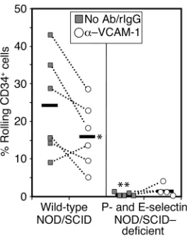
P- and E-selectins are required for human CD34+cell rolling
in BM microvessels and homing to the BM compartment. Pre-vious studies have suggested important roles for P- and E-selectins and VCAM-1 in mouse progenitor homing to the BM, and that each pathway (VCAM-1/
β1 integrin and selectin/mucin ligands) contributes equally to recruitment of mouse HPCs (15–18). To evaluate the role of endothelial selectins and VCAM-1 in the initial interactions of human CD34+cells with
BM microvessels, we backcrossed the P- and E-selectin mutations into the NOD/SCID background and used an mAb (MK 2.7) to inhibit VCAM-1 function. CD34+
cells were purified from MNCs prepared from mPB samples and fluorescently labeled with CFSE. Mice were preinjected with either an anti–VCAM-1 Ab (MK 2.7) or rat IgG control prior to the injection of CD34+
cells. Anti–VCAM-1 administration reduced CD34+
cell rolling by about 33% (P = 0.05) in the BM microvasculature of wild-type NOD/SCID mice, com-pared with IgG injection or no injection (Figure 3). Strikingly, CD34+cell rolling was drastically reduced
(∼95% reduction) in BM microvessels of P/E–/–
[image:6.576.93.256.56.157.2]NOD/SCID mice preinjected with either IgG or anti–VCAM-1 (Figure 3). These differences were not due to alterations in hemodynamics of the BM micro-circulation since there was no difference in the shear
Figure 2
[image:6.576.58.539.597.695.2]Human CD34+cell rolling and arrest in collecting venules and sinusoids of the BM in NOD/SCID mice. CFSE-labeled human CD34+cells puri-fied from mPB were injected into NOD/SCID mice, and the fractions of cells rolling or arrested in collecting venules or sinusoids were deter-mined by IVM analysis of videotapes. Bars represent mean ± SEM. The differences between Cv and sinusoids for the number of rolling and arrested cells are not significant (P= 0.14 and 0.6, respectively). n= 7 different human donors and the same number of mice.
Table 1
Hemodynamic characteristics of BM microvessels
Mice Source of No. of Vessel diameter (µm) Vmax(µm/s) WSR (s–1)
CD34 cells Cv S Mice Cv S Cv +S Cv S Cv +S Cv S Cv +S
NOD/SCID mPB 30 30 10 49 ± 3 28 ± 2A 38 ± 2 3,731 ± 390 1,578 ± 146A 2,655 ± 249 321 ± 40 245 ± 21 283 ± 23 CB 17 30 8 47 ± 3 32 ± 1A 40 ± 3 3,206 ± 233 1,625 ± 166A 2,762 ± 347 283 ± 22 216 ± 20A 297 ± 27 BM 15 18 7 54 ± 4 29 ± 2A 37 ± 2 3,681 ± 640 1,997 ± 242A 2,197 ± 174 309 ± 42 287 ± 37 240 ± 16 N/S WT mPB 18 23 5 42 ± 2 26 ± 1A 33 ± 2 3,050 ± 476 1,326 ± 147A 2,224 ± 296 295 ± 49 222 ± 26 272 ± 30 N/S PE–/– mPB 9 23 5 49 ± 3 30 ± 2A 35 ± 2 3,201 ± 561 1,555 ± 211A 2,018 ± 251 259 ± 41 252 ± 28 254 ± 23 N/S E–/– CB 19 14 7 41 ± 3 32 ± 1A 37 ± 2 1,873 ± 182 1,267 ± 159A 1,619 ± 134 234 ± 28 208 ± 29 223 ± 20
rate between the two groups of mice (Table 1). These results indicate that, in contrast to murine progenitors (16, 17), the initial interactions of human CD34+cells
are largely dependent on endothelial selectins that are constitutively expressed in the BM tissue and that VCAM-1 plays a partial role in this activity.
To determine whether the defect in the initial inter-actions of human CD34+cells in P/E–/–NOD/SCID
mice prevents extravasation into the BM compart-ment (i.e., homing), unsorted MNCs from mPB were
injected into wild-type or P/E–/–NOD/SCID mice.
Homed human cells were detected by FACS analysis using species-specific Ab’s against CD34 and CD45 (Figure 4, a and b). Homing of CD34+/CD45+human
cells was reduced by approximately 90% in P/E–/–mice
compared with wild-type animals (Figure 4, c–e). Of note, CD45+CD34–lymphocytes also lodged in the
BM of NOD/SCID mice 2 hours after their injection (Figure 4c, lower region), suggesting that donor lym-phocytes also rapidly home to the recipient BM after transplantation. However, the numbers of lympho-cytes were only modestly reduced (39%, P= 0.02) in the BM of P/E–/–animals (Figure 4d). Taken together,
our intravital observations and the results from the short-term homing assays indicate that endothelial selectins are necessary for human CD34+cell rolling
on BM microvessels and homing into the BM com-partment of NOD/SCID mice.
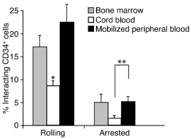
Defective interactions of CB-derived CD34+cells with BM
microvessels. Human HPCs can be routinely harvested for clinical transplantation from three different sources, including the BM, mPB, and CB. To evaluate differential homing mechanisms of human HPCs, we isolated CD34+cells from fresh samples obtained from
these three sources. Fluorescently labeled CD34+cells
were injected via a carotid artery catheter, and their interactions in the contralateral midparietal BM were recorded. Detailed analyses of videotapes revealed that the mean percentages of rolling and arrested cells in NOD/SCID BM microvessels were similar between mPB- and BM-derived CD34+cells (Figure 5).
Howev-er, the interactions of CB-derived CD34+cells were
sig-nificantly altered (∼50% and ∼60% reduction for rolling and arrested cells, respectively) compared with those derived from adult sources (Figure 5). Hemodynamic parameters were similar among the three groups (Table 1). These results suggest a defect in the capacity of neonate-derived CD34+ cells to interact with the
[image:7.576.103.235.52.220.2]endothelium of BM microvessels.
Figure 3
[image:7.576.93.488.521.627.2]Role of P- and E-selectins and VCAM-1 in the initial interactions of human CD34+ cells with BM microvessels. Fluorescently labeled human CD34+cells purified from mPB were injected into P/E–/–or wild-type mice backcrossed into the NOD/SCID background. Mice were injected via carotid catheter with control rat IgG (or no Ab in two experiments) and then with labeled CD34+cells. After 15 minutes of recording, fluorescent cells were completely cleared from the circula-tion. The same mouse was then injected with anti–VCAM-1 and a sec-ond bolus of fluorescent CD34+cells. Horizontal bars represent mean values. Each dot represents a single experiment before (filled squares) or after anti–VCAM-1 injection (open circles). *P= 0.05 by paired Stu-dent ttest; **P= 0.003 by unpaired Student ttest compared with IgG group in wild-type animals.
Figure 4
Neonate-derived CD34+cell binding to soluble P-selectin,
but not soluble E-selectin, is impaired. Since endothelial selectins are required for rolling of CD34+cells
(Fig-ure 3), the lower rolling numbers of CB CD34+cells
suggested a reduced expression or function of selectin ligands on neonatal progenitor cells. To evaluate this possibility, we tested the ability of CB- and mPB-derived CD34+cells to bind P-selectin or E-selectin in
a fluid-phase assay. Mononuclear cells were isolated from fresh mPB and CB samples and stained with anti-CD34 and the human P-selectin–IgG chimera in the presence and absence of divalent cations. Eight individual donor pair samples were thus prepared in
parallel and evaluated by FACS analysis. As shown in Figure 6a, the geometric mean of fluorescence of P-selectin–IgG binding among CD34+cells was 59%
lower in CB than in mPB (P= 0.008). Binding was spe-cific since it was abrogated in divalent cation–chelat-ed samples (Figure 6, a and b). Analysis of flow cytom-etry scattergrams revealed that while the vast majority (90% ± 1%) of mPB CD34+cells bound P-selectin–IgG,
approximately one-third of CB-derived CD34+cells
(34% ± 3%) did not bind soluble P-selectin (Figure 6b), suggesting a defect in expression or function of P-selectin ligand on a subpopulation of CB CD34+
cells. This finding was not exclusive to the human P-selectin chimeric construct since similar results were obtained with murine P-selectin–IgM (data not shown). To evaluate the function of E-selectin ligands, we analyzed the binding of a murine E-selectin–IgM chimera to purified adult and neonatal CD34+cells.
In contrast to P-selectin binding, only 60–70% of CD34+cells bound E-selectin–IgM, but there was no
significant difference in binding between CB and mPB (68% ± 2% and 62% ± 6% for CB and mPB CD34+
cells, respectively; n= 6, P= 0.33). These data indicate that function or expression of P-selectin ligands, but not E-selectin ligands, is significantly altered on neonate-derived CD34+cells.
Because PSGL-1 has been reported to be the main functional receptor for P-selectin in BM-derived CD34+
cells (24), we analyzed its expression. Using two differ-ent mAb’s against PSGL-1 (PSL-275 and KPL1), we found that most adult and neonatal CD34+ cells
[image:8.576.87.277.52.190.2]expressed this antigen (94% ± 2% in CB and 97% ± 1% in mPB, n = 5) (Figure 6c). To investigate whether PSGL-1 is the main functional P-selectin ligand on mPB and CB CD34+cells, MNCs were stained with
Figure 5
[image:8.576.69.527.472.622.2]Comparative analysis of the interactions of CD34+cells with BM microvessels in NOD/SCID mice. Fluorescently labeled CD34+cells from steady-state human BM, mPB, or CB were injected into NOD/SCID mice, and the numbers of cells interacting (rolling or arrested) with the BM microvasculature were determined by analysis of video recordings from fluorescence intravital microscopy experi-ments. n = 7–10 mice and human donors. *P< 0.02 compared with BM and mPB. **P= 0.03.
Figure 6
anti-CD34 Ab and incubated with either function-blocking (KPL1) or non-function-blocking (PSL-275) mAb against PSGL-1 prior to evaluating soluble P-selectin binding. Preincubation of cells with KPL1, but not with PSL-275, almost completely inhibited P-selectin–IgG binding in both CB- and mPB-derived CD34+cells
(Fig-ure 6d). The normal expression of PSGL-1 glycoprotein on CB CD34+ cells, together with the evidence of
abnormal function in a subset of CD34+cells, suggests
defective posttranslational modifications of the PSGL-1 protein in a subpopulation of CB CD34+cells.
The PSGL-1/P-selectin pathway plays a major role in the ini-tial interactions of CD34+cells with BM microvessels. To assess
whether PSGL-1 mediates CD34+cell rolling in vivo, we
treated fluorescently labeled mPB CD34+ cells with
anti–PSGL-1 or control IgG and evaluated their behav-ior in the BM microcirculation after intracarotid injec-tion. As shown in Figure 7a, the number of rolling CD34+cells was significantly lower (∼56% reduction) in
the group treated with anti–PSGL-1. To further test whether the reduced rolling capacity of CB-derived CD34+cells might be due to defective PSGL-1, we
test-ed the individual roles of PSGL-1, P-selectin, and E-selectin using E–/– mice backcrossed in the
NOD/SCID background and inhibitory Ab’s against P-selectin and PSGL-1. We found that the reduction observed earlier (Figure 3) in CB CD34+cell rolling
com-pared with cells derived from mPB was similar in wild-type NOD/SCID and E–/–NOD/SCID mice (Figure 7b).
However, inhibition of P-selectin function in both wild-type and E–/–NOD/SCID mice greatly reduced (by 74%
and 68%, respectively) the number of rolling CB CD34+
cells in the BM microvasculature (Figure 7b). In addi-tion, blockage of PSGL-1 function on CB CD34+cells
inhibited their interactions in both wild-type and E-selectin–deficient mice (68% and 55% reduction, respectively). Inhibition using an anti–E-selectin Ab (clone 9A9) produced similar results to E-selectin defi-ciency (n= 2, data not shown). These data indicate that the PSGL-1/P-selectin pathway plays a major role in ini-tial interactions and strongly suggest that the reduced ability of CB CD34+ cells to interact with the BM
microvessels is due to altered P-selectin binding. Neonatal CD34+cells defective in P-selectin binding are
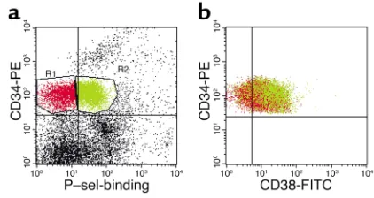
enriched in primitive CD34+CD38lo/–cells. CD34+cells
rep-resent a heterogeneous population of progenitors with various degrees of hematopoietic maturation. The lack or dim expression of CD38 in human CD34+cells is
considered to be a surrogate marker of their primitive nature (25). To further characterize the population of CB-derived CD34+cells that do not bind P-selectin–IgG,
we analyzed P-selectin chimera binding and CD38 expression among CD34+cells. As shown in Figure 8,
CD34+cells that did not bind P-selectin–IgG (Figure 8a,
region in red) contained 23% ± 3% CD38lo/–cells,
com-pared with only 9% ± 1% CD34+cells that bound
solu-ble P-selectin (Figure 8b, region in green; n= 8 different donors, P= 0.0006). Thus, CD34+progenitors that do
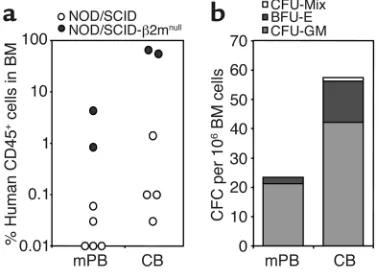
not bind P-selectin are enriched in primitive HPCs. Engraftment of NOD/SCID mice by CB CD34+cells is not
compromised. To determine whether the defect of CB CD34+cell rolling might influence their engraftment
into NOD/SCID mice, CD34+cells were purified from
CB and healthy mPB donors and transplanted into sublethally irradiated (350–375 cGy) NOD/SCID ani-mals or NOD/SCID mice deficient in β2 microglobu-lin. One month after transplantation, mice were sacrificed to assess the engraftment levels of human-derived progenitors. As shown in Figure 9, there was a trend toward higher engraftment levels in the CB group compared with the mPB group, as measured by the frequency of human CD45+cells and human
[image:9.576.58.276.55.200.2]pro-genitors in the BM compartment. These data are
Figure 7
[image:9.576.315.526.523.635.2]Role of P-selectin and PSGL-1 in CD34+cell interactions with BM microvessels. Fluorescently labeled CD34+cells from mPB (a) or CB (b) were preincubated with anti–PSGL-1 (black bars) or isotype-matched control Ab and injected into wild-type or E–/–NOD/SCID mice that had been treated with anti–P-selectin (white bars) or isotype-matched control Ab (gray bars). The number of cells interacting with the BM microvasculature was determined by analysis of video record-ings from fluorescence intravital microscopy experiments. *P< 0.02 compared with control Ab; **P= 0.06 compared with control Ab; #P< 0.02 compared with mPB control Ab group shown in a.
Figure 8
consistent with those of Wang et al., who found a higher level of engraftment of CB MNCs than of either BM or mPB MNCs (26), and suggest that CB CD34+
cells can compensate for their reduced initial interac-tions with the BM endothelium.
Discussion
The BM microvasculature differs from that of other tis-sues in several aspects. For example, the BM compart-ment does not contain capillaries but is endowed with a rich sinusoidal network. The fenestrated endotheli-um lining BM sinusoids promotes the release of bil-lions of blood cells each day. Yet hematopoietic pro-genitors and mature blood cells (for example, B lymphocytes) likely reenter the BM under homeostatic conditions, and such trafficking is critical during hematopoietic development and BM transplantation. Because collecting venules mediate the recruitment of mature leukocytes during inflammation, we initially analyzed whether human HPCs interacted preferen-tially with sinusoids or collecting venules. We found that the two vessel types recruited circulating progeni-tors equally well. It is interesting that a high frequency of cell arrest was mapped within two vessel diameters of venular junctions, suggesting preferential endothe-lial areas for the recruitment of CD34+cells. It is
possi-ble that these hotspots reflect differential endothelial adhesion molecule or chemokine expression in areas of nonlaminar blood flow. For example, alterations in shear stress can upregulate ICAM-1 and VCAM-1 expression (27). Although the numbers of rolling and arrested HPCs were similar between sinusoids and col-lecting venules, the majority of HPCs may in fact use sinusoids for BM homing, since the overall surface area of BM sinusoids is likely larger (28).
Strikingly, human CD34+cell rolling was virtually
absent in BM microvessels lacking P- and E-selectins. Endothelial selectins play a critical function in the recruitment of mature leukocytes (7, 8), but only a par-tial role (50% reduction) for P- and E-selectins was found in murine progenitor cell rolling (17) and hom-ing (16) in the BM. Near-complete inhibition of HPC homing was achieved by blocking VCAM-1 function in selectin-deficient mice in both assay systems. In con-trast to the studies with murine progenitors, we found that both rolling of human CD34+ cells on BM
microvessels and homing to the NOD/SCID mouse BM were almost completely dependent on the expres-sion of P- and E-selectin. Even if VCAM-1 inhibition significantly reduced the number of rolling cells, our results suggest that VCAM-1 cannot compensate when endothelial selectins are absent.
It is important to note that although the human α4 integrin (VLA-4) and human PSGL-1 have been shown to bind mouse VCAM-1 and mouse P-selectin, respectively, we cannot exclude the possibility that a difference in the affinities of human adhesion mole-cules for their respective murine counterreceptors may have influenced the results in this xenogeneic model. Our data are in agreement with those of Peled and colleagues, who found that a density of immobi-lized P- or E-selectin lower than the density of immo-bilized VCAM-1 could mediate human CD34+ cell
tethering under physiological flow in vitro (29), sug-gesting that human CD34+cells preferentially
inter-act with endothelial selectins. However, blockade of the VLA-4 integrin (the ligand of VCAM-1) signifi-cantly inhibited the migration of human CD34+cells
into fetal sheep BM (30).
It is likely that VLA-4 integrin, a versatile receptor that can mediate both cell-cell (via VCAM-1) and cell-matrix (via fibronectin) interactions (31), operates at each step involved in HPC homing, including rolling, arrest, and migration. The modest effect of VCAM-1 inhibition in our studies argues that VLA-4 may act primarily at the latter steps of the homing cascade (arrest and migra-tion), in which fibronectin plays important roles. Because selectin interactions with their ligands produce slow rolling and can induce signaling, it is also conceiv-able that the interactions of HPCs with endothelial selectins may enhance VLA-4’s affinity by increasing the contact of HPCs with immobilized chemokines on the endothelial surface, or perhaps by selectin ligand–medi-ated “inside-out” signaling (29, 32).
[image:10.576.78.270.56.193.2]Blockade of adhesion molecules involved in the inter-action of cells with extracellular matrix molecules (ECMs) can also alter progenitor behavior in vivo. For example, homing to the BM of β1 integrin–deficient murine progenitors is severely compromised (33), and blockade of either one of the two major β1 integrins (α4 and α5) on human CB progenitors was shown to inhibit migration through ECM and engraftment in NOD/SCID mice (34). These results, together with those of our colleagues, highlight the importance of
Figure 9
two critical and largely nonoverlapping pathways for human HPC homing in which the initial interactions are mediated by endothelial selectins, while β1 inte-grins are largely involved in the subsequent steps.
Since human hematopoietic progenitors can be obtained from three different sources for transplanta-tion, we sought to comparatively evaluate whether the hematopoietic milieu from which CD34+cells were
harvested might influence their behavior in vivo. Using our intravital microscopy system, we found that the number of rolling neonate-derived CD34+cells in the
BM of NOD/SCID mice was significantly reduced compared with those of adult CD34+cells obtained
either from steady-state BM or mPB. Because endothe-lial selectins mediate HPC rolling (Figure 3), we assessed the function of selectin ligands on CD34+
cells. Binding of soluble P-selectin was reduced in a subset of neonatal CD34+cells compared with binding
in HPCs derived from adult subjects.
A defect in the posttranslational processing of PSGL-1 protein was suggested by two observations: PSGL-1 is expressed on all CB CD34+cells (Figure 6c),
but a subset of these cells do not bind P-selectin; and PSGL-1 is the sole P-selectin ligand on CD34+cells
obtained from either steady-state BM (24), neonatal blood (ref. 29 and this study), or mPB (Figure 6d). Our results suggest that the reduced interactions of CB-derived CD34+cells are due to surface expression of
nonfunctional PSGL-1, since the PSGL-1/P-selectin pathway plays a major role in CD34+cell rolling in vivo.
Although the molecular defect responsible for defective PSGL-1 function in the subset of CB-derived CD34+
cells remains to be defined, specific posttranslational modifications in the PSGL-1 polypeptide have been well described for mature leukocytes, including sialyla-tion, fucosylasialyla-tion, and tyrosine sulfation (13).
Interestingly, Mariscalco and colleagues found that neonatal neutrophil rolling on monolayers of Chinese hamster ovary (CHO) cells transfected with P-selectin was reduced compared with that of adult neutrophils (35). Further studies are needed to characterize PSGL-1’s defect(s) in both mature and immature neonatal cells. It is also noteworthy that P-selectin was shown to be absent in endothelial cells of newborn rat mesentery and decreased in newborn human mesen-tery (36). It is thus tempting to speculate that the P-selectin/PSGL-1 adhesion pathway is not fully func-tional during fetal life and may become operative only in the postnatal period.
Since the fraction of neonatal CD34+cells that does
not bind P-selectin is enriched in primitive CD34+CD38lo/–cells, our findings may have important
implications for clinical BM transplantation. Neona-tal blood has been used as a source of HPCs in more than 1,500 cases of transplantation thus far (37). Although CB-derived progenitors offer several advan-tages over those harvested from adult sources, includ-ing wide availability, ease of harvest, and lower inci-dence of graft-versus-host disease, the limited number
of cells recovered per unit has restricted transplanta-tion mostly to individuals with low body weights (i.e., children) (22). Significant delays in platelet and myeloid engraftment have been reported in CB cell transplantation (22, 38). It is unclear, however, whether this is due to low numbers of transplanted HPCs or a defect in homing. Our results indicate that although homing of CB CD34+cells is impaired, their
engraftment is at least as high as that of adult CD34+
cells. Thus, these data raise the possibility that the defect in the initial interactions of CB CD34+cells in
the BM microcirculation may be compensated for by other “youthful” characteristics of neonatal cells. For example, CB progenitors have a greater proliferation and expansion capacity (26, 39) and a greater propor-tion of immature CD34+CD38– cells than do their
adult counterparts (40). In support of the present studies, while our manuscript was under review, Szil-vassy et al. reported on a discrepancy between homing and engraftment of fetal liver progenitor cells (reduced homing and higher engraftment of fetal HPCs com-pared with BM- and mPB-derived HPCs) (41). Greater insights into the mechanisms and regulation of PSGL-1’s posttranslational modifications in neonatal cells may thus allow us to optimize the initial interac-tions of CB-derived stem cells with endothelial cells, and to enhance their lodgement in the BM. Our results indeed underscore the enormous potential of CB cells as a source of hematopoietic stem cells.
Acknowledgments
We thank Steve Fruchtman, Luis Isola, and the Stem Cell Lab staff for help in procuring human adult hematopoietic samples, and the Labor and Delivery team at Mount Sinai Medical Center for assistance in obtaining neonatal blood. We thank Robert S. Schaub, John Lowe, Barry Wolitzky, and Masaru Okabe for pro-viding reagents. We also thank Seunghee Kim-Schulze for help in the initial experiments and Yoshio Kataya-ma for helpful discussions. These studies were funded in part by a scholarship from the American Society of Hematology and the National Institutes of Health (R01 DK-56638) to P.S. Frenette.
1. Jacobson, L.O., Marks, E.K., Robson, M.J., Gaston, E., and Zirkle, R.E. 1949. The effect of spleen protection on mortality following X-radiation.
J. Lab. Clin. Med.34:1538–1543.
2. Lorenz, E., Uphoff, D., Reid, T.R., and Shelton, E. 1951. Modification of irradiation injury in mice and guinea pigs by bone marrow injections.
J. Natl. Cancer Inst.12:197–201.
3. Barnes, D.W.H., Corp, M.J., Loutit, J.F., and Neal, F.E. 1956. Treatment of murine leukaemia with x-rays and homologous bone marrow. Pre-liminary communication. Br. Med. J.2:626–627.
4. Thomas, E.D. 1995. History, current results, and research in marrow transplantation.Perspect. Biol. Med.38:230–237.
5. Frenette, P.S., and Wagner, D.D. 1996. Adhesion molecules—Part II: Blood vessels and blood cells. N. Eng. J. Med.335:43–45.
6. Kansas, G.S. 1996. Selectins and their ligands: current concepts and con-troversies. Blood.88:3259–3287.
7. Frenette, P.S., Mayadas, T.N., Rayburn, H., Hynes, R.O., and Wagner, D.D. 1996. Susceptibility to infection and altered hematopoiesis in mice deficient in both P- and E-selectins. Cell. 84:563–574.
9. Moore, K.L., et al. 1992. Identification of a specific glycoprotein ligand for P-selectin (CD62) on myeloid cells. J. Cell Biol.118:445–456. 10. Frenette, P.S., et al. 2000. P-Selectin glycoprotein ligand 1 (PSGL-1) is
expressed on platelets and can mediate platelet-endothelial interactions in vivo. J. Exp. Med.191:1413–1422.
11. Zannettino, A.C., et al. 1995. Primitive human hematopoietic progeni-tors adhere to P-selectin (CD62P). Blood.85:3466–3477.
12. Tracey, J.B., and Rinder, H.M. 1996. Characterization of the P-selectin ligand on human hematopoietic progenitors. Exp. Hematol.
24:1494–1500.
13. Moore, K.L. 1998. Structure and function of P-selectin glycoprotein lig-and-1. Leuk. Lymphoma.29:1–15.
14. Springer, T.A. 1995. Traffic signals on endothelium for lymphocyte recir-culation and leukocyte emigration. Annu. Rev. Physiol.57:827–872. 15. Papayannopoulou, T., Craddock, C., Nakamoto, B., Priestley, G.V., and
Wolf, N.S. 1995. The VLA4/VCAM-1 adhesion pathway defines con-trasting mechanisms of lodgement of transplanted murine hemopoiet-ic progenitors between bone marrow and spleen. Proc. Natl. Acad. Sci. USA.92:9647–9651.
16. Frenette, P.S., Subbarao, S., Mazo, I.B., von Andrian, U.H., and Wagner, D.D. 1998. Endothelial selectins and vascular cell adhesion molecule-1 promote hematopoietic progenitor homing to bone marrow. Proc. Natl. Acad. Sci. USA.95:14423–14428.
17. Mazo, I.B., et al. 1998. Hematopoietic progenitor cell rolling in bone marrow microvessels: parallel contributions by endothelial selectins and VCAM-1. J. Exp. Med.188:465–474.
18. Vermeulen, M., et al. 1998. Role of adhesion molecules in the homing and mobilization of murine hematopoietic stem and progenitor cells.
Blood.92:894–900.
19. Shultz, L.D., et al. 1995. Multiple defects in innate and adaptative immunologic function in NOD/LtSz-scid mice. J. Immunol.
154:180–191.
20. Dick, J.E., Bhatia, M., Gan, O., Kapp, U., and Wang, J.C. 1997. Assay of human stem cells by repopulation of NOD/SCID mice. Stem Cells.
15(Suppl. 1):199–203; discussion 204–207.
21. Rocha, V., et al. 2001. Comparison of outcomes of unrelated bone mar-row and umbilical cord blood transplants in children with acute leukemia. Blood.97:2962–2971.
22. Rubinstein, P., et al. 1998. Outcomes among 562 recipients of placental-blood transplants from unrelated donors. N. Engl. J. Med.339:1565–1577. 23. Maly, P., et al. 1996. The alpha(1,3)fucosyltransferase Fuc-TVII controls leukocyte trafficking through an essential role in L-, E-, and P-selectin ligand biosynthesis. Cell.86:643–653.
24. Levesque, J.P., et al. 1999. PSGL-1-mediated adhesion of human hematopoietic progenitors to P-selectin results in suppression of hematopoiesis.Immunity.11:369–378.
25. Terstappen, L.W., Huang, S., Safford, M., Lansdorp, P.M., and Loken, M.R. 1991. Sequential generations of hematopoietic colonies derived from single nonlineage-committed CD34+CD38- progenitor cells. Blood.
77:1218–1227.
26. Wang, J.C., Doedens, M., and Dick, J.E. 1997. Primitive human hematopoietic cells are enriched in cord blood compared with adult bone marrow or mobilized peripheral blood as measured by the quanti-tative in vivo SCID-repopulating cell assay. Blood.89:3919–3924. 27. Walpola, P.L., Gotlieb, A.I., Cybulsky, M.I., and Langille, B.L. 1995.
Expression of ICAM-1 and VCAM-1 and monocyte adherence in arter-ies exposed to altered shear stress. Arterioscler. Thromb. Vasc. Biol.15:2–10. 28. Lichtman, M.A. 1981. The ultrastructure of the hemopoietic
environ-ment of the marrow: a review. Exp. Hematol.9:391–410.
29. Peled, A., et al. 1999. The chemokine SDF-1 stimulates integrin-mediat-ed arrest of CD34+cells on vascular endothelium under shear flow.
J. Clin. Invest.104:1199–1211.
30. Zanjani, E.D., Flake, A.W., Almeida-Porada, G., Tran, N., and Papayannopoulou, T. 1999. Homing of human cells in the fetal sheep model: modulation by antibodies activating or inhibiting very late acti-vation antigen-4-dependent function. Blood.94:2515–2522.
31. Masumoto, A., and Hemler, M.E. 1993. Multiple activation states of VLA-4. Mechanistic differences between adhesion to CS1/fibronectin and to vascular cell adhesion molecule-1. J. Biol. Chem.268:228–234. 32. Lorenzon, P., et al. 1998. Endothelial cell E- and P-selectin and vascular
cell adhesion molecule-1 function as signaling receptors. J. Cell Biol.
142:1381–1391.
33. Potocnik, A.J., Brakebusch, C., and Fassler, R. 2000. Fetal and adult hematopoietic stem cells require beta1 integrin function for colonizing fetal liver, spleen, and bone marrow. Immunity.12:653–663.
34. Peled, A., et al. 2000. The chemokine SDF-1 activates the integrins LFA-1, VLA-4, and VLA-5 on immature human CD34(+) cells: role in
transendothelial/stromal migration and engraftment of NOD/SCID mice. Blood.95:3289–3296.
35. Mariscalco, M.M., Tcharmtchi, M.H., and Smith, C.W. 1998. P-Selectin support of neonatal neutrophil adherence under flow: contribution of L-selectin, LFA-1, and ligand(s) for P-selectin. Blood.91:4776–4785. 36. Lorant, D.E., Li, W., Tabatabaei, N., Garver, M.K., and Albertine, K.H.
1999. P-selectin expression by endothelial cells is decreased in neonatal rats and human premature infants. Blood.94:600–609.
37. Gluckman, E. 2001. Hematopoietic stem-cell transplants using umbili-cal-cord blood. N. Engl. J. Med.344:1860–1861.
38. Barker, J.N., et al. 2001. Survival after transplantation of unrelated donor umbilical cord blood is comparable to that of human leukocyte antigen-matched unrelated donor bone marrow: results of a antigen-matched-pair analy-sis. Blood.97:2957–2961.
39. Mayani, H., and Lansdorp, P.M. 1998. Biology of human umbilical cord blood-derived hematopoietic stem/progenitor cells. Stem Cells.16:153–165. 40. Cardoso, A.A., et al. 1993. Release from quiescence of CD34+
CD38-human umbilical cord blood cells reveals their potentiality to engraft adults. Proc. Natl. Acad. Sci. USA.90:8707–8711.