1512
NOVEL ALGORITHMS FOR RESOLUTION ENHANCEMENT
OF IMAGES
1RENU SHARMA, 2MADHU JAIN
1Assistant Professor, AKGEC, Department of Electronics & Communication Engineering, AKGEC, Gzbd, India
2Assistant Professor, JIIT., Department of Electronics & Communication Engineering, JIIT, Noida, India
E-mail: 1[email protected], 2[email protected]
ABSTRACT
In this paper, three new algorithms have been proposed for resolution enhancement of different gray scale images. These algorithms were based on dual tree complex wavelet transform (DTCWT). Firstly, a reference image was converted to low resolution image. In the next step, this low resolution image was decomposed using DTCWT. Due to that, high sub-band and low sub-band images had been generated. High sub-band image was further processed for sharpness enhancement. Gaussian filter and Fast non local mean (NLM) filter were further used for generating super-resolution image. Contrast limited adaptive histogram equalization (CLAHE) was also used in two of the proposed algorithms. Results were simulated on MATLAB software. Qualitative analysis proves that the visual quality of the input image is improved. Quantitative analysis was also carried out in terms of peak signal-to-noise ratio (PSNR). The simulation results show that proposed algorithms are better than existing ones.
Keywords: Resolution Enhancement, Contrast Enhancement, DTCWT, Gaussian Filter, Fast NLM Filter.
1. INTRODUCTION
Signal processing is used to extract the useful information from a signal and transform the signal in to desired form [1-37]. Some important types of signals are electrocardiogram (ECG),
electroencephalogram, seismic signal, speech signal, image signal, power signal, video signals etc. Digital image processing has a broad range of applications such as remote sensing, image and data storage for transmission in business applications, medical imaging, acoustic imaging, Forensic sciences and industrial automation [1-37].
Enhancing resolution of images plays vital role in improving the visual quality of the image [1-12, 17]. In the process of magnifying an image, its clarity should not degrade. Information should be intact in the image after magnification. The quality of the gray scale image should be improved to use in various applications. One example is the case of synthetic aperture radar (SAR) imaging in which satellites are used to capture images for research purpose [12]. These images must be of great quality to extract minute information from them.
Other example is of biomedical imaging where finest quality of the image is required [3]. Severe noise may degrade the quality of the image which must be removed by appropriate filtering operation. There are different types of wavelet transform present in literature [1, 3, 5-13, 14-16]. These types of transform are distinguished depending on the nature of the input signal. It may also differ depending on one dimensional or two dimensional nature of the input signal.
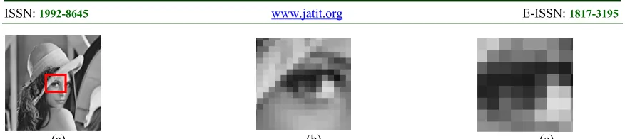
In common parlance, resolution improvement can be best viewed when the image is magnified, as shown in Fig. 1 and Fig. 2. In Fig. 1(a), red colored rectangle shows the region which is magnified and tested for resolution enhancement. Fig. 2(a) shows the enhanced version of Fig. 1(a) image using DTCWT algorithm. As it may be observed that magnification of such image has not degraded the quality of the image in Fig. 2(b) & Fig. 2(c).
2. DIGITAL IMAGE PROCESSING
computer-1513 readable format. Now days, digital cameras are available which were launched by Canon, Nikon, Sony etc. that can easily convert the image into compatible mode. This format may be one of the formats such as tiff, bmp, jpeg, png etc. Digital images are generated by the conversion of continuous signals in digital format. Major aspect that deals with digital image processing is that, most of the real-time applications use image processing techniques. This requires improvement of visual aspect of the image much higher than the original image.
Every branch of science that deals with the research and development scenario or even other sub-disciplines collects images from surrounding and universe for processing purpose. Digital image processing is an important tool for evaluating the information present in images. Images captured using digital camera, are easy to process using computer technology. To assess the information present in digital images, there are certain areas which lie under image processing. Image enhancement, image restoration, image segmentation, image inpainting are some of the areas under which processing of digital images is done.
Image enhancement is further distributed in subsections which include resolution enhancement, contrast enhancement, noise reduction, edge preservation etc. Images considered for such type of processing may vary with the applications. Medical images are readily used for enhancement, to improving the visual quality of the image. Image restoration is the process of compensating or reducing the noise present in image. Noise in any medical image may introduce due to several reasons such as patient’s motion, low illumination of light in case of radiographic image. So, noise reduction or removal of artifacts present in those images need to be removed. Image segmentation is another class of method under digital image processing. Under image segmentation, input image is divided into subsections based on pixels. This is done by transforming the image into appropriate mode. As far as image inpainting is concerned, image is being inpainted for recovering the scratch present in an image.
3. LITERATURE SURVEY
Hasan Demirel et.al [1] proposed a technique for improving the resolution using
Discrete wavelet transform (DWT) [21-24] and stationary wavelet transform (SWT). In this paper, high frequency subband image obtained by DWT and input image was interpolated for resolution enhancement. SWT was used to enhance the edges. SWT decompose the image to generate high frequency subband image. This image is used to modify the estimated high frequency subband image obtained by DWT. Resulting images were combined to generate high resolution image. Hasan Demirel et.al [12] proposed a technique based on Complex wavelet transform (CoWT) for satellite images. CoWT is preferred because of directional selectivity property. HE Si-hua et.al [2] proposed a technique for improving the resolution of the image using fractal coding. Results showed that such type of coding method kept the image details intact. Also, reconstructed image showed small error. Debesh Jha et. al [13] proposed DTCWT based algorithm for distinguishing Alzheimer's disease (AD) effected human brain and healthy human brain. Proposed algorithm is divided into three stages namely, DTCWT which was used for feature extraction, reduction of feature dimension using principal component analysis (PCA) and lastly classification between normal brain and AD affected brain was accomplished using feed-forward artificial neural network (ANN).
Muhammad Zafar Iqbal et. al [3] has improved resolution and contrast of medical images using DTCWT for brain magnetic resonance imaging (MRI), chest and ribs X-Ray images. He used non local mean (NLM) filter [7] for better result but it increased the computational complexity of the algorithm. In the proposed work the complexity of algorithm is reduced by applying fast NLM filter. Here, three algorithms are proposed which gives lower complexity as well as high efficiency as compared to other existing algorithms [1-10]. This paper is organized as follows. Section 2 presents introduction of digital image processing. Section 3 describes the literature survey. Section 4 presents the application of resolution enhancement. Section 5 describes the details of proposed algorithm. Section 6 explains the parameters used for quantitative analysis. In Section 7 simulated results are discussed. The conclusions are given in section 8. Future scope is mentioned in section 9.
4. APPLICATION OF RESOLUTION
ENHANCEMENT
1514 a). It is required for displaying standard definition
video on high definition TV sets.
b). To convert grainy and old video material in digital format.
c). Low resolution (75 DPI) to high resolution (600 or 1200 DPI).
d). In medical imaging, for enhancing the visual quality of CT scan, MRI, PET images etc. e). In the field of forensics, high resolution images
are required to recognize criminal face.
5. PROPOSED ALGORITHM
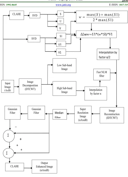
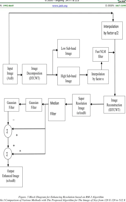
In this paper three different algorithms are proposed for resolution enhancement namely RM-1(DTCWT-Gaussian-CLAHE), RM-2(DTCWT-Fast NLM-CLAHE) and RM-3(DTCWT-RM-2(DTCWT-Fast NLM). RM-1 and RM-2 algorithms are proposed for resolution as well as contrast enhancement. While RM3 proposed only for resolution enhancement. The block diagram of RM-1 algorithm is shown in Fig. 3. Resolution enhancement of the input low resolution (LR) image of dimension AxB is performed by decomposing it using DTCWT. One level of decomposition produce 16 sub-bands images out of which 4 images are low sub-band images. Remaining 12 images are high sub-band which contains directional information. Further, high sub-band images are interpolated using Lanczos interpolation by factor α. Also for contrast enhancement, singular value decomposed (SVD) is used. SVD decompose the image in three different matrices namely, S, U and V. Matrix U and V are orthogonal matrix and S is a diagonal matrix. Contrast limited adaptive histogram equalization (CLAHE) is also used to enhance the contrast [21-22] of the LR image. Then, a weighting function is calculated to generate new image and it is interpolated by factor α/2. To increase the computational speed of the proposed algorithm fast non local mean (NLM) filter is used. Then these images are combined and reconstructed using inverse DTCWT. This will generate a super resolution image of dimension αA x αB. Further, Gaussian filter is used for filtering any noise component present in the super resolution image. Then finally, resolution enhanced image is produced. Block diagram of proposed RM2 algorithm is shown in Fig. 4. Initial procedure of enhancing resolution and contrast of the LR image is same as explained for RM1 algorithm. After super resolution image is generated, further visual quality is improved using combination of filters.
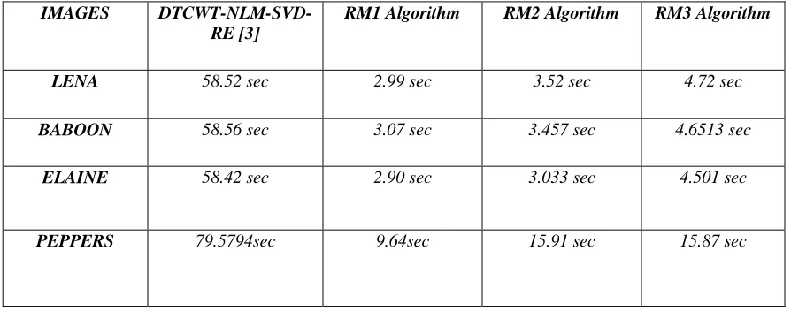
These filters include median and Gaussian filters. Now this super-resolution image of dimension αA x αB is passed through median filter. Output of median filter is passed through two low pass Gaussian filters. These are used to generate low pass component which are subtracted from the output of median filter. Resulting image is again added to the output of median filter to improve visual quality of image. To increase its contrast CLAHE algorithm is used. Finally enhanced image is generated with dimension αA x αB. Proposed RM3 algorithm is shown in Fig. 5. In this LR image is not passed through CLAHE and SVD. Remaining steps are same as that of RM2 algorithm.
6. PERFORMANCE METRIC
Quantitative analysis is done in this section to show the comparison of the proposed algorithm with the state-of-art techniques. Certain parameters are evaluated which are summarized below to compute the quality of the image quantitatively.
6.1 Peak Signal-To-Noise Ratio (PSNR)
This is the utmost important and immensely popular parameter for showing the actual improvement of the algorithm. This is evaluated by the mathematical formula given by Eqn. 1
2
10log
MAX
IPSNR
MSE
(1)where MAXI is the maximum intensity of the reconstructed image and MSE is the mean square error between reference image and output enhanced image. PSNR is evaluated in terms of decibels (dB). Ideal value for this is 30 to 50 dB for 8 bits per pixel case.
6.2 Mean Square Error (MSE)
MSE is used to compute the error between the reference (high resolution image) image and output enhanced image. Major criteria while evaluating the peak signal-to-noise ratio (PSNR) and MSE is that both the images should be of same dimension and same class. MSE is computed using Eqn. 2
2 1 1 0 01
( , )
( , )
N M i jMSE
X i j
Y i j
M N
(2)1515 MSE should lie between 0 and 1. Generally, 0 means least error between the images and 1 means the maximum error.
7. RESULTS AND DISCUSSION
Standard images [1, 4] are collected to build the data set. These images are originally of size 512 x 512. For converting them to low resolution image, they are down-sampled by factor 4 which generates actual input image of size 128 x 128. Results are simulated using MATLAB 2014b version on i5 core processor. Fig. 6-9 shows the images which result from various algorithms and are used for comparison. Fig. 6(a) shows the original image of Lena having dimension 512 x 512. Fig. 6(b) shows the down-sampled image of dimension 128x128. Fig. 6(c) shows the result for using DTCWT-NLM-SVD-RE [3] algorithm which enhances the resolution as well contrast of the image. Fig. 6(d).shows the result of using RM2 algorithm which reduces the computational complexity. Fig. 6(e) shows the image generated by using RM1 algorithm. Fig. 6(f) shows the result of using proposed algorithm which is RM3 algorithm that enhances the resolution of the image. Fig. 7 shows the similar results for the Baboon image. Fig. 8 shows the results for the Elaine image and Fig. 9 shows the result for using the Peppers image. It is clearly seen from the images that results are more improved using proposed algorithm. For the quantitative analysis calculation of PSNR (dB) and MSE is done which is shown in Table I
Table I shows the comparison of the proposed algorithm (i.e., RM1, RM2 and RM3) with existing algorithms [1] and [3]. PSNR (dB) is calculated for all the test images. It is observed that RM3 algorithm provides better PSNR value for all the images. Table II shows the comparison of the processing time. It may be observed that RM1 reduced the processing time as compared to [3].
8. CONCLUSION
DTCWT based three different algorithms are proposed in this paper. RM1 and RM2 algorithms are used to improve the resolution as well as contrast of the input image. RM3 algorithm is used for improving the resolution of the input image. To show the quantitative analysis of the proposed algorithms images are collected namely, Lena, Elaine, Baboon and Peppers. Comparative result is shown in Table I and Table II. Table I shows the comparison of PSNR (dB) value of the proposed work and other algorithms. Table II
shows the comparison of processing time for proposed algorithms with current state of art. It is seen that for all the images, RM3 algorithm provides better value for PSNR (dB) whereas RM1 algorithm provides better result for processing time. Due to Fast NLM [24], processing time is reduced and it provides better results for resolution enhancement.
9. FUTURE SCOPE
This paper used DTCWT for improving the resolution of the image. Other wavelet transforms such as curvelet transform, contourlet transform can also be used in behalf of DTCWT. This may provide better results. For reducing the computational complexity, other filters can also be used.
REFERENCES
[1] Hasan Demirel and Gholamreza Anbarjafari, “Image resolution enhancement by using discrete and stationary wavelet decomposition”, IEEE Transactions on Image Processing, Vol. 20, No. 5, 2011,pp.
1458-1460.
[2] HE Si-hua and WU Zhen, “Method of single image super-resolution enhancement based on fractal Coding”, 3rd International Conference on Computer Science and Network Technology, 2013, pp 1034-1036.
[3] Muhammad Zafar Iqbal, Abdul Ghafoor, Adil Masood Siddiqui, Muhammad Mohsin Riaz and Umar Khalid, “Dual-tree complex wavelet transform and SVD based medical image resolution enhancement”, Elsevier, Signal processing, Vol. 10, 2014, pp
430-437.
[4] A. Temizel and T. Vlachos, “Wavelet domain image resolution enhancement using cycle-spinning”, IEEE, Electronics Letter,
Vol. 41, No. 3, 2005, pp. 119-121.
[5] Muhammad Zafar Iqbal, Abdul Ghafoor and Adil Masood Siddiqui, “Satellite image resolution enhancement using dual-tree complex wavelet transform and non local means”, IEEE Geoscience and remote sensing letters, Vol. 10, No. 3, 2013, pp.
451-455.
[6] W. L. Lee, C.C. Yang, H.T. Wu and M.J. Chen, “Wavelet-based interpolation scheme for resolution enhancement of medical images”, J. Signal Process. Syst. 55, 2009,
pp. 251-265.
1516 “Wavelet transform and non local means based super resolution”, International Symposium on Computer and Communications, Vol. 17, 2012.pp. 119-124.
[8] Hasan Demirel, G. Anbarjafari and S. Izadpanahi, “Improved motion-based localized super resolution technique using discrete wavelet transform for low resolution video enhancement”, proc. 17th Eur. Signal Process. Conf., Glasgow, Scotland, 2009,.
pp. 1097-1101.
[9] A. Temizel, “Image resolution enhancement using wavelet domain hidden Markov tree and coefficient sign estimation”, Proc. Int. Conf. Image Process., Vol. 5, 2007, pp.
381-384.
[10] A. Temizel and T. Vlachos, “Image resolution upscaling in the wavelet domain using directional cycle spinning” J. Electron. Imag., Vol. 14, No. 4, 2005.
[11] S. Zhao, H. Han, and S. Peng, “Wavelet domain HMT-based image super resolution”
Proc. IEEE Int. Conf. Image Process., Vol.
2, Sep. 2003, pp. 933–936.
[12] Hasan Demriel and Gholamreza Anbarjafari, “Satellite image Resolution enhancement using complex wavelet transform”, IEEE Geoscience and remote sensing letters, Vol.
7, No. 1, 2010, pp 123-126.
[13] Debesh Jha, Ji-In Kim and Goo-Rak Kwon, “Diagnosis of Alzheimer’s Disease Using Dual-Tree Complex Wavelet Transform, PCA, and Feed-Forward Neural Network”.
Journal of Healthcare Engineering, Hindawi, pp 1-13.
[14] R. C. Gonzalez and R. E. Woods, Digital Image Processing, 3rd Edition, Prentice Hall, NJ, 2008.
[15] R. Yan, L. Shao, Y. Lin, “Nonlocal Hierarchical dictionary learning using wavelets for image denoising”, IEEE Trans. Image Process. 22, 2013, pp. 4689-4698.
[16] S. Mallat, “A Wavelet Tour of Signal”
Processing, 2nd ed. New York: Academic,
1999.
[17] Madhu Jain, Maneesha Gupta and N. K. Jain, “The Design of the IIR Differintegrator and its application in Edge Detection,”
Journal of Information Processing Systems,
Vol. 10, No.2, 2014, pp. 223-239.
[18] Dinh-Hoan Trinh, Marie Luong, Francoise Dibos, Jean-Marie Rocchisani, Canh-Duong Pham and Truong Q. Nguyen, “Novel example-based method for super-resolution and denoising of medical images”, IEEE
Transactions on image processing, Vol. 23,
No. 4, 2014, pp. 1882-1895.
[19] Hasan Demirel and Gholamreza Anbarjafari, “Pose Invariant Face recognition using probability distribution in different color channels”, IEEE signal processing letters, Vol. 15, pp. 537-540, 2008.
[20] Jingyu Yang, Yao Wang, Wenli Xu and Qionghai Dai, “Image coding using dual-tree discrete wavelet transform”, IEEE transactions on image processing, Vol. 17,
No. 9, pp. 1555-1569, 2008.
[21] Yue Fang, Yifan Wang, Cuifang Kuang and Xu Liu, “Enhancing the resolution and contrast in CW-STED microscopy”, Elsevier, Optics communication, Vol. 322,
pp. 169-174, 2014.
[22] Md Belayet Hossain, Khin Wee Lai, Belinda Pingguan-Murphy, Yan Chai Hum, Maheza Irna Mohd Salim and Yih Miin Liew, “Contrast enhancement of ultrasound imaging of the knee joint cartilage for early detection of knee osteoarthritis”, Elsevier, Biomedical signal processing and control,
Vol. 13, pp. 157-167, 2014.
[23] A. Buades, B. Coll and J. M. Morel, “A review of denoising algorithm with new one”, SIAM Journal on Multiscale Modeling and Simulation, Vol. 4, No. 2, pp. 495-530,
2005.
[24] Jerome Darbon, Alexandre Cunha, Tony F. Chan, Stanley Osher and Grant J. Jensen, “Fast nonlocal filtering applied to electron cryomicroscopy, IEEE International Symposium on Biomedical Imaging”, pp. 1331-1334, 2008.
[25] Maneesha Gupta, Madhu Jain and B. Kumar, “Novel Class of Stable Wideband Recursive Digital Integrators and Differentiators,” IET Signal Processing, vol.4, No.5, pp.560–566,
2010.
[26] Maneesha Gupta, Madhu Jain and B. Kumar, “Recursive Wideband Digital Integrator and Differentiator,” International Journal of Circuit Theory and Applications (IJCTA),
vol.39, No.7, pp.775–782, 2011.
[27] Madhu Jain, Maneesha Gupta and Nitin Jain, “Linear Phase Second Order Recursive Digital Integrators and Differentiators,”
Radio engineering, Vol. 21, No. 2, pp. 712 -
717, 2012.
[28] Maneesha Gupta, Madhu Jain and B. Kumar, “Wideband Digital Integrator and Differentiator,” IETE Journal of Research,
1517 [29] Madhu Jain, Maneesha Gupta and Nitin Jain,
“Analysis and Design of Digital IIR Integrators and Differentiators using Minimax and Pole, Zero and Constant Optimization Methods,” ISRN Electronics, Vol. 2013, pp. 1 - 14, 2013.
[30] Madhu Jain, Maneesha Gupta and Nitin Jain, “Design of Half Sample Delay Recursive Digital Integrators using Trapezoidal Integration Rule”, International Journal of Signal & Imaging Systems Engineering, Vol.
9, No. 2, pp. 126 - 134, 2016
[31] Maneesha Gupta, Madhu Jain and B. Kumar, “Wideband digital integrator”, in the proceedings of IEEE International Conference on Multimedia, Signal Processing and Communication technologies (IMPACT 2009), pp. 107-109, March 2009, India.
[32] Madhu Jain, Maneesha Gupta and Nitin Jain, “A new fractional order recursive digital integrator using continued fraction expansion” ”, in the proceedings of 2010 IEEE India International Conference on Power Electronics (IICPE 2010), pp. 1-6, January 2011, India.
[33] Maneesha Gupta, Madhu Jain and B. Kumar, “Design of a novel Digital Integrator”, in the proceedings of International Conference on Aerospace Electronics, Communications and Instrumentation (ASECI 2010), pp. 191-193, January 2010, India.
[34] Bhabatosh Chanda And Dwijest Dutta Majumder, “Digital Image Processing And Analysis”, 2002.
[35] A. K. Jain, “Fundamentals Of Digital Image Processing”, Englewood Cliffs, Nj: Prentice Hall, 2009.
[36] R.M. Haralick, and L.G. Shapiro, “Computer And Robot Vision”, Vol-1, Addison Wesley, Reading, Ma, 2012.
[37] Zhou Wang, lan Conrad Bovik’s Jain, “Image Quality Assessment:From Error Visibility To Structure Similarity” IEEE Transaction On Image Processing, Vol.13
1518
[image:7.612.87.525.69.167.2]
(a) (b) (c)
Figure. 1 Lena 512 x 512 Image, (a) original Image, (b) 3 times magnification of red colored eye region, (c) 4 times magnification of red colored eye region
[image:7.612.104.516.323.710.2]
(a) (b) (c)
Figure 2 Lena 512 x 512 Image, (a) Enhanced Image, (b) 3 times magnification of red colored eye region, (c) 4 times magnification of red colored eye region.
Figure. 3 Block Diagram for Enhancing Resolution and Contrast based on RM-1Algorithm
SVD
Image Decomposition
(DTCWT) Interpolation
by factor α High Sub-band
Image
Image Reconstruction
(IDTCWT) Input
Image (AxB)
Low Sub-band Image
Fast NLM filter
Interpolation
by factor α/2
1*( * 1)* 1
LLnew U
w S
V
U
Gaussian
Filter
V
S1
U1
V1
Super Resolution
Image (αAxαB) Output
Enhanced Image (αAxαB)
CLAHE SVD S
max( ) max( 1)
2* max( 1)
S
S
w
S
1519
Figure. 4 Block Diagram for Enhancing Resolution and Contrast based on RM-2 Algorithm
Image Decomposition
(DTCWT) Input
Image
(AxB) Interpolation
by factor α High Sub-band
Image
1*( * 1)* 1
LLnew U
w S
V
V
Fast NLM filter Low Sub-band
Image
Interpolation by
factor α/2
V1
Super Resolution
Image (αAxαB)
U1
m ax( )
m ax( 1)
2 * m ax( 1)
S
S
w
S
Image Reconstruction
(IDTCWT) U
∑
SVD
∑
SVD
S1
Median
Filter
S
Output Enhanced Image
(αAxαB) CLAHE
CLAHE
Gaussian Filter
Gaussian Filter
_
+
+
1520
Figure. 5 Block Diagram for Enhancing Resolution based on RM-3 Algorithm
Table I Comparison of Various Methods with The Proposed Algorithm for The Image of Size from 128 X 128 to 512 X 512.
Output
Enhanced Image
(αAxαB)
Median
Filter
Image
Reconstruction
(IDTCWT)
Gaussian
Filter
Gaussian
Filter
∑
∑
Super
Resolution
Image
(αAxαB)
Interpolation
by factor α
High Sub-band
Image
Input
Image
(AxB)
Image
Decomposition
(DTCWT)
Fast NLM
filter
Low Sub-band
Image
Interpolation
by factor α/2
_
+
1521
PARAMETER PSNR (dB)
IMAGES
METHODS
Lena Baboon Elaine Peppers
DWT-SWT [1] 34.8200 23.8700 35.0100 33.0600
DTCWT-NLM-SVD-RE [3] 25.6054 25.4233 33.2323 25.1677
RM1 25.6162 25.2700 30.9711 24.8013
RM2 31.6556 28.4781 28.2221 33.5452
[image:10.612.83.530.348.524.2]RM3 38.7207 34.8921 37.9261 41.1749
Table II Comparison of Processing Time with The Proposed Algorithms for the Image of Size from 128 X 128 to 512 X 512.
IMAGES DTCWT-NLM-SVD-RE [3]
RM1 Algorithm RM2 Algorithm RM3 Algorithm
LENA 58.52 sec 2.99 sec 3.52 sec 4.72 sec
BABOON 58.56 sec 3.07 sec 3.457 sec 4.6513 sec
ELAINE 58.42 sec 2.90 sec 3.033 sec 4.501 sec
1522
[image:11.612.86.534.114.204.2]
(a) (b) (c) (d) (e) (f)
Figure. 6 Simulated Result of 512 x 512 Lena Image, (a) Original 512 x 512 Image, (b) Input 128 x 128 Image, (c) DTCWT-NLM-SVD-RE Result, (d) RM2 Result, (e) RM1 Result, (f) RM3 Result
[image:11.612.85.529.270.353.2]
(a) (b) (c) (d) (e) (f)
Figure. 7 Simulated Result of 512 x 512 Baboon Image, (a) Original 512 x 512 Image, (b) Input 128 x 128 Image, (c) DTCWT-NLM-SVD-RE Result, (d) RM2 Result, (e) RM1 Result, (f) RM3 Result
(a) (b) (c) (d) (e) (f)
Figure. 8 Simulated Result of 512 x 512 Elaine Image, (a) Original 512 x 512 Image, (b) Input 128 x 128 Image, (c) DTCWT-NLM-SVD-RE Result, (d) RM2 Result, (e) RM1 Result, (f) RM3 Result
(a) (b) (c) (d) (e) (f)
[image:11.612.81.518.420.505.2] [image:11.612.82.520.563.649.2]