Background
Idiopathic infl ammatory myopathies (IIMs), also known as myositis, are a group of rheumatic disorders clinically characterized by muscle weakness, leading to disability, decreased quality of life, and a reduced life expectancy. Although this is a relatively rare disease, our under-standing of risk factors and the underlying immuno-pathogenesis has increased substantially in recent years. (See reviews by Betteridge et al. [1], Chinoy et al. [2], and Rayavarapu et al. [3] published in 2011.)
Myositis shares many features with rheumatoid arthritis and systemic lupus erythematosus, namely as diff erent examples of disabling chronic infl ammatory syndromes, which can be reevaluated in the light of distinct genetic and environmental contributions [4]. Common traits between these rheumatic disorders include a major histo-compatibility complex (MHC) class II association, in-fl am matory cell infi ltrates in aff ected tissues, and the presence of predictive or disease activity-associated autoantibodies (or both). Taken together, these observations point to a central role for adaptive immune reactions in disease manifestation.
Th e spectrum of infl ammatory myopathies is getting broader, and the classifi cation criteria for the IIMs, designed by Tony Amato on behalf of the Muscle Study Group, proposed the following categories: (i) inclusion body myositis, (ii) polymyositis (PM), (iii) dermato myo-sitis (DM), (iv) non-specifi c myomyo-sitis, and (v) immune-mediated necrotizing myopathy [5]. Some IIMs share common histopathological features of leukocyte infi l tra-tion, preferentially T cells and macrophages in skeletal muscle tissue, whereas others display no or spare peri-vascular and perimysial infi ltrates. Novel studies of this latter group which are based on detailed immuno-pathology suggest that the predominant abnormal histo-logical feature is instead membrane attack complex (MAC) deposition on the sarcolemma in both non-necrotic and non-necrotic muscle fi bers [5,6]. Many patients have manifestations besides in the muscles, such as in the lungs (mostly PM), skin (DM), and sometimes in the joints. Additionally, some patients display more than one rheumatic diagnosis, and systemic sclerosis is the most common connective tissue disease associated with IIM [7].
Both CD4+ and CD8+ T cells have been described to be present and active in patients with myositis. Th e presence of cytotoxic CD8+ T cells has been attributed to virus or intracellular bacterial infections, which would generate potent eff ector cells. CD8+ T cells are often subdivided on the basis of their diff erentiation level, fi rstly into naïve and activated/memory T cells; the latter subset can be further subdivided in three groups (central memory T cells (TCM), eff ector memory T cells (TEM) and TEMRA) on the basis of their surface expression of diff erent lymph node homing markers [8]. A summary of candidate infectious agents associated with myositis was published recently [9].
Th e presence of CD4+ T cells could also be associated with infectious agents, but in the context of myositis, it is more likely that these cells develop as a consequence of an autoimmune reaction [1]. Owing to how immune responses are orchestrated by CD4 cell-derived cyto-kines, CD4+ T cells are traditionally regarded as helper cells. Indeed, the most common way of subdividing CD4 T cells is based on secretion of specifi c cytokines, together with activity of so-called master transcription factors. In this fashion, CD4 T cells can be subdivided
Abstract
T cells of both the CD4 and CD8 lineage are commonly found in aff ected tissues of patients with idiopathic infl ammatory myopathies, but understanding the contribution of these cells to immunopathogenesis remains challenging. Given recent advances in identifying more myositis-associated autoantibodies and their putative targets, we suggest that studies on autoreactive T cells targeting those autoantigens are one way forward. Another (so far, more frequently used) approach comes from studies on eff ector T cells in the context of myositis. This review summarizes recent advances and current hypotheses in both of these contexts.
© 2010 BioMed Central Ltd
T cells in myositis
Vivianne Malmström*, Paulius Venalis and Inka Albrecht
R E V I E W
*Correspondence: [email protected]
Rheumatology Unit, Dept of Medicine, L8:04, Karolinska Institutet, Karolinska University Hospital Solna, SE-17176 Stockholm, Sweden
into diff erent T helper subsets such as Th 1, Th 2, Th 9, Th 17, and Th 22 and regulatory T (Treg) cells [10].
How-ever, in recent years, it has become clear that CD4+
T cells can also diff erentiate into cytotoxic eff ector cells reminiscent of CD8 cells and natural killer (NK) cells [11]. Such cells have been named CD4+CD28null T cells and fall outside of the classic T helper subsets. Th ey represent terminally diff er en tiated cells, which in addition to being potent inter feron-gamma (IFNγ) and tumor necrosis factor (TNF) producers have acquired many NK-related receptors and cytotoxic capacity by expressing both perforin and gran zymes [12].
It is well established that T cells can be found at all of the diff erent sites of disease manifestations in patients with myositis. But the importance of the presence of these cells is still a matter of debate, as is their antigen specifi city. In recent years, an increasing number of myositis-associated and myositis-specifi c autoantibodies have been identifi ed. Th e targets of those autoantibodies might represent diff erent candidate autoantigens [1]. Clearly, such data implicate antigens that could also be studied with regard to T-cell function. Below, we will discuss T cells in the three major aff ected tissues in patients with myositis rather than in subgroups according to the classic disease sub-entities PM, DM, and IBM.
Muscle-infi ltrating T cells
Th e main manifestations, shared by all three subsets of myositis, are proximal muscle weakness and muscle fatigue. Some patients have persistent cellular infi ltrates, which are associated with sustained muscle weakness.
How do the fi rst T cells migrate to muscle?
Th ere have been ample eff orts in dissecting how and
which T cells (and other infl ammatory cells) migrate into muscle. Here, chemokines govern the migration of leuko-cytes to sites of infl ammation (Figure 1), and several studies have addressed this issue and demonstrated
expres sion of the α-chemokines CXCL9 and CXCL10
and the β-chemokines CCL2, CCL3, CCL4, CCL19, and
CCL21 in IIM muscle. Th e chemokines can be produced
by infi ltrating infl ammatory cells but potentially also by muscle fi bers themselves. Th e reason why muscle fi bers would express chemokines could be infection, trauma, and genetic predisposition.
Although many studies have investigated mRNA from muscle biopsies, making it diffi cult to elucidate the cellular source of the chemokines, there are data that IIM muscle fi bers themselves can produce chemokines. In this context, CCL2 expression has been demonstrated by immuno-histo chemical staining of muscle biopsies [13,14] and in myoblast cell cultures in which co-stimulation with IL1β leads to elevated CCL2 mRNA levels [15]. CCL2 is a
chemoattractant for CCR2- and CCR4-express ing cells,
including monocytes, memory T cells, and dendritic cells. Additional chemokines are interesting in this context, such as CCL3 and CCL4, which are chemo attractants for macrophages and T cells, and CCL3 is a potent regulator
of Th 1-committed T cells. Indeed, a signifi cant
upregulation of CCL3 had been demonstrated in IBM myofi bers. Both CCL3 and CCL4 are present in muscle-infi ltrating mononuclear cells in DM, PM, and IBM [16].
Similarly to the traffi c via high endothelial venules into lymph nodes, CCR7-CCL19 interaction has been sug-gest ed to contribute to amplifying/sustaining T-cell traffi c to sites of infl ammation. Also, muscle fi bers positive for this chemokine and infi ltrating lymphocytes positive for the receptor have been demonstrated [17]. Further dissection for extranodal lymphoid microstructures has been performed, and indeed such structures can be found [18]. Th e existance of such lymph node structures indicates that lymphocyte activation and diff erentiation could take place within the muscle, and there is support from studies of B cells, plasma cells, and immunoglobulin sequences that plasma cell diff erentiation can take place in this location [19].
A common observation in biopsies of aff ected muscle from patients with IIM is the focal distribution of the in-fl ammatory infi ltrates. Th e reasons behind this have not yet been delineated, but it is tempting to speculate that migration of the fi rst cells into muscle is a rare event. Once a few infl ammatory cells have entered the muscle, a feedback loop is started because of chemokine produc-tion by the infl ammatory cells.
Which T cells are found in aff ected muscles?
[image:2.612.315.549.88.232.2]A long-standing dogma has been that CD8 cells are more common in PM and IBM but that CD4 cells are more pronounced in DM. T-cell receptor (TCR) profi ling by performing CDR3 spectratyping of the TCR Vβ chains
Figure 1. Chemokines regulate the migration of infi ltrating cells. Stressed or damaged muscle fi bers secrete chemokines, which will attract infl ammatory cells. The infi ltrating cells will also secrete chemokines attracting even more cells and creating a loop. DC, dendritic cell; Th1, T helper 1.
Infection trauma injury chemokines Memory
T cells (CD4+, CD8+) Th1 cells
Macro phages
DCs Monoc
for CD8+ T cells in PM and IBM demonstrated that CD8 T cells (potentially autoreactive) are clonally expanded and can persist for years [20-22]. A strong bias of Vβ expression in the IBM muscle as compared with the corresponding blood further suggests that the T cells are clonally expanded in situ, or are specifi cally recruited to the muscle, and may be driven by muscle-specifi c auto-antigens [20]. However, within the Vβ subfamilies, there was a high variability in clonal restriction between patients for both PM [23,24] and IBM [25,26], possibly suggesting the presence of several local autoantigens and epitope spreading. For CD4+ T cells, in contrast, CDR3 spectratyping has revealed much more polyclonal patterns [21]. Th is, together with the observation that many muscle-infi ltrating cells express perforin or gran-zyme B or both, could be seen as evidence for an exclu-sively cytotoxic CD8 T cell-mediated immuno patho logy. How ever, CD4 T cells have been less studied, but it has been demonstrated that CD4+ T cells can be a major part of the muscle infi ltrate [12], so more studies are needed to fully evaluate this. Moreover, today we know that
CD4+CD28null T cells predominate the infi ltrate in
aff ected muscle and those cells express cytotoxic eff ector substances [12,27], allowing us to reevaluate older fi nd-ings in a new context.
FOXP3+ Treg cells are critical cells in maintaining immune homeostasis and preventing autoimmune and chronic infl ammatory disease. Th erefore, it is appealing to suspect that patients with IIM may have a numerical defi cit in Treg cells. Th is was, however, not the case [28], and similar data exist for other rheumatic diseases. An alternative hypothesis is that the Treg cells are func-tionally defi cient or that the infl ammatory milieu does not allow Treg cell suppression as indicated in rheu-matoid arthritis [29]. During investigations of biopsies before and after glucocorticoid therapy, both the overall T-cell count and the Treg cell count were reduced after therapy whereas persistent CD4+CD28null T cells could be observed [30].
How could T cells perpetuate local disease?
Cytokine staining of muscle biopsies has demonstrated only modest levels of T cell-derived cytokines. However, since many cytokines function directly on neighboring cells, this could be due in part to rapid consumption. Another interesting eff ector function is the perforin/ granzyme B axis and its eff ect on muscle fi bers (Figure 2). We recently started to address whether CD28null T cells could directly interact with muscle fi bers by an autolo-gous co-culture system, and we have preliminary data in support of this [31]. A granzyme attack normally would be expected to induce apoptosis in the aff ected cell, but one needs to remember that muscle fi bers are multi-nucleated cells and, as such, may not undergo classic
apoptosis [32]. Instead, one could think that the common observation of regenerating fi bers could be a result of such an insult. Such regenerating fi bers also express high levels of Jo-1, one of the candidate auto antigens [33]. In the context of CD28null T cells, an additional concern is their longevity, and this subset is the reason why persis-tent infi ltrates are sometimes seen even after aggressive therapy [30].
Skin-infi ltrating T cells
DM is the IIM subtype that aff ects both skin and muscle. Historically, this disease has been looked upon as more CD4-driven than PM and IBM. Granzyme B-expressing T cells have been found in other rheumatic diseases with skin involvement (such as systemic lupus erythematosus) but were found to be low in DM [34]. Also, the degree of
FOXP3+ cells has been reported to be low [35,36]. In
contrast, type I IFN appears strong in aff ected skin of patients with DM [37] and could lead to accumulation of
CXCR3+ lymphocytes. Indeed, enrichment of CXCR3+
cells has been reported in DM skin [38]. Another interesting T-cell population, CXCR5+ T helper cells, has been studied in peripheral blood of patients with juvenile DM and found to display Th 2 and Th 17 activities and to be associated with disease activity [39].
Lung involvement in myositis
Interstitial lung disease (ILD) is a heterogeneous group of non-infectious lung disorders characterized by infl am ma-tory cell infi ltration and interstitial fi brosis. It remains one of the greatest contributors to morbidity and mortality in myositis [40] and is associated with a poor quality of life for patients with myositis. Myositis-asso ciated ILD is closely linked to the appearance of auto antibodies raised against anti-histidyl tRNA synthetase (Jo1) and the
so-called anti-synthetase syndrome [41]. In anti-Jo-1+
[image:3.612.316.547.88.192.2]patients, there are a few reports on T lympho cytes with specifi city toward this particular autoantigen (reviewed in [42]). Additionally, T cells from bronchoalveolar lavage
Figure 2. Phenotype and eff ector functions of muscle-infi ltrating T cells. Perforin and granzyme B-positive cells are abundant in mononuclear cell infi ltrates in aff ected muscle. Not only CD8+ but
also CD4+CD28null T cells express these eff ector molecules. IFN-γ, interferon-gamma; Th1, T helper 1.
CD8+ CD28null CD4+ granzyme cells Th1 IFN-γ
fl uid from patients with myositis-asso ciated ILD showed a strong bias toward Vbeta3 T-cell receptor expression as compared with healthy controls, suggesting a specifi c role for T cells in the development of ILD [43,44].
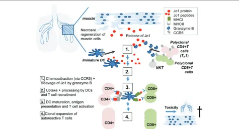
Since ILD often precedes myositis symptoms, it is suggested that the autoimmune reaction might start in the lungs (Figure 3). Indeed, it has been demonstrated that the Jo1 antigen is highly expressed in the lung com-pared with other organs [33]. Interestingly, a proteo-lytically sensitive conformation of Jo-1 is found in the lungs and leads to cleavage by granzyme B [45]. Granzyme B generates unique fragments of this auto-antigen. It is suggested that these fragments are taken up by immature dendritic cells, which get activated, mature into professional antigen-presenting cells, and stimulate CD4+ T cells, initiating downstream immune cascades. Both CD8+ T cells and CD4+CD28null T cells may play a role in cytotoxicity in both the muscle and the lungs. Th ey could contribute not only to destruction of target tissues in myositis but also to initiation of autoimmunity through cleavage of Jo1 mediated by granzyme B.
Th e trigger for the initiation of the autoimmune res-ponse has not yet been clarifi ed. On the one hand, it is hypothesized that the infl ammatory cascade might be initiated by an infection with an as-yet-unidentifi ed virus (for example, Coxsackie, infl uenza, HIV, hepatitis C
virus, and cytomegalovirus) [42]. Th ose viruses may enter the respiratory tract, where they may lead not only to cell death and an associated release of Jo-1 antigen into the extracellular space but also to a modifi cation of this enzyme. Another possibility is genetic predisposition to reduced apoptotic clearance or increased apoptosis in conjunction with environmental stimuli such as smoking [46]. Th e release of Jo1 and its special conformations into the extracellular milieu are believed to trigger not only activation of T cells by antigen-presenting cells but also migration of cells expressing CCR5, including den dritic cells and T cells [47], especially Th 1 cells that were shown to express predominantly CCR5 [48]. Th is may explain an infi ltration of mostly Th 1 cells in the lung of patients with myositis-associated ILD [49]. In addition, it was shown that there is a disease-specifi c association between Jo-1, ILD, and serum levels of CXCL9 and CXCL10 [50], two IFN-γ inducible chemokines attracting CXCR3-express-ing cells, includCXCR3-express-ing NK and Th 1 cells, further enhancing
the infi ltration of pro-infl ammatory Th 1 cells and
emphasizing a role for these cells in disease pathogenesis.
T cell-independent autoantibodies?
[image:4.612.69.545.89.346.2](B-cell activating factor) and IL-21 [51]. Moreover, high BAFF levels have been found in the circulation of anti-Jo-1+ patients and an even higher concentration of BAFF is associated with myositis-associated ILD [52]. Th is leads to a higher appearance of B cells and plasma cells that possibly could locally produce autoantibodies. Th e autoantibodies are believed to build immune complexes with Jo-1 fragments and nucleic acid released from dying cells, which may activate plasmacytoid dendritic cells (pDCs). Th ese cells are the major source of type I IFN, and IFN-α can be detected in muscle, skin, and peripheral
blood of anti-Jo-1+ myositis patients where pDCs are
highly enriched [53-55]. IFN-α released by pDCs in turn upregulates expression of BAFF, creating a positive feedback loop and ultimately leading to a break of tolerance. In addition, IFN-α upregulates expression of intercellular adhesion molecule (ICAM) on lung epi the-lial cells, enhancing recruitment of even more infl am ma-tory cells and further amplifying the infl ammama-tory cascade, and furthermore may contribute toward CD28 downregulation and thereby the appearance of the CD28null phenotype [56].
Since the association between autoimmune IIM and ILD was initially described, 35 years ago, a great deal of knowledge has been added, especially regarding diag-nosis and therapy of myositis-associated ILD. Th e initiat-ing trigger and detailed pathogenesis of this disease remain to be elucidated. Having a closer look at how myositis-associated ILD is initiated and what major key players are involved at what time point not only will improve our understanding of disease mechanisms but also may reveal therapeutic possibilities.
How to continue dissecting T cells in myositis?
Our understanding of T-cell function and regulation is continually growing. Transferring this knowledge to clinical settings can allow the identifi cation of new biomarkers. But to truly understand the contribution of T cells to myositis, we will need focused studies in which patient material is fi rst stratifi ed for autoantibodies and HLA type and in which autoreactive T cells are charac-ter ized in detail.
A third, indirect, way of increasing our understanding of T cells in this disorder involves studies before and after
diff erent treatment regimes. Assessing T-cell eff ector functionality at baseline and after a given time of therapy can also reveal T-cell involvement. Ultimately, we may be able to predict which patients with myositis have a disease infl uenced by T cells and which do not and thereby pave the way for individual ized treatment strategies.
Abbreviations
BAFF, B-cell activating factor; DM, dermatomyositis; IBM, inclusion body myositis; IFN-γ, interferon-gamma; IIM, idiopathic infl ammatory myopathy; IL, interleukin; ILD, interstitial lung disease; NK, natural killer; pDC, plasmacytoid dendritic cell; PM, polymyositis; TCR, T-cell receptor; Th, T helper; Treg, regulatory T.
Competing interests
The authors declare that they have no competing interests.
Published: 28 December 2012
References
1. Betteridge ZE, Gunawardena H, McHugh NJ: Novel autoantibodies and clinical phenotypes in adult and juvenile myositis.Arthritis Res Ther 2011, 13:209.
2 . Chinoy H, Lamb JA, Ollier WE, Cooper RG: Recent advances in the immunogenetics of idiopathic infl ammatory myopathy.Arthritis Res Ther
2011, 13:216.
3 . Rayavarapu S, Coley W, Nagaraju K: An update on pathogenic mechanisms of infl ammatory myopathies.Curr Opin Rheumatol 2011, 23:579-584. 4 . Chinoy H, Li CK, Platt H, Fertig N, Varsani H, Gunawardena H, Betteridge Z,
Oddis CV, McHugh NJ, Wedderburn LR, Ollier WE, Cooper RG; UK Adult Onset Myositis Immunogenetic Consortium and UK Juvenile Dermatomyositis Research Group: Genetic association study of NF-kappaB genes in UK Caucasian adult and juvenile onset idiopathic infl ammatory myopathy.
Rheumatology (Oxford) 2012, 51:794-799.
5 . Hoogendijk JE, Amato AA, Lecky BR, Choy EH, Lundberg IE, Rose MR, Vencovsky J, de Visser M, Hughes RA: 119th ENMC international workshop: trial design in adult idiopathic infl ammatory myopathies, with the exception of inclusion body myositis, 10-12 October 2003, Naarden, The Netherlands.Neuromuscul Disord 2004, 14:337-345.
6 . Stenzel W, Goebel HH, Aronica E: Immune-mediated necrotizing myopathies- a heterogeneous group of diseases with specifi c myopathological features.Neuropathol Appl Neurobiol 2012, 38:632-646. 7 . Troyanov Y, Targoff IN, Tremblay JL, Goulet JR, Raymond Y, Senecal JL: Novel
classifi cation of idiopathic infl ammatory myopathies based on overlap syndrome features and autoantibodies: analysis of 100 French Canadian patients.Medicine (Baltimore) 2005, 84:231-249.
8 . Sallusto F, Lenig D, Forster R, Lipp M, Lanzavecchia A: Two subsets of memory T lymphocytes with distinct homing potentials and eff ector functions.Nature 1999, 401:708-712.
9 . Gan L, Miller FW: State of the art: what we know about infectious agents and myositis.Curr Opin Rheumatol 2011, 23:585-594.
1 0. Noelle RJ, Nowak EC: Cellular sources and immune functions of interleukin-9.Nat Rev Immunol 2010, 10:683-687.
1 1. Namekawa T, Wagner UG, Goronzy JJ, Weyand CM: Functional subsets of CD4 T cells in rheumatoid synovitis.Arthritis Rheum 1998, 41:2108-2116. 12 . Fasth AE, Dastmalchi M, Rahbar A, Salomonsson S, Pandya JM, Lindroos E,
Nennesmo I, Malmberg KJ, Söderberg-Nauclér C, Trollmo C, Lundberg IE, Malmström V: T cell infi ltrates in the muscles of patients with dermatomyositis and polymyositis are dominated by CD28null T cells.
J Immunol 2009, 183:4792-4799.
13. Ba ird GS, Montine TJ: Multiplex immunoassay analysis of cytokines in idiopathic infl ammatory myopathy.Arch Pathol Lab Med 2008, 132:232-238. 14. De Paepe B, Creus KK, De Bleecker JL: Chemokine profi le of diff erent
infl ammatory myopathies refl ects humoral versus cytotoxic immune responses.Ann N Y Acad Sci 2007, 1109:441-453.
15. Ma rino M, Scuderi F, Provenzano C, Scheller J, Rose-John S, Bartoccioni E: IL-6 regulates MCP-1, ICAM-1 and IL-6 expression in human myoblasts.
J Neuroimmunol 2008, 196:41-48.
16. Sc hmidt J, Barthel K, Wrede A, Salajegheh M, Bahr M, Dalakas MC: Interrelation of infl ammation and APP in sIBM: IL-1 beta induces accumulation of beta-Autoimmune Basis of Rheumatic Diseases
This article is part of a series on Myositis, edited by Ingrid Lundberg, which can be found online at
http://arthritis-research.com/series/myositis
This series forms part of a special collection of reviews covering major autoimmune rheumatic diseases, available at:
amyloid in skeletal muscle.Brain 2008, 131 (Pt 5):1228-1240. 17. Ta teyama M, Fujihara K, Misu T, Itoyama Y: CCR7+ myeloid dendritic cells
together with CCR7+ T cells and CCR7+ macrophages invade CCL19+ nonnecrotic muscle fi bers in inclusion body myositis.J Neurol Sci 2009, 279:47-52.
18. Lop ez De Padilla CM, Vallejo AN, Lacomis D, McNallan K, Reed AM: Extranodal lymphoid microstructures in infl amed muscle and disease severity of new-onset juvenile dermatomyositis.Arthritis Rheum 2009, 60:1160-1172. 19. Sal ajegheh M, Pinkus JL, Amato AA, Morehouse C, Jallal B, Yao Y, Greenberg SA:
Permissive environment for B-cell maturation in myositis muscle in the absence of B-cell follicles.Muscle Nerve 2010, 42:576-583.
20. Sal ajegheh M, Rakocevic G, Raju R, Shatunov A, Goldfarb LG, Dalakas MC: T cell receptor profi ling in muscle and blood lymphocytes in sporadic inclusion body myositis.Neurology 2007, 69:1672-1679.
21. Hof bauer M, Wiesener S, Babbe H, Roers A, Wekerle H, Dornmair K, Hohlfeld R, Goebels N: Clonal tracking of autoaggressive T cells in polymyositis by combining laser microdissection, single-cell PCR, and CDR3-spectratype analysis.Proc Natl Acad Sci U S A 2003, 100:4090-4095.
22. Munt zing K, Lindberg C, Moslemi AR, Oldfors A: Inclusion body myositis: clonal expansions of muscle-infi ltrating T cells persist over time.Scand J Immunol 2003, 58:195-200.
23. Benve niste O, Chérin P, Maisonobe T, Merat R, Chosidow O, Mouthon L, Guillevin L, Flahault A, Burland MC, Klatzmann D, Herson S, Boyer O: Severe perturbations of the blood T cell repertoire in polymyositis, but not dermatomyositis patients.J Immunol 2001, 167:3521-3529.
24. O’Hanl on TP, Dalakas MC, Plotz PH, Miller FW: Predominant TCR-alpha beta variable and joining gene expression by muscle-infi ltrating lymphocytes in the idiopathic infl ammatory myopathies.J Immunol 1994, 152:2569-2576. 25. Fyhr I M, Moslemi AR, Lindberg C, Oldfors A: T cell receptor beta-chain
repertoire in inclusion body myositis.J Neuroimmunol 1998, 91:129-134. 26. Dimitr i D, Benveniste O, Dubourg O, Maisonobe T, Eymard B, Amoura Z, Jean L,
Tiev K, Piette JC, Klatzmann D, Herson S, Boyer O: Shared blood and muscle CD8+ T-cell expansions in inclusion body myositis.Brain 2006, 129 (Pt 4):986-995.
27. Pandya JM, Fasth AE, Zong M, Arnardottir S, Dani L, Lindroos E, Malmstrom V, Lundberg IE: Expanded T cell receptor Vbeta-restricted T cells from patients with sporadic inclusion body myositis are proinfl ammatory and cytotoxic CD28null T cells.Arthritis Rheum 2010, 62:3457-3466.
28. Waschbis ch A, Schwab N, Ruck T, Stenner MP, Wiendl H: FOXP3+ T regulatory cells in idiopathic infl ammatory myopathies.J Neuroimmunol 2010, 225:137-142.
29. Herrath J, Muller M, Amoudruz P, Janson P, Michaelsson J, Larsson PT, Trollmo C, Raghavan S, Malmstrom V: The infl ammatory milieu in the rheumatic joint reduces regulatory T-cell function.Eur J Immunol 2011, 41:2279-2290. 30. Loell IM , Pandya J, Raghavan S, Zong M, Malmstrom V, Lundberg IE: Persisting
CD28(null) T cells, but not regulatory T cells, in muscle tissue of myositis patients after immunosuppressive therapy.Arthritis Rheum 2011, 63:S86-S86. 31. Venalis P, Pandya J, Stache V, Nader G, Malmstrom V, Lundberg IE, Fasth A:
CD28null T cells from myositis patients are cytotoxic to autologous muscle cells in vitro.Arthritis Rheum 2011, 63:S323-S324.
32. Nagaraju K, Casciola-Rosen L, Rosen A, Thompson C, Loeffl er L, Parker T, Danning C, Rochon PJ, Gillespie J, Plotz P: The inhibition of apoptosis in myositis and in normal muscle cells.J Immunol 2000, 164:5459-5465. 33. Casciola-Ro sen L, Nagaraju K, Plotz P, Wang K, Levine S, Gabrielson E, Corse A,
Rosen A: Enhanced autoantigen expression in regenerating muscle cells in idiopathic infl ammatory myopathy.J Exp Med 2005, 201:591-601.
34. Grassi M, C apello F, Bertolino L, Seia Z, Pippione M: Identifi cation of granzyme B-expressing CD-8-positive T cells in lymphocytic infl ammatory infi ltrate in cutaneous lupus erythematosus and in dermatomyositis.Clin Exp Dermatol
2009, 34:910-914.
35. Solomon GJ, Magro CM: Foxp3 expression in cutaneous T-cell lymphocytic infi ltrates.J Cutan Pathol 2008, 35:1032-1039.
36. Antiga E, Kr etz CC, Klembt R, Massi D, Ruland V, Stumpf C, Baroni G, Hartmann M, Hartschuh W, Volpi W, Del Bianco E, Enk A, Fabbri P, Krammer PH, Caproni M, Kuhn A: Characterization of regulatory T cells in patients with
dermatomyositis.J Autoimmun 2010, 35:342-350.
37. Wenzel J, Sch midt R, Proelss J, Zahn S, Bieber T, Tuting T: Type I interferon-associated skin recruitment of CXCR3+ lymphocytes in dermatomyositis.
Clin Exp Dermatol 2006, 31:576-582.
38. Caproni M, To rchia D, Cardinali C, Volpi W, Del Bianco E, D’Agata A, Fabbri P: Infi ltrating cells, related cytokines and chemokine receptors in lesional skin
of patients with dermatomyositis.Br J Dermatol 2004, 151:784-791. 39. Morita R, Sch mitt N, Bentebibel SE, Ranganathan R, Bourdery L, Zurawski G,
Foucat E, Dullaers M, Oh S, Sabzghabaei N, Lavecchio EM, Punaro M, Pascual V, Banchereau J, Ueno H: Human blood CXCR5(+)CD4(+) T cells are counterparts of T follicular cells and contain specifi c subsets that diff erentially support antibody secretion.Immunity 2011, 34:108-121. 40. Cottin V, Thiv olet-Bejui F, Reynaud-Gaubert M, Cadranel J, Delaval P, Ternamian
PJ, Cordier JF: Interstitial lung disease in amyopathic dermatomyositis, dermatomyositis and polymyositis.Eur Respir J 2003, 22:245-250. 41. Mammen AL: Aut oimmune myopathies: autoantibodies, phenotypes and
pathogenesis.Nat Rev Neurol 2011, 7:343-354.
42. Connors GR, Ch ristopher-Stine L, Oddis CV, Danoff SK: Interstitial lung disease associated with the idiopathic infl ammatory myopathies: what progress has been made in the past 35 years?Chest 2010, 138:1464-1474.
43. Chino Y, Murat a H, Goto D, Matsumoto I, Tsutsumi A, Sakamoto T, Ohtsuka M, Sekisawa K, Ito S, Sumida T: T cell receptor BV gene repertoire of lymphocytes in bronchoalveolar lavage fl uid of polymyositis/dermatomyositis patients with interstitial pneumonitis.Int J Mol Med 2006, 17:101-109.
44. Englund P, Wah lstrom J, Fathi M, Rasmussen E, Grunewald J, Tornling G, Lundberg IE: Restricted T cell receptor BV gene usage in the lungs and muscles of patients with idiopathic infl ammatory myopathies.Arthritis Rheum 2007, 56:372-383.
45. Levine SM, Rab en N, Xie D, Askin FB, Tuder R, Mullins M, Rosen A, Casciola-Rosen LA: Novel conformation of histidyl-transfer RNA synthetase in the lung: the target tissue in Jo-1 autoantibody-associated myositis.Arthritis Rheum 2007, 56:2729-2739.
46. Chinoy H, Adim ulam S, Marriage F, New P, Vincze M, Zilahi E, Kapitány A, Gyetvai A, Ekholm L, Novota P, Remakova M, Charles P, McHugh NJ, Padyukov L, Alfredsson L, Vencovsky J, Lundberg IE, Danko K, Ollier WE, Cooper RG: Interaction of HLA-DRB1*03 and smoking for the development of anti-Jo-1 antibodies in adult idiopathic infl ammatory myopathies: a European-wide case study.Ann Rheum Dis 2012, 71:961-965.
47. Howard OM, Dong HF, Yang D, Raben N, Nagaraju K, Rosen A, Casciola-Rosen L, Härtlein M, Kron M, Yang D, Yiadom K, Dwivedi S, Plotz PH, Oppenheim JJ: Histidyl-tRNA synthetase and asparaginyl-tRNA synthetase, autoantigens in myositis, activate chemokine receptors on T lymphocytes and immature dendritic cells.J Exp Med 2002, 196:781-791.
48. Loetscher P, Ugu ccioni M, Bordoli L, Baggiolini M, Moser B, Chizzolini C, Dayer JM: CCR5 is characteristic of Th1 lymphocytes.Nature 1998, 391:344-345. 49. Kurasawa K, Nawa ta Y, Takabayashi K, Kumano K, Kita Y, Takiguchi Y, Kuriyama T,
Sueishi M, Saito Y, Iwamoto I: Activation of pulmonary T cells in corticosteroid-resistant and -sensitive interstitial pneumonitis in dermatomyositis/polymyositis.Clin Exp Immunol 2002, 129:541-548. 50. Richards TJ, Egge been A, Gibson K, Yousem S, Fuhrman C, Gochuico BR, Fertig
N, Oddis CV, Kaminski N, Rosas IO, Ascherman DP: Characterization and peripheral blood biomarker assessment of anti-Jo-1 antibody-positive interstitial lung disease.Arthritis Rheum 2009, 60:2183-2192.
51. Mackay F, Schneid er P: Cracking the BAFF code.Nat Rev Immunol 2009, 9:491-502.
52. Krystufkova O, Va llerskog T, Helmers SB, Mann H, Putova I, Belacek J, Malmstrom V, Trollmo C, Vencovsky J, Lundberg IE: Increased serum levels of B cell activating factor (BAFF) in subsets of patients with idiopathic infl ammatory myopathies.Ann Rheum Dis 2009, 68:836-843.
53. Lopez de Padilla CM, Vallejo AN, McNallan KT, Vehe R, Smith SA, Dietz AB, Vuk-Pavlovic S, Reed AM: Plasmacytoid dendritic cells in infl amed muscle of patients with juvenile dermatomyositis.Arthritis Rheum 2007, 56:1658-1668. 54. Shrestha S, Wersh il B, Sarwark JF, Niewold TB, Philipp T, Pachman LM: Lesional and nonlesional skin from patients with untreated juvenile dermatomyositis displays increased numbers of masT cells and mature plasmacytoid dendritic cells.Arthritis Rheum 2010, 62:2813-2822.
55. Lundberg IE, Helme rs SB: The type I interferon system in idiopathic infl ammatory myopathies.Autoimmunity 2010, 43:239-243.
56. Borthwick NJ, Lowd ell M, Salmon M, Akbar AN: Loss of CD28 expression on CD8(+) T cells is induced by IL-2 receptor gamma chain signalling cytokines and type I IFN, and increases susceptibility to activation-induced apoptosis.
Int Immunol 2000, 12:1005-1013.
doi:10.1186/ar4116