Potravinarstvo Slovak Journal of Food Sciences vol. 14, 2020, p. 164-169 https://doi.org/10.5219/1338 Received: 11 February 2020. Accepted: 2 April 2020. Available online: 28 April 2020 at www.potravinarstvo.com © 2020 Potravinarstvo Slovak Journal of Food Sciences, License: CC BY 3.0
ISSN 1337-0960 (online)
MASTITIS PATHOGENS AND SOMATIC CELL COUNT IN EWES MILK
Kristína Tvaro
ž
ková, Vladimír Tan
č
in, Michal Uhrin
č
a
ť
, Luká
š
Hleba, Lucia Ma
č
uhová
ABSTRACTThe aim of this study was to determine the occurrence of pathogens in selected group of ewes and the relationship between somatic cell count (SCC) and the presence of pathogens. The experiment was carried out on a dairy farm, where predominantly breed was a Tsigai. Sampling was carried out in monthly intervals as part of the milk recording test day from February to July 2019. A total of 303 ewes were included in the survey, during the milk recording test day. The ewes with SCC ≥1000 × 103 cells.mL-1 were selected for further sampling at half udder level. Based on SCC the ewes were
divided into five groups: <200 ×103; ≥200 <400 × 103; ≥400 <600 × 103; ≥600 <1000 × 103; ≥1000 × 103 cells.mL-1. The
first group of SCC contained 33.9% of milk samples, the second 14.1% of samples, the third 5.7% of samples, the fourth 6.2% and the fifth 40.1% of samples. The most common pathogens were coagulase negative staphylococci (CNS). The most frequent CNS was Staphylococcus (S.) simulans (24.4%). S. aureus was identified in 5.3% of bacteriological positive samples. Almost 70% of ewes with bacteriological positive samples were repeated identified the presence of pathogens during tested period. SCC ≥500 × 103 cells.mL-1 were detected in 92.5% bacteriological positive milk samples. The
presence of pathogens increased SCC in milk (p <0.001) as compared to samples free of pathogens.In conclusion, the SCC ≥500 × 103 cells.mL-1 could be important for detection of subclinical mastitis at half udder level in dairy ewes.
Keywords: ewes; mastitis; somatic cell count; SCC; pathogens INTRODUCTION
Mastitis is big healthy, economic and welfare problem in dairy animals. The main cause of increase SCC in milk of ewes is intramammary infection (Souza et al., 2012). Thus, SCC in milk can be used as indicator for diagnostic of subclinical mastitis (Tvarožková et al., 2019; Olechnowicz and Jaskowski, 2005; Leitner, Silanikove and Merin, 2008; Zigo et al., 2019). However, the physiological level of SCC in ewe’s milk is still under discussion despite the researches. The results of researches point to the need to set a limit for physiological level of SCC in raw ewe’s milk in relation to mastitis (Persson et al., 2017). Our preliminary results also support that high SCC are caused by presence of pathogens (Uhrinčať et al., 2019). CNS are the most common pathogens isolated from milk samples of ewes (Holko et al., 2019; Zigo et al., 2014; Zafalon et al., 2018).
Scientific hypothesis
The hypothesis of this article is that high SCC in milk is caused by presence of mastitis pathogens. The aim of this study was the evaluation of occurrence of pathogens in milk of ewes and the possible relation of pathogens with SCC.
MATERIAL AND METHODOLOGY
The experiment was carried out on farm with Tsigai ewes as dominantly kept breed, together with Lacaune and Improved Valachian (303 dairy ewes in farm). The ewes were on pasture during the day and received concentrates in amounts of 200 g per day during milking.
The milk sampling was performed once a month during morning milking as a part of regular milk recording test day from February to July 2019 (February, March, May, June, July). SCC was determined using a Somacount 150 (Bentley Czech, USA). The ewes with SCC ≥1000 × 103 cells.mL-1 at any time during the regular sampling
period were selected for further sampling at half udder level three days later. All these ewes were sampled again always on third day after further regular recording test days during whole lactation period even if they had low SCC at regular sampling. The milk samples were collected at half udder level and analysed on SCC and presence of pathogens. Thus 95 ewes (407 milk samples) without symptoms of clinical mastitis were selected into study. For the bacteriological cultivation and the presence of pathogens the milk samples were colltableected by discarding first squirts of milk and subsequently cleaning of the teat end with 70% alcohol and approximately 5 mL of milk from each udder halves was taken in sterile tube.
The inoculum of each sample of milk was inoculated onto blood agar (Oxoid LTD, Hamshire, UK). All plates were incubated aerobically at 37 °C and evaluated after 24 hours. The all plates were re-evaluated after another 24 hours incubation. Colonies were identified on basis of cells morphology, Gram staining, type of hemolysis, the activities of catalase (3% H2O2, Merck, Darmstadt,
Germany) esculin hydrolysis and cytochrome oxidase C (Bactident Oxidase, Merck). Presumptive Staphylococcus aureus were detected with the clumping factor test (DiaMondiaL Staph Plus Kit, Germany). Esculin positive streptococci were cultivated on modified Rambach agar to identification Streptococcus uberis or Enterococcus sp. according to Watts, Salmon and Yancey (1993). Lancefield serotyping (DiaMondiaL Strept Kit, Germany) was used to characterize esculin negative streptococci. The species of gram-negative rods were identified used by MALDI-TOF MS (Bruker Daltonics, Bremen, Germany). Contagious pathogens (Staphylococcus aureus, Streptococcus agalactiae) were classified as positive if one or more colony-forming unit (CFU) were found. Other pathogens were classified as positive if at least five CFU were found. Samples were classified as contaminated if three and more pathogens were isolated from one milk samples and growth of contagious pathogens were not identified.
The identification to species level by applying MALDI-TOF MS (Bruker Daltonics, Bremen, Germany). Briefly, fresh colony material was spotted by direct transfer method on to MALDI-TOF MS target plate, allowed to dry at room temperature and overlaid with 1 µL of matrix solution (saturated solution α-cyano-4-hydrosy-cinnamic acid in 50% acetonitrile and 2.5% trifluoroacetic acid) and allowed to dry at room temperature. Before the matrix solution was added 1 µL of 70% formic acid to the bacterial spot and allowed to dry for direct transfer-formic acid method. A loopful of bacterial colony was suspended in 300 µL distilled water and 900 µL ethanol was added for the protein extraction. The supernatant was discarded after centrifugation of cell suspension at 17,000 × g for 2 min. The centrifugation was repeated and the remaining ethanol was discarded. After dried the pellet was resuspended in 5 to 50 µL formic acid-water (70:30) in depending on the size of pellet and an equal volume of acetonitrile was added finally. 1 µL of the supernatant was transferred to the MALDI-TOF MS target plate after centrifugation at 17,000 × g for 2 min and allowed to dry before applying 1 µL of matrix solution. The MALDI Biotyper software version 2.0 (Bruker Daltonics) was used for bacterial identification.
Statistical analysis
Samples on the basis of SCC at half udder level were divided into following five SCC groups for evalutation of the distribution of milk samples: first <200 × 103 cells.mL-1; second ≥200 <400 × 103 cells.mL-1; third
≥400 <600 × 103 cells.mL-1; fourth ≥600 <1000 ×
103 cells.mL-1; fifth ≥1000 × 103 cells.mL-1 (Using Excel,
Microsoft, USA). The distribution of samples according SCC group was done by Microsoft Excel. Somatic cell
score was used for statistical evaluation (SCS) and SCS was calculated according formula:
SCS = LOG2 (SCC/100000) + 3
For statistical evaluation the data were divided according month of sampling where five groups of samples were created: Feburary, March, May, Juni and July. The samples also were divided into 9 pathogens group differed by presence of pathogens (1st group – major (S. aureus,
Str. agalactiae), 2nd – minor (environmental pathogens
other than CNS), 3rd – S. simulans, 4th – S. schleiferi, 5th –
S. caprae, 6th – S. epidermidis, 7th – S. chromogenes, 8th –
other CNS. Control group (9th group) consists from
samples free of pathogens. Obtained data were processed by Microsoft Excel program and statistically evaluated by SAS/ 8.2 (2002). The model was tested by using Fisher’s F-test. Differences between the levels of the effects were tested by Scheffe’s multiple range test for studied trait. Data are presented as LSmeans ± standard error for evaluation of somatic cells the following model was used:
y= Xβ + Zu+e
y – was the measurements for somatic cell counts
β – the fixed effects of months, pathogens e – random error, assuming e ~ N (0, I δ2e)
X, Z – incidence matrices for fixed effects and random cow effect, resp.
RESULTS AND DISCUSSION
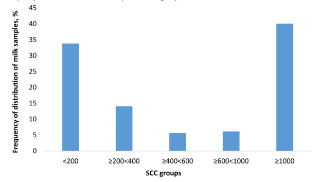
When evaluating the entire observation period tested ewes on udder half level the first group of SCC contained 33.9% of samples, the second 14.1% of samples, the third 5.7% of samples, the fourth 6.2% and the fifth 40.1% of samples (Figure 1). In our previous works we presented higher percentage (from 58.9% to 71.8%) of ewes in group <200 × 103 cells.mL-1 under usual test day sampling for
determination of physiological levels of SCC in healthy udder (Tančin et al., 2017a; Tvarožková et al., 2018). The proposed physiological threshold of SCC for diagnosis of mastitis in Sarda sheep was determined by Caboni et al. (2017) at 265 × 103 cells.mL-1. Zafalon et
al. (2016) detected the value of SCC >400 × 103 cells.mL-1
for diagnose of subclinical mastitis in flocks. Earlier proposed value of SCC for the diagnosis of mastitis was 500 × 103 cells.mL-1(Nunes et al., 2008). Low percentage
of samples in group <200 × 103 cells.mL-1 in present work
could be explained by sampling schedule, where these samples were colected from the ewes with high SCC at udder level three days before. Thus some health problems of udder should be expected as it is presented later in article by cultivation of milk sample on pathogens presence. In ewes with SCC above 400 × 103 cells.mL-1
three or more months during lactation there were 5.6 to 7.5-fold higher probability of a subclinical mastitis in compared with ewes with SCC below above limit(Spanu et al., 2011). Berthelot et al. (2006) reported 15% occurrence of subclinical mastitis if SCC in flock was over 650 × 103 cells.mL-1.
Figure 1 Frequency of distribution of milk samples in SCC groups.
Table 1 The occurrence of pathogens in months of observation.
Pathogen February March May Jun July
Enterococcus faecalis 2 1 3 - - Micrococcus luteus - - - - 1 S. aureus 1 2 4 1 - S. capitis 1 2 - - - S. caprae 2 3 2 1 20 S. cohni - - - - 1 S. epidermidis 3 7 3 3 5 S. felis - - 1 - - S. haemolyticus 1 - - - - S. chromogenes 5 4 1 - - S. lentus - - - - 1 S. piscifermentans - - - 4 - S. sciuri 1 - - - - S. schleiferi - - 8 21 - S. simulans 7 6 15 3 2 S. warneri - - 1 - - Str. pluranimalium 1 1 - - - 0 5 10 15 20 25 30 35 40 45 <200 ≥200<400 ≥400<600 ≥600<1000 ≥1000 Fr eq uen cy o f d istr ib uti on o f m ilk sa m pl es, % SCC groups
Total 407 examined milk samples tested on presence of mastitis pathogens were as negative classified 63.1% of milk samples, out of these samples 75.9% had SCC below 500 × 103 cells.mL-1. 36.1% of samples were classified as
bacteriological positive and 0.7% of milk samples were classified as contaminated. Only 7.5% of bacteriological positive samples had SCC below 500 × 103 cells.mL-1.
Two pathogens were identified in 2.7% of bacteriological positive samples. In 67.9% of ewes with bacteriological positive samples there were repeated detected the presence of pathogens during tested period. Thus pathogens could persist in udder throughout whole lactation. Also up to 21.1% from these ewes had infected both udder halves pathogens repeatedly.
Important group of samples are those in fifth group – with very high SCC (Figure 1). In fifth SCC group there were almost 80% bacteriological positive samples, which indicated the reason of high SCC. Also even 92.5% bacteriological positive samples had SCC ≥500 × 103 cells.mL-1. We detected significant lower SCS in milk
of ewes without (4.03 ±0.12) than with the presence of any pathogens (p <0.001). There was no effect of different pathogens on SCS which ranged from 6.68 ±0.41 to 8.11 ±0.63. Also no effect of month of sampling on SCS was found out. Different pathogens could differently influence SCC (Abu Baker Idriss et al., 2013; Bagnicka et al., 2011). Kiossis et al. (2007) used level of SCC of
≥500 × 103 cells.mL-1 and bacteriological positive milk
samples for the diagnose of subclinical mastitis. Significantly higher SCC in bacteriological positive samples as compared with bacteriological negative samples found out Ozenc et al. (2011) in their study. Świderek et al. (2016) reported that milk samples without bacteria had the lowest average SCC. Also, Persson et al. (2017) detected significantly higher SCC for udder halves with intramammary infection compared to udder halves without bacterial findings.
From major pathogens only S. aureus was identified in 5.3% of bacteriological positive samples (Table 1). Other contagious pathogens were not found out in tested group of ewes. Low frequency of contagious pathogens in milk was also presented in our previous work (Tančin et al. 2017b; Holko et al. 2018) or abroad works (Ergűn et al. 2009; Kern et al. 2013). Zigo et al. (2011) detected S. aureus in 9.3% of positive samples. Moroni et al. (2007) isolated S. aureus in 8.4% of infected milk samples. S. aureus was determined in 6.2% of subclinical mastitis cases (Queiroga, 2017). The most frequent pathogens isolated from the milk samples were CNS. Also high occurrence of CNS was reported in study Zigo et al. (2014) and Vasileiou et al. (2018). The most frequent CNS was S. simulans (24.6%) followed by S. schleiferi (21.6%), S. caprae (20.9%) (Table 1). From CNS found in farm had the highest occurrence S. epidermidis (36.3%) and S. caprae (21.3%) (Pilipčincová et al., 2010). Rahman et al. (2016) showed that the most dominant CNS were S. epidermidis, S. xylosus and S. chromogenes. Vasiľ et al. (2018) investigated that the most frequent CNS were S. epidermidis (24.3%), S. schleiferi (16.6%) and S. chromogenes (15.3%). Enterococcus faecalis and Streptococcus (Str.) pluranimalium was determined also (Table 1). Zigo et al. (2017) determined the incidence of Enterococcus faecalis in 6.1% of positive samples. Grazia
Puggioni et al. (2019) detected 29.4% the occurrence of Enterococcus faecalis in their study.
CONCLUSION
CNS were the most common group of pathogens in milk followed by increased SCC in milk. Staphylococcus (S.) simulans (24.4%) was the most frequent CNS in milk samples. From contagious pathogens was identified S. aureus in 5.3% of bacteriological positive samples. The presence of mastitis pathogens during tested period were repeated detected in 67.9% of ewes with bacteriological positive samples. 92.5% bacteriological positive milk samples had SCC ≥500 × 103 cells.mL-1. The high SCC
≥500 × 103 cells.mL-1 and bacteriological positive milk
samples from udder halves may be useful criterion for detection of subclinical mastitis and poosible use for selecting ewes for dry treatment or culling.
REFERENCES
Abu Baker Idriss, S. E., Tančin, V., Foltys, V., Kirchnerová, K., Tančinová, D., Vršková, M. 2013. Relationship between mastitis caqusative pathogens and somatic cell counts in milk of dairy cows. Potravinarstvo, vol. 7, no. 1, p. 207-212.
https://doi.org/10.5219/304
Bagnicka, E., Winnicka, A., Jóźwik, A., Rzewuska, M., Strzałkowska, N., Kościuczuk, E., Prusak, B., Kaba, B., Horbańczuk, J., Krzyżewski, J. 2011. Relationship between somatic cell count and bacterial pathogens in goat milk. Small
Ruminant Research, vol. 100, no. 1, p. 72-77.
https://doi.org/10.1016/j.smallrumres.2011.04.014
Berthelot, X., Lagriffoul, G., Concordet, D., Barillet, F., Bergonier, D. 2006. Physiological and pathological thresholds of somatic cell counts in ewe milk. Small Ruminant Research,
vol. 62, no. 1-2, p. 27-31.
https://doi.org/10.1016/j.smallrumres.2005.07.047
Caboni, P., Manis, C., Ibba, I., Contu, M., Coroneo, V., Scano, P. 2017. Compositional profile of ovine milk with a high somatic cell count: A metabolomics approach.
International Dairy Journal, vol. 69, p. 33-39.
https://doi.org/10.1016/j.idairyj.2017.02.001
Ergűn, Y., Aslantas, O., Dogruer, G., Kirecci, E., Saribay, M. K., Ates, C. T., Ulku, A., Demir, C. 2009. Prevalence and etiology of subclinical mastitis in Awasi dairy ewes in southern Turkey. Turkish Journal of Veterinary & Animal
Science, vol. 33, no. 6, p. 477-483.
Grazia Puggioni, G., M., Tedde, V., Uzzau, S., Dore, S., Liciardi, M., Cannas, E. A., Pollera, C., Moroni, P., Bronzo, V., Addis, M., F. 2019. Relationship of Late Lactation Milk Somatic Cell Count and Cathelicidin withIntramammary Infection in Small Ruminants. Pathogens, vol. 9, no. 1, 37 p.
https://doi.org/10.3390/pathogens9010037
Holko, I., Tančin, V., Tvarožková, K., Supuka, P., Supuková, A. 2018. Udder Pathogens Isolated from Sheep Milk in Slovakia. XLVIII. Lenfeldovy a Höklovy dny. Brno:
Veterinární a farmaceutická univerzita, p. 169-172. ISBN
978-80-7305-808-1.
Holko, I., Tančin, V., Tvarožková, K., Supuka, P., Supuková, A., Mačuhová, L. 2019. Occurrence and antimicrobial resistance of common udder pathogens isolated from sheep milk in Slovakia. Potravinarstvo Slovak Journal
of Food Sciences, vol. 13, no. 1, p. 258-261.
https://doi.org/10.5219/1067
Kern, G., Traulsen, I., Kemper, N., Krieter, J. 2013. Analysis of somatic cell counts and risk factors associated with occurrence of bacteria in ewes of different primary
purposes. Livestock Science, vol. 157, no. 2-3, p. 597-604.
https://doi.org/10.1016/j.livsci.2013.09.008
Kiossis, E., Brozos, C. N., Petridou, E., Boscos, C. 2007. Program for the control of subclinical mastitis in dairy Chios breed ewes during lactation. Small Ruminant Research, vol.
73, no. 1-3, p. 194-199.
https://doi.org/10.1016/j.smallrumres.2007.01.021
Leitner, G., Silanikove, N., Merin, U. 2008. Estimate of milk and curd yield loss of sheep and goats with intramammary infection and its relation to somatic cell count.
Small Ruminant Research, vol. 74, no. 1-3, p. 221-225.
https://doi.org/10.1016/j.smallrumres.2007.02.009
Moroni, P., Pisoni, G., Varisco, G., Boettcher, P. 2007. Effect of intramammary infection in Bergamasca meat sheep on milk parameters and lamb growth. Journal of Dairy
Research, vol. 74, no. 3, p. 340-344.
https://doi.org/10.1017/S0022029907002506
Nunes, G. R., Blagitz, M. G., Freitas C. F., Souza, F. N., Ricciardi, M., Stricagnolo, C. R., Sanches, B. G. S., Azedo, M. R., Sucupira, M. C. A., Della Libera, A. M. M. P. 2008. Avaliação de indicadores inflamatórios no diagnóstico da mamite ovina (Evaluation of the indicators of inflammation in the diagnosis of ovine mastitis). Arquivos do Instituto
Biológico, vol. 75, p. 271-281.
Olechnowicz, J., Jaskowski, J. M. 2005. Komórki somatyczne mleka owczego (Somatic cells in sheep milk).
Medycyna Weterynaryjna, vol. 61, no. 2, p. 136-141.
Ozenc, E., Seker, E., Baki Acar, D., Birdane, M. K., Darbaz, I., Dogan, N. 2011. The importance of staphylococci and threshold value of somatic cell count for diagnosis of subclinical mastitis in Pirlak sheep at mid-lactation.
Reproduction in Domestic Animals, vol. 46, no. 6, p. 970-974.
https://doi.org/10.1111/j.1439-0531.2011.01768.x
Persson, Y., Nyman, A. K., Söderquist, L., Tomic, N., Persson Waller, K. 2017. Intramammary infections and somatic cell count in meat and pelt producing ewes with clinically healthy udders. Small Ruminant Research, vol. 156, p. 66-72. https://doi.org/10.1016/j.smallrumres.2017.09.012
Pilipčincová, I., Bhide, M., Dudriková, E., Trávniček, M. 2010. Genotypic Characterization of Coagulase-negative Staphylococci Isolated from Sheep Milk in Slovakia. Acta
Veterinaria Brno, vol. 79, no. 2, p. 269-275.
https://doi.org/10.2754/avb201079020269
Queiroga, M. C. 2017. Prevalence and aetiology of sheep mastitis in Alentejo regions of Portugal. Small Ruminant
Research, vol. 153, p. 123-130.
https://doi.org/10.1016/j.smallrumres.2017.06.003
Rahman, B., Ownagh, A., Mardani, K., Ardebil, F. F. 2016. Prevalence and molecular characterization of staphylococci isolated from sheep with subclinical mastitis in West-Azerbaijan province, Iran. Veterinary Research Forum, vol. 7, no. 2, p. 155-162. Available at: https://www.ncbi.nlm.nih.gov/pmc/articles/PMC4959344/
Souza, F. N., Blagitz, M. G., Penna, C. F. A. M., Della Libera, A. M. M. P., Heinemann, M. B., Cerqueira, M. M. O. P. 2012. Somatic cell count in small ruminants: Friend or foe?
Small Ruminant Research, vol. 107, no. 2-3, p. 65-75.
https://doi.org/10.1016/j.smallrumres.2012.04.005
Spanu, C., Berger, Y. M., Thomas, D. L., Ruegg, P. L. 2011. Impact of intramammary antimicrobial dry treatment and teat sanitation on somatic cell count and intamammary infection in ewes. Small Ruminant Research, vol. 97, no. 1-3,
p. 139-145.
https://doi.org/10.1016/j.smallrumres.2011.03.005
Świderek, W. P., Charon, K. M., Winnicka, A., Gruszczyńska, J., Pierzchała, M. 2016. Physiological
Threshold of Somatic Cell Count in Milk of Polish Heath Sheep and Polish Lowland Sheep. Annals of Animal Science, vol. 16, no. 1, p. 155-170. https://doi.org/10.1515/aoas-2015-0071
Tančin, V., Baranovič, Š., Uhrinčať, M., Mačuhová, L., Vršková, M., Oravcová, M. 2017a. Somatic cell count in raw ewes milk in dairy practice: frequency of distribution and possible effect on milk yield and composition. Mljekarstvo,
vol. 67, no. 4, p. 253-260.
https://doi.org/10.15567/mljekarstvo.2017.0402
Tančin, V., Holko, I., Vršková, M., Uhrinčať, M., Mačuhová, L. 2017b. Relationship between presence of mastitis pathogens and somatic cell count in milk of ewes.
XLVII. Lenfeldovy a Höklovy dny. Brno: Veterinární a
farmaceutická univerzita, p. 230-233. ISBN
978-80-7305-793-0.
Tvarožková, K., Tančin, V., Uhrinčat, M., Hleba, L., Mačuhová, L. 2019. Evaluation of the health status of udder ewes during lactation: somatic cells and pathogens. XLIX. Lenfeldovy a Höklovy dny. Brno: Veterinární a farmaceutická
univerzita, p. 307-312. ISBN 978-80-7305-828-9.
Tvarožková, K., Tančin, V., Uhrinčat, M., Mačuhová, L., Toman, R., Tunegová, M. 2018. Evaluation of somatic cells in milk of ewes as possible physiological level. Acta
Fytotechnica et Zootechnica, vol. 21, no. 4, p. 149-151.
https://doi.org/10.15414/afz.2018.21.04.149-151
Uhrinčať, M., Tančin, V., Tvarožková, K., Mačuhová, L., Vršková, M., Ptáček, M., Holko, I. 2019. The electrical conductivity of sheep'š milk and the possibility of mastitis detection. Potravinarstvo Slovak Journal of Food Sciences, vol.13, no. 1, p. 562-565. https://doi.org/10.5219/1074
Vasiľ, M., Elečko, J., Farkašová, Z., Zigo, F. 2018. Development of resistance to antibiotics in bacteria Staphylococcus sp. Isolated from milk samples in the sheep breedings on east of Slovakia. Potravinarstvo Slovak Journal
of Food Sciences, vol. 12, no. 1, p. 273-278.
https://doi.org/10.5219/904
Vasileiou, N. G. C., Gougoulis, D. A., Riggio, V., Ioannidi, K. S., Chatzopoulos, D. C., Mavrogianni, V. S., Petinaki, E., Fthenakis, G. C. 2018. Association of subclinical mastitis prevalence with sheep breeds in Greece. Journal of Dairy
Research, vol. 85, no. 3, p. 317-320.
https://doi.org/10.1017/S0022029918000407
Watts, J. L., Salmon, S. A., Yancey, Jr. R. J. 1993. Use of modified Rambach agar to differentiate Streptococcus uberis from other mastitis streptococci. Journal of Dairy Science,
vol. 76, no. 6, p. 1740-1743.
https://doi.org/10.3168/jds.S0022-0302(93)77506-2
Zafalon, L. F., Mascarenhas Santana, R. C., Esteves, S. N., Fim Júnior, G. A. 2018. Somatic cell count in the diagnosis of subclinical mastitis in sheep of different breeds. Semina:
Ciências Agrárias, vol. 39, no. 4, p. 1555-1564.
https://doi.org/10.5433/1679-0359.2018v39n4p1555
Zafalon, L. F., Mascarenhas Santana, R. C., Pilon, L. E., Fim Júnior, G. A. 2016. Diagnosis of subclinical mastitis in Santa Ines and Morada Nova sheep in south eastern Brazil.
Tropical Animal Health and Production, vol. 48, p. 697-972.
https://doi.org/10.1007/s11250-016-1046-1
Zigo, F., Elečko, J., Farkašová, Z., Zigová, M., Vasiľ, M., Ondrašovičová, S., Kudělková, L. 2019. Preventive methods in reduction of mastitis pathogens in dairy cows. Journal of
Microbiology, Biotechnology and Food Sciences, vol. 9, no.
1, p. 121-126. https://doi.org/10.15414/jmbfs.2019.9.1.121-126
Zigo, F., Vasiľ, M., Elečko, J., Lapin, M., Farkašová, Z. 2014. Production of enterotoxins of Staphylococcus sp.
isolated from samples of sheep milk. Potravinarstvo, vol. 8, no. 1, p. 92-96. https://doi.org/10.5219/361
Zigo, F., Vasiľ, M., Kadáši, M., Elečko, J., Farkašová, Z. 2011. Bacteria Staphylococcus sp. izolated from mastitis of sheep and their enterotoxigenic properties. Potravinarstvo, vol. 5, no. 4, p. 70-72. https://doi.org/10.5219/171
Zigo, F., Vasiľ, M., Takáč, L., Elečko, J., Tomko, M., Chripková, M. 2017. Mastitis pathogens isolated from samples of milk in dairy sheep and their resistance.
International Journal of Scientific Research, vol. 6, no. 8, p.
298-300.
Acknowledgments:
This work was supported by the Ministry of Education Science Research and Sports of the Slovak Republic/the Slovak Research and Development Agency (Projects No. APVV-15-0072) and KEGA 039SPU-4/2019 “Modernization of practical education of hygiene and prevention in animal production”.
Contact address:
Kristína Tvarožková, Slovak University of Agriculture in Nitra, Faculty of Agrobiology and Food Resources, Department of Veterinary Science, Tr. A. Hlinku 2, 949 76 Nitra, Slovakia, Tel.: +421944385272,
E-mail: [email protected]
ORCID: https://orcid.org/0000-0003-4989-6138
*Vladimír Tančin, Slovak University of Agriculture in Nitra, Faculty of Agrobiology and Food Resources, Department of Veterinary Science, Tr. A. Hlinku 2, 949 76 Nitra, Slovakia, NPPC-Research Institute for Animal Production Nitra, Hlohovecká 2, 95141 Lužianky Slovakia, Tel.: +421903546401,
E-mail: [email protected]
ORCID: https://orcid.org/0000-0003-2908-9937
Michal Uhrinčať, NPPC-Research Institute for Animal Production Nitra, Hlohovecká 2, 95141 Lužianky, Slovakia, Tel.: +421376546656,
E-mail: [email protected]
ORCID: https://orcid.org/0000-0002-5378-617X
Lukáš Hleba, Slovak University of Agriculture in Nitra, Faculty of Biotechnology and Food Sciences, Department of Microbiology, Tr. A. Hlinku 2, 949 76 Nitra, Slovakia, Tel.: +421 37 641 5811,
E-mail: [email protected]
ORCID: https://orcid.org/0000-0001-8244-6548
Lucia Mačuhová, NPPC-Research Institute for Animal Production Nitra, Hlohovecká 2, 95141 Lužianky, Slovakia, Tel.: +4213765466571,
E-mail: [email protected]
ORCID: https://orcid.org/0000-0002-0000-0000 Corresponding author: *