Methyl 4-chloro-3-nitrobenzoate
Bo-Nian Liu,aShi-Gui Tang,bHao-Yuan Li,aYe-Ming Xua and Cheng Guoa*
a
College of Science, Nanjing University of Technology, Xinmofan Road No. 5, Nanjing 210009, People’s Republic of China, andbCollege of Life Sciences and Pharmaceutical Engineering, Nanjing University of Technology, Nanjing 210009, People’s Republic of China
Correspondence e-mail: [email protected]
Received 19 November 2007; accepted 16 December 2007
Key indicators: single-crystal X-ray study;T= 293 K; mean(C–C) = 0.004 A˚; Rfactor = 0.045;wRfactor = 0.142; data-to-parameter ratio = 13.9.
In the title compound, C8H6ClNO4, the molecules are linked
by C—H O interactions to form a chain parallel to theaaxis. The chains are further connected by slipped – stacking between symmetry-related benzene rings, with a centroid-to-centroid distance of 3.646 (2) A˚ and an interplanar distance of 3.474 A˚ , resulting in an offset of 1.106 A˚.
Related literature
For related literature, see: de Souzaet al.(2006); Jin & Xiao (2005); Spiniello & White (2003); Jo¨nssen et al. (2004); Andrews & Ladlow (2003).
Experimental
Crystal data
C8H6ClNO4
Mr= 215.59
Triclinic,P1 a= 7.338 (1) A˚ b= 7.480 (1) A˚ c= 9.715 (2) A˚
= 98.39 (3) = 94.89 (3)
= 118.95 (3)
V= 454.1 (2) A˚3
Z= 2
MoKradiation
= 0.41 mm1 T= 293 (2) K 0.400.100.10 mm
Enraf–Nonius CAD-4 diffractometer
Absorption correction: scan (Northet al., 1968) Tmin= 0.854,Tmax= 0.961
1918 measured reflections
1773 independent reflections 1389 reflections withI> 2(I) Rint= 0.019
3 standard reflections every 200 reflections intensity decay: none
Refinement
R[F2> 2(F2)] = 0.046 wR(F2) = 0.142
S= 1.12 1773 reflections
128 parameters
H-atom parameters constrained max= 0.21 e A˚
3
min=0.24 e A˚
3
Table 1
Hydrogen-bond geometry (A˚ ,).
D—H A D—H H A D A D—H A
C5—H5 O2i
0.93 2.47 3.272 (3) 145
Symmetry code: (i)xþ1;y;z.
Data collection: CAD-4 Software (Enraf–Nonius, 1989); cell refinement: CAD-4 Software; data reduction: XCAD4 (Harms & Wocadlo,1995); program(s) used to solve structure: SHELXS97 (Sheldrick, 1997); program(s) used to refine structure:SHELXL97 (Sheldrick, 1997); molecular graphics: ORTEPIII (Burnett & Johnson, 1996), ORTEP-3 for Windows (Farrugia, 1997) and PLATON (Spek, 2003); software used to prepare material for publication:SHELXL97.
The authors thank the Center for Testing and Analysis, Nanjing University, for support.
Supplementary data and figures for this paper are available from the IUCr electronic archives (Reference: DN2295).
References
Andrews, S. P. & Ladlow, M. (2003).J. Org. Chem.68, 5525–5533.
Burnett, M. N. & Johnson, C. K. (1996).ORTEPIII. Report ORNL-6895. Oak Ridge National Laboratory, Tennessee, USA.
Enraf–Nonius (1989).CAD-4 Software. Version 5.0. Enraf–Nonius, Delft, The Netherlands.
Farrugia, L. J. (1997).J. Appl. Cryst.30, 565.
Harms, K. & Wocadlo, S. (1995).XCAD4. University of Marburg, Germany. Jin, L.-F. & Xiao, F.-P. (2005).Acta Cryst.E61, o1237–o1238.
Jo¨nssen, D., Warrington, B. H. & Ladlow, M. (2004).J. Comb. Chem.6, 584– 595.
North, A. C. T., Phillips, D. C. & Mathews, F. S. (1968).Acta Cryst.A24, 351– 359.
Sheldrick, G. M. (1997). SHELXL97 and SHELXS97. University of Go¨ttingen, Germany.
Souza, M. V. N. de, Vasconcelos, T. R. A., Wardell, S. M. S. V., Wardell, J. L., Low, J. N. & Glidewell, C. (2006).Acta Cryst.C62, o295–o298.
Spek, A. L. (2003).J. Appl. Cryst.36, 7–13.
Spiniello, M. & White, J. M. (2003).Org. Biomol. Chem.1, 3094–3096. Structure Reports
Online
supporting information
Acta Cryst. (2008). E64, o456 [doi:10.1107/S1600536807067219]
Methyl 4-chloro-3-nitrobenzoate
Bo-Nian Liu, Shi-Gui Tang, Hao-Yuan Li, Ye-Ming Xu and Cheng Guo
S1. Comment
Some derivatives of benzoic acid are important chemical materials. We report here the crystal structure of the title
compound, (I).
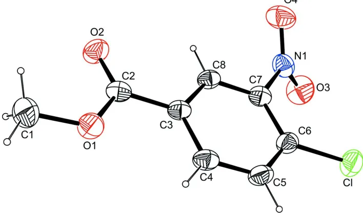
In compound (I) the nitro group is twisted with respect to the phenyl ring making a dihedral angle of 45.4 (1)° (Fig. 1).
Similar twisted conformations are observed in related structures where the aryl ring bears nitro and halide adjacent to
each other (de Souza et al., 2006; Spiniello & White, 2003), whereas a planar conformation is observed in other case (Jin
& Xiao, 2005).
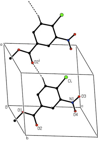
The molecules of (I) are linked by C—H···O interactions to form a chain parallel to the a axis (Table 1, Fig. 2). The
chains are further connected by slippest π–π stacking between symmetry related phenyl rings with a centroit to centroid
distance Cg1···Cg1i (Symmetry code: (i) 1 - x, 1 - y, 1 - z) of 3.646 (2) Å and an interplanar distance of 3.474 Å resulting
in an offset of 1.106 Å.
S2. Experimental
4-Chloro-3-nitrobenzoic acid (35.0 g, 0.174 mol) was suspended in methanol (150 ml) and cooled to 0°. Concentrated
sulfuric acid (15 ml) was slowly added with stirring, and then the mixture was heated at reflux for 17 h. Upon cooling to
room temperature, a precipitate formed, which was collected by filtration and washed with cold methanol (2*50 ml) and
hexane (2*50 ml) to afford the methyl ester as a white solid (31.8 g, 85%) (Andrews & Ladlow, 2003; Jönssen et al.,
2004). Pure compound (I) was obstained by crystallizing from methanol. Crystals of (I) suitable for X-ray diffraction
were obstained by slow evaporation of an methanol solution.
S3. Refinement
All H atoms were placed geometrically and treated as riding on their parent C atoms with C—H = 0.93 Å (Caromatic)
Figure 1
The molecular structure of (I), showing the atom-labelling scheme. Displacement ellipsoids are drawn at the 30%
Figure 2
Partial packing view of (I) showing the formation of the chain through C—H···O hydrogen bonds indicated as dashed
lines. H atoms not involved in hydrogen bondings have been omitted for clarity. [Symmetry code: (i) 1 + x, y, z]
Methyl 4-chloro-3-nitrobenzoate
Crystal data
C8H6ClNO4 Mr = 215.59 Triclinic, P1 Hall symbol: -P 1
a = 7.338 (1) Å
b = 7.480 (1) Å
c = 9.715 (2) Å
α = 98.39 (3)°
γ = 118.95 (3)°
V = 454.1 (2) Å3 Z = 2
F(000) = 220
Dx = 1.577 Mg m−3
Mo Kα radiation, λ = 0.71073 Å Cell parameters from 25 reflections
Box, colourless
Data collection
Enraf–Nonius CAD-4 diffractometer
Radiation source: fine-focus sealed tube Graphite monochromator
ω/2θ scans
Absorption correction: ψ scan (North et al., 1968)
Tmin = 0.854, Tmax = 0.961
1918 measured reflections
1773 independent reflections 1389 reflections with I > 2σ(I)
Rint = 0.019
θmax = 26.0°, θmin = 2.2° h = 0→9
k = −9→8
l = −11→11
3 standard reflections every 200 reflections intensity decay: none
Refinement
Refinement on F2
Least-squares matrix: full
R[F2 > 2σ(F2)] = 0.046 wR(F2) = 0.142 S = 1.12 1773 reflections 128 parameters 0 restraints
Primary atom site location: structure-invariant direct methods
Secondary atom site location: difference Fourier map
Hydrogen site location: inferred from neighbouring sites
H-atom parameters constrained
w = 1/[σ2(F
o2) + (0.0588P)2 + 0.2181P]
where P = (Fo2 + 2Fc2)/3
(Δ/σ)max = 0.001
Δρmax = 0.21 e Å−3
Δρmin = −0.24 e Å−3
Special details
Geometry. All e.s.d.'s (except the e.s.d. in the dihedral angle between two l.s. planes) are estimated using the full covariance matrix. The cell e.s.d.'s are taken into account individually in the estimation of e.s.d.'s in distances, angles and torsion angles; correlations between e.s.d.'s in cell parameters are only used when they are defined by crystal symmetry. An approximate (isotropic) treatment of cell e.s.d.'s is used for estimating e.s.d.'s involving l.s. planes.
Refinement. Refinement of F2 against ALL reflections. The weighted R-factor wR and goodness of fit S are based on F2,
conventional R-factors R are based on F, with F set to zero for negative F2. The threshold expression of F2 > σ(F2) is used
only for calculating R-factors(gt) etc. and is not relevant to the choice of reflections for refinement. R-factors based on F2
are statistically about twice as large as those based on F, and R- factors based on ALL data will be even larger.
Fractional atomic coordinates and isotropic or equivalent isotropic displacement parameters (Å2)
x y z Uiso*/Ueq
C1 −0.2034 (5) 0.0805 (6) 0.0613 (3) 0.0722 (9) H1A −0.3435 −0.0080 0.0779 0.108* H1B −0.1832 0.0171 −0.0248 0.108* H1C −0.1857 0.2143 0.0536 0.108* C2 −0.0585 (4) 0.1907 (4) 0.3056 (3) 0.0454 (6) C3 0.1131 (4) 0.2225 (4) 0.4166 (3) 0.0417 (6) C4 0.2701 (4) 0.1747 (4) 0.3866 (3) 0.0502 (7)
H4 0.2672 0.1180 0.2941 0.060*
C5 0.4285 (4) 0.2108 (4) 0.4925 (3) 0.0536 (7)
H5 0.5306 0.1769 0.4709 0.064*
H8 0.0146 0.3344 0.5771 0.051* Cl 0.63808 (12) 0.33572 (14) 0.75859 (9) 0.0731 (3) N1 0.2849 (4) 0.4450 (4) 0.8022 (2) 0.0569 (6) O1 −0.0490 (3) 0.1079 (3) 0.1782 (2) 0.0591 (5) O2 −0.1910 (3) 0.2373 (3) 0.3279 (2) 0.0626 (6) O3 0.4525 (4) 0.5930 (4) 0.8665 (2) 0.0838 (7) O4 0.1169 (4) 0.3786 (4) 0.8445 (2) 0.0801 (7)
Atomic displacement parameters (Å2)
U11 U22 U33 U12 U13 U23
C1 0.075 (2) 0.089 (2) 0.0466 (17) 0.0412 (19) 0.0021 (15) 0.0024 (15) C2 0.0449 (14) 0.0383 (13) 0.0488 (15) 0.0175 (11) 0.0148 (11) 0.0063 (11) C3 0.0406 (13) 0.0343 (12) 0.0489 (14) 0.0170 (10) 0.0144 (11) 0.0086 (10) C4 0.0533 (15) 0.0490 (15) 0.0560 (16) 0.0298 (13) 0.0225 (13) 0.0108 (12) C5 0.0475 (15) 0.0572 (16) 0.0691 (18) 0.0328 (13) 0.0226 (13) 0.0198 (14) C6 0.0412 (13) 0.0462 (14) 0.0633 (17) 0.0207 (12) 0.0122 (12) 0.0191 (12) C7 0.0466 (14) 0.0388 (13) 0.0491 (15) 0.0187 (11) 0.0126 (11) 0.0085 (11) C8 0.0399 (13) 0.0363 (12) 0.0532 (15) 0.0199 (10) 0.0147 (11) 0.0084 (10) Cl 0.0564 (5) 0.0836 (6) 0.0821 (6) 0.0363 (4) 0.0021 (4) 0.0278 (4) N1 0.0682 (16) 0.0606 (15) 0.0451 (13) 0.0349 (13) 0.0112 (12) 0.0106 (11) O1 0.0626 (12) 0.0725 (13) 0.0444 (10) 0.0400 (11) 0.0080 (9) −0.0024 (9) O2 0.0584 (12) 0.0832 (15) 0.0574 (12) 0.0468 (11) 0.0118 (9) 0.0049 (10) O3 0.0794 (16) 0.0785 (16) 0.0661 (15) 0.0294 (13) −0.0085 (12) −0.0115 (12) O4 0.0839 (17) 0.0938 (18) 0.0647 (14) 0.0446 (14) 0.0324 (13) 0.0128 (12)
Geometric parameters (Å, º)
C1—O1 1.450 (4) C4—H4 0.9300
C1—H1A 0.9600 C5—C6 1.375 (4)
C1—H1B 0.9600 C5—H5 0.9300
C1—H1C 0.9600 C6—C7 1.402 (4)
C2—O2 1.207 (3) C6—Cl 1.725 (3)
C2—O1 1.324 (3) C7—C8 1.370 (4)
C2—C3 1.486 (4) C7—N1 1.471 (3)
C3—C8 1.380 (4) C8—H8 0.9300
C3—C4 1.402 (3) N1—O3 1.215 (3)
C4—C5 1.376 (4) N1—O4 1.220 (3)
O1—C1—H1A 109.5 C6—C5—H5 119.6
O1—C1—H1B 109.5 C4—C5—H5 119.6
H1A—C1—H1B 109.5 C5—C6—C7 118.1 (3)
O1—C1—H1C 109.5 C5—C6—Cl 118.7 (2)
C8—C3—C2 118.5 (2) C3—C8—H8 120.0 C4—C3—C2 122.8 (2) O3—N1—O4 125.0 (3) C5—C4—C3 120.7 (2) O3—N1—C7 117.8 (2)
C5—C4—H4 119.6 O4—N1—C7 117.1 (2)
C3—C4—H4 119.6 C2—O1—C1 116.9 (2)
C6—C5—C4 120.8 (2)
Hydrogen-bond geometry (Å, º)
D—H···A D—H H···A D···A D—H···A
C5—H5···O2i 0.93 2.47 3.272 (3) 145