c-Abl regulates YAP
Y357
phosphorylation to
activate endothelial atherogenic responses to
disturbed flow
Bochuan Li, … , Yi Zhu, Ding Ai
J Clin Invest.
2019;
129(3)
:1167-1179.
https://doi.org/10.1172/JCI122440
.
Local flow patterns determine the uneven distribution of atherosclerotic lesions. This
research aims to elucidate the mechanism of regulation of nuclear translocation of
Yes-associated protein (YAP) under oscillatory shear stress (OSS) in the atheroprone phenotype
of endothelial cells (ECs). We report here that OSS led to tyrosine phosphorylation and
strong, continuous nuclear translocation of YAP in ECs that is dependent on integrin a5b1
activation. YAP overexpression in ECs blunted the anti-atheroprone effect of an integrin
a5b1–blocking peptide (ATN161) in
Apoe
–/–mice. Activation of integrin a5b1 induced
tyrosine, but not serine, phosphorylation of YAP in ECs. Blockage of integrin a5b1 with
ATN161 abolished the phosphorylation of YAP at Y
357induced by OSS. Mechanistic
studies showed that c-Abl inhibitor attenuated the integrin a5b1–induced YAP tyrosine
phosphorylation. Furthermore, the phosphorylation of c-Abl and YAP
Y357was significantly
increased in ECs in atherosclerotic vessels of mice and in human plaques versus normal
vessels. Finally, bosutinib, a tyrosine kinase inhibitor, markedly reduced the level of
YAP
Y357and the development of atherosclerosis in
Apoe
–/–mice. The c-Abl/YAP
Y357pathway serves as a mechanism for the activation of integrin a5b1 and the atherogenic
phenotype of ECs in response to OSS, and provides a potential therapeutic strategy for
atherogenesis.
Research Article
Vascular biology
Find the latest version:
Introduction
Atherosclerosis has become one of the major causes of death worldwide because of its severe complications, such as acute myo-cardial infarction and stroke (1). Vascular endothelial cell (EC) activation is an initial step of atherosclerosis. ECs are responsive to various chemical and mechanical stimuli, including tumor necrosis factor α, lipopolysaccharide, and shear stress. EC activa-tion triggers inflammatory responses to increase the expression of intercellular adhesion molecule 1 (ICAM-1) and vascular adhesion molecule 1 (VCAM-1), and the subsequent local recruitment of immune cells (2). As the interface between circulating blood and the vessel wall, ECs are constantly exposed to mechanical forces generated by blood flow. Atherosclerosis preferentially develops at branches and curvatures in the arterial tree where flow is dis-turbed. In contrast, the flow in straight parts of the arteries gener-ates laminar shear stress (LSS), which is atheroprotective (3).
The Hippo pathway plays an evolutionarily conserved funda-mental role in controlling organ size (4). The core components of the mammalian Hippo pathway contain kinase cascade of mam-malian Ste20-like kinases 1/2 (Mst1/2) and large tumor suppres-sor 1/2 (Lats1/2). Mst1/2, in complex with a regulatory protein salvador (Sav1), phosphorylates and activates Lats1/2, which also forms a complex with a regulatory protein Mps-one-binding (Mob1) (5). The transcription coactivator Yes-associated protein
(YAP) is a major downstream effector of the Hippo pathway (6). When the Hippo signaling pathway is inactivated, YAP and its paralog TAZ translocate into the nucleus and activate gene tran-scription by binding to the TEAD family and other trantran-scription factors (4). The subcellular localization of YAP, which is important for its transcriptional activity, is regulated by its phosphorylation status. Phosphorylation at S127 of YAP leads to its direct binding
to 14-3-3 and retention in cytoplasm (4, 7). Phosphorylation of YAP S397 (S381) by Lats1/2 promotes YAP ubiquitination-dependent
degradation and reduces its nuclear localization (8). In addition, phosphorylation of YAP at Y357 increases its stability and in turn
upregulates its activity (9). Recently, YAP was identified to be cru-cial in ECs in different biological processes, including angiogen-esis (10) and atherosclerosis (11, 12). Moreover, the suppression of YAP in response to LSS plays an important role in maintaining vessel homeostasis in ECs (11, 12). However, the regulation of YAP by OSS and the underlying mechanism is largely unknown.
Integrins, a diverse family of cell-surface heterodimeric pro-teins that facilitate cell–extracellular matrix (ECM) adhesion (13), are associated with endothelial activation (14–16). We and other groups identified integrin α5β1 as a membrane mechanical sensor responsive to OSS and with a proinflammatory and atheroprone role in ECs (17–19). Additionally, fibronectin, as an endogenous ligand that activates integrin α5β1 (20), can amplify the effect of oscillatory flow on EC activation (19). In contrast, an integrin α5 inhibitor peptide, ATN161, retards the development of atheroscle-rosis (17). However, the signal transduction underlying integrin activation remains unknown.
Nonreceptor protein tyrosine kinases (NRTKs), which trans-fer phosphate groups to tyrosine residues on protein substrates in the cytoplasm, are important components of signaling pathways
Local flow patterns determine the uneven distribution of atherosclerotic lesions. This research aims to elucidate the mechanism of regulation of nuclear translocation of Yes-associated protein (YAP) under oscillatory shear stress (OSS) in the atheroprone phenotype of endothelial cells (ECs). We report here that OSS led to tyrosine phosphorylation and strong, continuous nuclear translocation of YAP in ECs that is dependent on integrin α5β1 activation. YAP overexpression in ECs blunted the anti-atheroprone effect of an integrin α5β1–blocking peptide (ATN161) in Apoe–/– mice. Activation of integrin
α5β1 induced tyrosine, but not serine, phosphorylation of YAP in ECs. Blockage of integrin α5β1 with ATN161 abolished the phosphorylation of YAP at Y357 induced by OSS. Mechanistic studies showed that c-Abl inhibitor attenuated the integrin α5β1–
induced YAP tyrosine phosphorylation. Furthermore, the phosphorylation of c-Abl and YAPY357 was significantly increased
in ECs in atherosclerotic vessels of mice and in human plaques versus normal vessels. Finally, bosutinib, a tyrosine kinase inhibitor, markedly reduced the level of YAPY357 and the development of atherosclerosis in Apoe–/– mice. The c-Abl/YAPY357
pathway serves as a mechanism for the activation of integrin α5β1 and the atherogenic phenotype of ECs in response to OSS, and provides a potential therapeutic strategy for atherogenesis.
c-Abl regulates YAP
Y357
phosphorylation to activate
endothelial atherogenic responses to disturbed flow
Bochuan Li, Jinlong He, Huizhen Lv, Yajin Liu, Xue Lv, Chenghu Zhang, Yi Zhu, and Ding AiTianjin Key Laboratory of Metabolic Diseases; Key Laboratory of Immune Microenvironment and Disease (Ministry of Education); Collaborative Innovation Center of Tianjin for Medical Epigenetics and Department of Physiology and Pathophysiology, Tianjin Medical University, Tianjin, China.
Authorship note: BL and JH contributed equally to this work.
Conflict of interest: The authors have declared that no conflict of interest exists.
License: Copyright 2019, American Society for Clinical Investigation.
Submitted: May 23, 2018; Accepted: January 3, 2019.
The Journal of Clinical Investigation
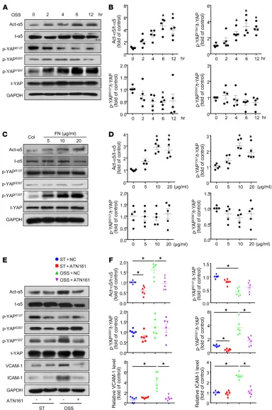
to mediate OSS-induced EC activation (17), and fibronectin is a major endogenous ligand of integrin α5β1 (22), we detected YAP nuclear translocation in ECs seeded on fibronectin-coated plates. As shown in Figure 1, E and F, both integrin α5β1 activation and YAP nuclear translocation were induced by fibronectin, as evi-denced by immunofluorescent staining. Western blot analysis also indicated that YAP nuclear translocation was increased with fibronectin treatment (Figure 1, G and H).
To further test whether the regulation of YAP in response to OSS depends on integrin α5β1, we used ATN161, an integrin
α5β1–blocking peptide, in OSS-treated ECs. Pretreatment with ATN161 did not alter the YAP subcellular localization in HUVECs under static conditions, but completely blocked the OSS-induced YAP nuclear translocation (Figure 1, I and J). Moreover, in vivo evidence was obtained by examining the subcellular distribution of YAP in aortas of WT and Itga5 heterozygous knockout (α5+/–)
mice. As shown in Figure 1K, YAP exhibited nuclear localization in the inner curvature of the aortic arch (AA) but not the outer cur-vature of the AA or the thoracic aorta (TA) of WT mice. The flow pattern–dependent difference of YAP cellular distribution was abat-ed in α5+/– mice (Figure 1K). Thus, integrin α5β1 is essential in flow-
induced YAP subcellular localization in vivo.
EC-specific YAP overexpression blunts the atheroprotective effect of integrin α5β1 blockage. Next, we asked whether YAP
over-expression could abolish the beneficial effects of integrin α5β1 inhibition in vivo. To answer this question, we crossbred loxp-stop-loxp YAP-transgenic mice with Tie2-Cre mice to generate
Tie2-Cre+/–YAPflox/– mice (EC-YAPtg), and generated EC-specific YAP-
overexpression mice on an Apoe–/– background (EC-YAPtgApoe–/–).
We treated those mice with or without an integrin-blocking pep-tide ATN161. After 4 weeks of the Western-type diet (WTD), Oil Red O staining of aortas revealed that, in comparison with the scramble peptide, ATN161 significantly decreased the total ath-erosclerotic area in aortas of YAPfloxApoe–/– mice (Figure 2, A and
B). Aortic root staining showed that ATN161 reduced the lesion area, lipid deposition, and macrophage infiltration, but had min-imal effects on collagen fiber or vascular smooth muscle cell con-tent (Figure 2, C–E and Supplemental Figure 1, A–D; supplemental material available online with this article; https://doi.org/10.1172/ JCI122440DS1). In contrast, the overexpression of YAP in ECs of mice aggravated atherosclerosis, as compared with YAPfloxApoe–/–
mice (Figure 2, A–E). ATN161 treatment failed to prevent the development of atherosclerosis in EC-YAPtg Apoe–/– mice (Figure 2,
A–E). The levels of plasma triglyceride and cholesterol and blood pressure did not change among the groups (Figure 2F and Supple-mental Figure 1E). These results indicate that YAP is an important downstream effector of integrin α5β1 in EC activation.
OSS induced phosphorylation of YAP at Tyr357 through integrin
α5β1. Because the cellular localization of YAP was regulated by
its protein phosphorylation modification, we determined the lev-els of YAP phosphorylation at Ser127 (4), Ser397 (8), and Tyr357 (9) under OSS. OSS significantly increased the level of p-YAPY357,
while decreasing that of p-YAPS127 without altering p-YAPS397
(Fig-ure 3, A and B). Simultaneously, the OSS activation of integrin α5β1 showed a similar time dependency as the p-YAPY357 (Figure 3, A and
B). Fibronectin, as an agonist of integrin α5β1, dose-dependently upregulated the protein levels of both active-integrin α5β1 and (21). NRTKs are divided into 9 main families according to their
structures. Several kinases from different families, including focal adhesion kinase (FAK) and c-Src, mediate the signal transduction of integrins (22). c-Abl has been shown to be colocalized with inte-grin α5 and activated in cells subjected to fibronectin treatment (23) or fluid shear stress (24). Furthermore, with the emerging importance of NRTKs in neurodegenerative diseases, chronic myeloid leukemia, and carcinogenesis, tyrosine kinase inhibitors are being used in the clinic (21). Although a spleen tyrosine kinase inhibitor, fostamatinib, has been found to attenuate atherogenesis in mice (25), there is a paucity of information on the NRTKs as a potential target for treating atherosclerosis.
In this study, we investigated the regulation of YAP by OSS and the underlying mechanism in ECs. Our study provides both in vivo and in vitro evidence that activation of integrin α5β1 and its downstream kinase c-Abl mediates the OSS-induced YAP nuclear translocation via phosphorylating YAP at Y357. In
addi-tion, we have demonstrated that inhibition of c-Abl blunts the OSS-induced EC activation and the development of early-stage atherosclerosis.
Results
OSS increased YAP nuclear translocation via integrin α5β1. YAP,
as a novel mechanosensor (26), has been reported to promote EC activation in response to blood flow (12). To investigate the effects of OSS on the pattern of YAP nuclear localization, human umbilical vein ECs (HUVECs) were subjected to OSS (0.5 ± 4 dyn/cm2, 1 Hz) for different durations. YAP showed significant
nuclear localization beginning from 4 hours and sustained until 12 hours, as compared with static treatment (Figure 1, A and B). Western blot analysis further confirmed the increased nuclear localization of YAP at 6 hours after OSS stimulation (Figure 1, C and D). Given that integrin α5β1 has been previously reported
Figure 1. Oscillatory shear stress (OSS) increased Yes-associated protein (YAP) nuclear translocation via integrin α5β1. (A and B) Human umbilical vein endothelial cells (HUVECs) were exposed to OSS (0.5 ± 4 dyn/cm2)
The Journal of Clinical Investigation
tion of the fibronectin-induced p-YAPY357 protein by 88% (Figure
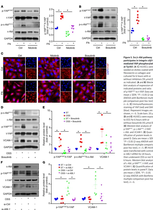
5B). Consistent with these findings, immunofluorescence staining of YAP indicates that its fibronectin-induced nuclear localization was blocked by Src/c-Abl inhibitors (Figure 5C and Supplemental Figure 2C). In addition, nilotinib or bosutinib treatment did not affect the proliferation or apoptosis of ECs (Supplemental Fig-ure 2, D–K). Moreover, bosutinib treatment or siRNA-mediated knockdown of ABL1 decreased OSS-induced upregulation of p-YAPY357 and VCAM-1 at protein level (Figure 5, D–G).
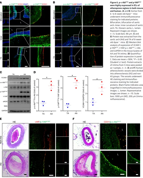
p-c-AblY412 and p-YAPY357 were highly expressed in ECs of
athero-prone regions in both mouse and human. We next determined the
expression of p-c-AblY412 and p-YAPY357 in atheroprone regions of
Apoe–/– mouse aortas. In the inner curvature and bifurcation of the
AA where the flow is disturbed, both p-c-AblY412 and p-YAPY357 were
expressed at higher levels in ECs. In contrast, in the TA, where the flow is laminar, both p-c-AblY412 and p-YAPY357 exhibited weak
colo-calization with the EC marker (Figure 6, A and B). Furthermore, the protein levels of p-c-AblY412, p-YAPY357, and the EC activation
marker VCAM-1 were significantly increased in the AA intima tis-sue lysates than those of TA intima (Figure 6, C and D). Consistent with these results in mice, the expression of both p-c-AblY412 and
p-YAPY357 in ECs of human coronary arteries was high in plaques
but not in normal vessels (Figure 6, E and F). Taken together, these findings indicate a predominant role of c-Abl in regulating YAP phosphorylation at Tyr357 to contribute to the development of flow-dependent atherosclerosis.
Bosutinib retarded both diet-induced and partial ligation– induced atherosclerosis in Apoe–/– mice. In light of our finding that
tyrosine kinase–mediated YAP phosphorylation might contribute to an atheroprone phenotype in ECs, bosutinib was injected into
Apoe–/– mice that were fed a WTD for 4 or 12 weeks. Bosutinib
sig-nificantly reduced the lesion area in aortas (Figure 7A and Supple-mental Figure 3A). Oil Red O staining of whole aortas indicated that the reduction mainly occurred in the AA, where the lesion area was greatly decreased (Figure 7, B–E and Supplemental Fig-ure 3, B–E). Lesion area and lipid deposition in aortic roots were also reduced by bosutinib (Supplemental Figure 3, F–K and Sup-plemental Figure 4, A–F), in parallel with the reduced levels of both p-c-AblY412 and p-YAPY357 (Supplemental Figure 3, L and M
and Supplemental Figure 4, G and H). In addition, blood pressure in Apoe–/– mice was not affected by long-term bosutinib treatment
(Supplemental Figure 3N). Next, we used Apoe–/– mice with partial
ligation to further test the effect of bosutinib. Consistent with the diet-induced model, bosutinib significantly reduced the develop-ment of atherosclerosis (Figure 7F). En face immunofluorescence staining of ligated carotid arteries showed that the levels of EC inflammatory markers VCAM-1 and ICAM-1 were abolished by bosutinib administration (Figure 7, G and H). These results indi-cate the atheroprotective effect of bosutinib on both early and advanced atherosclerosis.
Discussion
YAP has been identified as a mechanosensor of hemodynamic stress that contributes to EC activation in early-stage atheroscle-rosis (11, 12). However, the transduction signaling pathway link-ing shear stress and YAP is still unknown. Here, we found that under OSS, YAP nuclear translocation was partly mediated by p-YAPY357 (Figure 3, C and D). Meanwhile, fibronectin had little
effect on the phosphorylation of YAP at Ser127 or Ser397 (Figure 3, C and D). In contrast, blockage of integrin α5β1 with ATN161 mark-edly decreased the OSS-induced protein levels of active integrin
α5β1 and p-YAPY357, as well as the inflammatory markers (ICAM-1
and VCAM-1) (Figure 3, E and F). These results indicate that the OSS-induced EC proinflammatory phenotype was attributable, at least in part, to the integrin α5β1-YAPY357 pathway.
Tyr357 phosphorylation of YAP induced endothelial activation both in vitro and in vivo. To assess the functional importance of
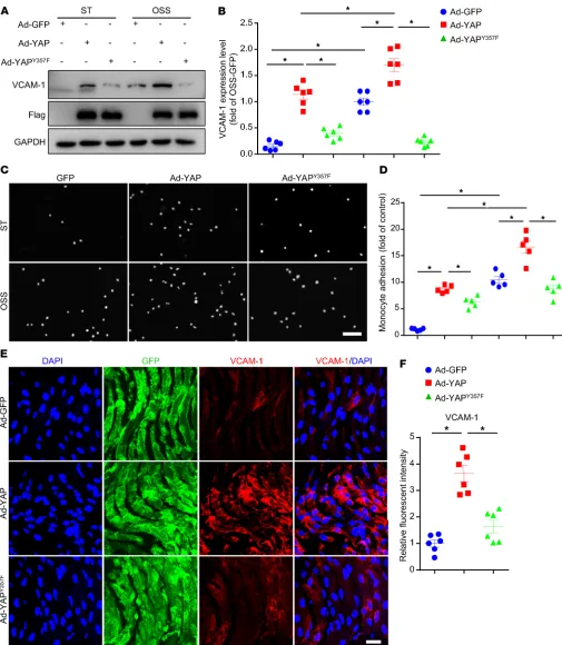
YAP phosphorylation at Y357, we constructed adenovirus for YAP with mutation of Tyr357 to a non–phospho mimetic variant (Ad-YAPY357F), and explored its function in vitro and in vivo. At
first, HUVECs were infected with Ad-GFP, Ad-YAP, or Ad-YAPY357F
and then applied with OSS. OSS-induced upregulation of VCAM-1 protein level, as well as the increased number of THP-1 cells adhering to HUVECs, were exacerbated by overexpression of WT YAP, but not by the loss-of-function mutant Ad-YAPY357F (Figure 4,
A–D). In vivo, the overexpression of YAPY357F increased the
expres-sion of VCAM-1 to a much lesser extent compared with those with Ad-YAP infection in left carotid artery endothelium of Apoe–/– mice
with partial ligation (Figure 4, E and F). Our data suggest that pY357 of YAP in ECs plays a detrimental role in OSS-induced EC activation and atherogenesis.
Src/c-Abl pathway participates in integrin α5β1–mediated YAP phosphorylation at Tyr357. We next explored which tyrosine kinase
was involved in OSS-induced YAPY357 phosphorylation in ECs. Because FAK was reported to mediate fibronectin-induced inte-grin α5β1 activation in smooth muscle cells (27), we first treated ECs with the FAK inhibitor PF-573228 (28) with or without fibronectin. PF-573228, which blocked fibronectin-mediated FAK phosphorylation at Tyr397, had no effect on p-YAPY357 or YAP
local-ization (Supplemental Figure 2, A and C). Next, we treated ECs with the Src family inhibitor (SU6656) and c-Abl inhibitor (nilo-tinib). SU6656 decreased p-YAPY357 by 19%, and nilotinib almost
abolished the fibronectin-induced phosphorylation of YAPY357
(Supplemental Figure 2B and Figure 5A). The dual inhibition of the c-Abl and Src families by the use of bosutinib led to the
reduc-Figure 2. EC specific YAP overexpression blunts the atheroprotective effect of integrin α5β1 blockage. EC-YAPtgApoe–/– and YAPfloxApoe–/– mice
were fed a WTD for 4 weeks, during which mice were intraperitoneally injected with scramble peptide (NC) or ATN161 (100 mg/kg) every 3 days. (A) Oil Red O staining of aortas. Scale bar: 4 mm. (B) Plaque area as a per-centage of total area. AA, aortic arch; TA, thoracic aorta; NS, not significant. Data are mean ± SEM. YAPfloxApoe–/– + NC (n = 8), YAPfloxApoe–/– + ATN161
(n = 7), EC-YAPtgApoe–/– + NC (n = 5), EC-YAPtgApoe–/– + ATN161 (n = 6).
*P < 0.05 (2-way ANOVA with Bonferroni multiple comparison post hoc test). (C–E) HE, Oil Red O, and Mac3 immunofluorescence staining of aortic roots. White dashed line indicates the size of plaque. Quantification of plaque size, Oil Red O–positive area in plaque and Mac3-positive area. Data are mean ± SEM. YAPfloxApoe–/– + NC (n = 8), YAPfloxApoe–/– + ATN161 (n = 7),
EC-YAPtgApoe–/– + NC (n = 5), EC-YAPtgApoe–/– + ATN161 (n = 6). *P < 0.05
(2-way ANOVA with Bonferroni multiple comparison post hoc test). NS, not significant. Scale bars: 100 μm. (F) Plasma levels of triglycerides (TG), total cholesterol (CHO), low-density lipoprotein cholesterol (LDL-C), and high-density lipoprotein cholesterol (HDL-C). Data are mean ± SEM. YAPfloxApoe–/– + NC (n = 13), YAPfloxApoe–/– + ATN161 (n = 12), EC-YAPtgApoe–/–
The Journal of Clinical Investigation
[image:7.585.109.493.53.631.2]R E S E A R C H A R T I C L E
Figure 3. OSS induced phosphorylation of YAP at Tyr357 through integrin α5β1. (A and B) HUVECs were exposed to OSS for different times, as indicated. Cells with static treatment (ST) were a control. Western blot analysis of protein levels of activated integrin α5 (Act-α5), total integrin α5 (t-α5), p-YAPS127,
p-YAPS397, p-YAPY357 and t-YAP. (B) Quantification of YAP phosphorylation and Act-α5 normalized with t-YAP and t-α5. Data are mean ± SEM, *P < 0.05
versus ST (1-way ANOVA with Bonferroni multiple comparison post hoc test), n = 5. (C and D) HUVECs were seeded on dishes coated with collagen (Col) or doses of fibronectin (FN) and cultured for 6 hours. Western blot analysis of protein level of activated integrin α5 (Act-α5), total integrin α5 (t-α5), p-YAPS127,
p-YAPS397, p-YAPY357, and t-YAP. (D) Quantification of YAP phosphorylation and Act-α5 normalized to t-YAP and t-α5. Data are mean ± SEM, *P < 0.05 (1-way
ANOVA with Bonferroni multiple comparison post hoc test), n = 5. (E) HUVECs were exposed to OSS or ST for 6 hours with or without pretreatment of ATN161 (10 μmol/l). Western blot analysis of activated integrin α5 (Act-α5), integrin α5 (t-α5), p-YAPS127, p-YAPS397, p-YAPY357, t-YAP, ICAM-1, and VCAM-1. (F)
Figure 4. Tyr357 phosphorylation of YAP induced endothelial activation in vitro and in vivo. (A and B) HUVECs were infected with indicated adenoviruses for 24 hours with or without exposure to OSS or ST for another 6 hours. Western blot analysis of protein level of VCAM-1. Data are mean ± SEM,
*P < 0.05 (2-way ANOVA with Bonferroni multiple comparison post hoc test), n = 6. (C and D) HUVECs were infected with the indicated adenovirus for 24 hours with or without OSS. THP-1 cells were labeled with fluorescence dye, then cell adhesion assay was performed. Representative images of adhesive cells. Scale bar: 500 μm. Cell number from 5 random fields with ×10 objective was counted in each well. The number of adhesive cells was normalized to that of HUVECs with Ad-GFP infection. Data are mean ± SEM, *P < 0.05 (2-way ANOVA with Bonferroni multiple comparison post hoc test), n = 5. (E) Male Apoe–/– mice underwent partial ligation of the carotid artery. During the ligation, carotid arteries were infused with the indicated adenovirus. Shows enface
The Journal of Clinical Investigation
R E S E A R C H A R T I C L E
a third-generation tyrosine kinase inhibitor of the Src/Abl path-way approved for adults with Philadelphia chromosome–positive chronic myeloid leukemia that is resistant and/or intolerant to prior therapy, and it has a more favorable hematologic toxicity profile than other tyrosine kinase inhibitors (40). Our results with animal models showed that bosutinib significantly reduced the lesion area in both diet- and ligation-induced atherosclero-sis. Of note, bosutinib almost completely inhibited lesion forma-tion in the AA but not TA in Apoe–/– mice; this strongly indicates
that bosutinib attenuated the OSS-induced atheroprone effect. Hence, bosutinib may be a potential therapeutical agent for ath-erosclerotic diseases.
In conclusion, we found that the phosphorylation of YAP at Y357 and the consequent YAP activation in ECs are highly rele-vant in mouse and human atherosclerosis. OSS- or fibronectin- mediated activation of integrin α5β1 and c-Abl serves as an import-ant upstream signal to facilitate the phosphorylation of YAP at Y357 (Figure 7I). Our findings reveal a novel YAP activation mechanism and provide evidence that the integrin α5β1/c-Abl/YAP pathway is a potential therapeutic target for early-stage atherosclerosis.
Methods
Antibodies and reagents. Antibodies against p-YAPY357 (ab62751),
t-YAP (ab52771), c-Abl (ab15130), p-c-AblY412 (ab47315), integrin α5
(ab150361), activated integrin α5 (ab72663), vWF (ab6994, ab11713), and CD31 (ab24590, ab28364) were from Abcam; t-YAP (catalog 14074), p-YAPS127 (catalog 4911), p-YAPS397(catalog 13619), VCAM-1
(catalog 39036 for IF), Ki67 (catalog 11882) and dylight 594 phalloi-din (catalog 12877) were from Cell Signaling Technology; and ICAM-1 (sc-7891), SMA (sc-53142), Mac3 (sc-20004), and VCAM-1 (sc-8304) (for Western blot analysis) were from Santa Cruz Biotechnology. Acti-vated integrin α5 for IF (MABT201) was from Merck. The in situ cell death detection kit, anti-rabbit and anti-mouse secondary antibodies, and fibronectin were from Sigma-Aldrich. Imatinib, nilotinib, and bosutinib were from Selleckchem. The integrin α5 inhibitor ATN161 (Ac-PHSCN-NH2) and scramble peptide (Ac-HSPNC-NH2) were syn-thesized with 98% peptide purity by ChinaPeptides.
Cell culture and shear stress experiments. Human umbilical vein endothelial cells (HUVECs) were isolated and cultured as described (17, 41). For flow experiments, confluent monolayers of HUVECs were seeded on glass slides, and a parallel plate flow system was used to impose oscillatory flow (0.5 ± 4 dyn/cm2). THP-1 cells (ATCC, catalog
TIB-202) were cultured with 1640 medium supplemented with 10% fetal bovine serum. The flow system was enclosed in a chamber held at 37°C and ventilated with 95% humidified air plus 5% CO2.
YAP recombinant adenovirus construction. Adenovirus express-ing green fluorescent protein (Ad-GFP) and Ad-Flag-tagged human YAP (NM_001282101, Ad-YAP) were from GeneChem, as previously reported (10). Ad-YAPY357F was constructed using site-directed
muta-genesis (GeneChem). HUVECs were infected with adenovirus at a multiplicity of infection (MOI) of 10, and no detectable cellular tox-icity was observed.
Western blot analysis. Cells or tissues were homogenized in cold RIPA lysis buffer supplemented with complete protease inhibitor cocktail and phosSTOP phosphatase inhibitor (Roche). Protein was resolved by SDS-PAGE and transferred to PVDF membrane (Bio-Rad). Target protein was detected by using a specific primary antibody
active integrin α5β1, which facilitates the activation of c-Abl and subsequent phosphorylation of YAP at Y357 in vivo and in vitro.
As a ligand of integrin α5β1, fibronectin also increased the phos-phorylation of YAP at Y357 via c-Abl. Use of a c-Abl inhibitor,
bosu-tinib, greatly attenuated the development of both surgical partial ligation and dietary hypercholesterolemia-induced atherosclero-sis in Apoe–/– mice.
Atherosclerotic plaques occur in a nonrandom manner and tend to develop in the vasculature locations with low shear stress and OSS. In addition, the deposited ECM also contributes to the preferential distribution of the atherosclerotic lesions, which is regulated by different flow patterns (29). For instance, the endo-thelium in atheroprotective regions resides on an ECM composed primarily of collagen IV and laminin (30), whereas fibronectin is preferentially deposited in atheroprone regions (31). Moreover, flow-induced tissue remodeling is highly dependent on ECM in atherosclerosis (32). pUR4, a fibronectin polymerization inhibitor, reduced the deposition of fibronectin in carotid arteries and sig-nificantly blocked flow-mediated accumulation of inflammatory cells and vascular remodeling (32). Besides the coupling of the external domain of integrins with the ECM to transduce signals into the cell, the conformational activation of integrin in respon-sive to ECMs is also induced by mechanical stimulation (33).
In this study, we found that the activation of integrin α5β1 in ECs involved specifically the phosphorylation of YAPY357 under
OSS, although OSS alters YAP phosphorylation at both Y357 and
S127. Consistent with this result, fibronectin increased YAP
phos-phorylation of only YAPY357, but not YAPS127. These findings suggest
that the activation of EC integrin α5β1 in response to atheroprone stimulations by both ECM fibronectin and OSS is dependent on YAPY357. Importantly, we demonstrated the relevance of YAPY357
to the atheroprone regions in 2 rodent atherosclerosis models and human plaques. Because of the apparent redundancy of the integrins responsive to these stimuli (34), further experiments are needed to investigate the selective activation of integrins and their distinct downstream signaling pathways.
Tyrosine kinase is required to link the phosphorylation of YAPY357 with the activation of integrin α5β1. Because of the
struc-tural diversity and varied catalytic domains of tyrosine kinases, the substrates of the tyrosine kinase family are distinguishable (35). According to previous reports, peptides with the motif YXXP are considered better substrates for c-Abl than other tyrosine kinases, and this is similar to the sequence surrounding Y357 (36). However, SU6656, a selective inhibitor for Src and other members of the Src family including Fyn and Yes (37), also partially blocked the fibronectin-induced YAPY357 phosphorylation. This might be
the result of the crosstalk between the c-Abl and Src families. c-Abl kinase activity could be upregulated by members of the Src fam-ily, and c-Abl can be directly phosphorylated by Fyn kinase (38). c-Abl also acts as a downstream effector of c-Src for growth factor– induced DNA synthesis (39). It is to be noted that c-Abl inhibitor (nilotinib) blocked integrin α5β1–induced YAPY357 phosphorylation
more markedly than did the Src inhibitor.
Given the interaction between these 2 pathways (37), we next used a dual Src/Abl kinase inhibitor and found that bosutinib profoundly reduced YAPY357 phosphorylation. Moreover, OSS-
Figure 5. Src/c-Abl pathway participates in integrin α5β1– mediated YAP phosphorylation at Tyr357. (A–C) HUVECs were seeded on dishes coated with fibronectin or collagen and cultured for 6 hours with or without inhibitors (10 μmol/l), as indicated. (A and B) Western blot analysis of expression of indicated proteins and ratio of p-YAPY357 to t-YAP. Data are
mean ± SEM, *P < 0.05 (2-way ANOVA with Bonferroni multi-ple comparison post hoc test), n = 6. (C) Immunofluorescence staining of YAP (red) and DAPI (blue). Represent images are shown, n = 6. Scale bars: 20 μm. (D and E) HUVECs were exposed to OSS for 6 hours with or without bosutinib (10 μmol/l). (D) Western blot analysis of p-YAPY357, p-c-AblY412, t-YAP,
c-Abl, and VCAM-1. (E) Quan-tification of protein levels in panel D. Data are mean ± SEM, *P < 0.05 (2-way ANOVA with Bonferroni multiple comparison post hoc test), n = 5. (F) HUVECs were transfected with control or ABL1 siRNA for 24 hours, then underwent OSS or not for 6 hours. Western blot analysis of c-Abl, p-YAPY357, t-YAP, and
The Journal of Clinical Investigation
[image:11.585.38.541.55.681.2]R E S E A R C H A R T I C L E
Figure 6. p-c-AblY412 and p-YAPY357 were highly expressed in ECs of atheroprone regions in both mouse and human. (A and B) Aortas from 6- to 8-week-old Apoe–/– mice
underwent immunofluorescence staining for indicated proteins. Bifurcation, bifurcation of aortic arch; Inner, inner curvature of aortic arch; TA, thoracic aorta; L, lumen. Represent images are shown, n = 6. Scale bars: 80 μm. (C and D) Protein was extracted from the aortic arch (AA) and TA of 8-week-old Apoe–/– mice. (C) Western blot
analysis of expression of VCAM-1, p-YAPY357, t-YAP, p-c-AblY412, c-Abl,
Figure 7. Bosutinib retarded both diet-induced and partial ligation–induced atherosclerosis in Apoe–/– mice. (A–E) Bosutinib (30 mg/kg) was injected every 3 days for 5 weeks into Apoe–/– mice,
which were fed a WTD from the second week. (A and B) Representative gross and Oil Red O staining of aortas. Scale bar: 1.5 mm (A); Scale bar: 4 mm (B). (C–E) Quantification of lesion area in panel B. AA, aortic arch; TA, thoracic aorta; NS, not significant. Data are mean ± SEM, *P < 0.05 (Student’s t test), n = 5–6. (F–H) Partial ligation was performed on left common carotid artery of Apoe–/– mice
The Journal of Clinical Investigation
R E S E A R C H A R T I C L E
Quantification of lipid levels. Blood samples were collected by tail bleeding into heparin-coated tubes. Plasma was separated by cen-trifugation. Total plasma cholesterol (CHO), triglycerides (TGs), low density lipoprotein cholesterol (LDL-C), and high-density lipoprotein cholesterol (HDL-C) levels were measured using kits from BioSino Bio-Technology and Science.
Human samples and staining. Human coronary arteries were obtained as we previously reported (42). Briefly, coronary arteries with or without atherosclerotic plaque were collected from the Verifi-cation Centre of Forensic Medicine affiliated with Shantou University. Immunofluorescence and HE staining were performed.
Statistics. Sample sizes were designed with adequate power according to the literature and our previous studies. No sample outliers were excluded. The experiments were not randomized and the investigators were not blinded to allocation during experi-ments and outcome assessment. The variance between the groups that were being statistically compared was similar. Data are mean ± SEM. Statistical analysis involved use of GraphPad Prism 6 by 2-tailed, unpaired Student’s t test, and 1-way ANOVA or 2-way ANOVA with Bonferroni multiple comparison post hoc test, as appropriate. In all experiments, P less than 0.05 was considered statistically significant.
Study approval. Studies using autopsy tissue were approved by the Ethics Committee of Tianjin Medical University. Family mem-bers of subjects provided written informed consent for autopsy. The investigation conformed to the Guide for the Care and Use of Laboratory Animals (National Academies Press, 2011). All animal protocols were reviewed and approved by the Animal Care and Use Committee at Tianjin Medical University.
Author contributions
YZ and DA designed research; BL, JH, HL, YL, XL, and CZ per-formed research; BL and JH analyzed data; and BL, JH, YZ, and DA wrote the paper.
Acknowledgments
This work was supported by grants from the Ministry of Science and Technology of China (2017YFC1307402, 2016YFC0903000) and the National Natural Science Founda-tion of China (91849105, 81730014, 81420108003, 81670388, 81870313). YZ is a fellow in the Jiangsu Collaborative Innovation Center for Cardiovascular Disease Translational Medicine. Address correspondence to: Yi Zhu or Ding Ai, Department of Physiology and Pathophysiology, Tianjin Medical Uni-versity, 22 Qixiangtai Rd, Tianjin, China, 300070. Phone: 86.22.83336665; Email: [email protected] (YZ). Phone: 86.22.83336591; Email: [email protected] (DA).
(1:1000). Bound antibodies were detected by horseradish peroxidase– conjugated secondary antibody (1:5000) and visualized by enhanced chemiluminescence (Cell Signaling Technology).
Cell adhesion assay. THP-1 cells were labeled with CellTrace cal-cein red-orange AM (Thermo Fisher Scientific, catalog C34851), then plated onto HUVEC plates at 2 × 10⁶ cells/well. After incubation for 60
minutes at 37°C, nonadherent cells were removed by washing 3 times with PBS. The numbers of stained adhering cells in 5 random fields were counted for each group under a fluorescence microscope.
Immunofluorescence staining. HUVECs, aortic root, or aorta sec-tions were fixed with 4% paraformaldehyde for 15 minutes. After per-meabilization/blocking in 0.05% Triton X-100 (in PBS) and 1% BSA for 30 minutes at room temperature, aortas were incubated at 4°C over-night in incubation buffer containing 1% BSA and primary antibodies (1:100) Mac3, α-SMA, activated-integrin α5, YAP1, vWF, p-YAPY357,
p-c-AblY412, VCAM-1, ICAM-1, and CD31. After being washed 3 times in
PBS, aortas were incubated with Alexa Fluor 488– or Alexa Fluor 594– conjugated secondary antibodies (1:200, Thermo Fisher Scientific) for 1 hour at room temperature. Fluorescent signals were detected using Leica confocal laser scanning microscopy.
Animal experiments. The 6-week-old male Apoe–/– mice (cata-log 002052) were purchased from the Jackson Laboratory. Itga5+/− mice and EC-YAPtg mice were generated as previously reported (12, 17). Briefly, we crossbred loxp-stop-loxp YAP-transgenic mice with Tie2-Cre mice to generate Tie2-Cre–mediated YAP transgenic mice (EC-YAPtg). All EC-YAPtg and Itga5+/− mice were bred in a C57BL/6
background. ATN161, scramble peptide, bosutinib, or dimethyl sulf-oxide was intraperitoneally injected every 3 days for 5 or 13 weeks, and then mice were fed a WTD (Research Diets, catalog D12109C) con-taining 40 kcal% fat, 1.25% cholesterol, and 0.5% cholic acid for 4 or 12 weeks from the second week.
Partial ligation of the carotid artery was performed as described (17). Apoe–/– mice were anesthetized by using isoflurane (2%–3%). The
left carotid artery was exposed by creating a ventral midline incision (4 mm–5 mm) in the neck. The left external carotid, internal carotid, and occipital arteries were ligated; the superior thyroid artery was left intact. Mice were monitored until recovery in a chamber on a heating pad after surgery. For bosutinib administration studies, bosutinib or dimethyl sulfoxide was intraperitoneally injected every 3 days for 3 weeks. Surgery was performed at the end of first week, and then mice were fed WTD for the last 2 weeks. For adenovirus infection studies, a single exposure of 5 × 10⁸ plaque-forming units of adenovirus was
lumi-nally delivered to the left carotid artery and kept inside for 40 minutes to allow for sufficient infection. The adenovirus solution was subse-quently removed and blood flow was restored. Mice were fed WTD immediately after the surgery for 1 week. Blood pressure of mice was monitored by a tail-cuff system (Softron, BP-98A). Each blood pressure value was averaged from 3 consecutive measurements per mouse.
1. Desjardins F, Balligand JL. Nitric oxide-depen-dent endothelial function and cardiovascular disease. Acta Clin Belg. 2006;61(6):326–334. 2. Humphrey JD, Dufresne ER, Schwartz MA.
Mechanotransduction and extracellular matrix homeostasis. Nat Rev Mol Cell Biol. 2014;15(12):802–812.
3. Davies PF. Hemodynamic shear stress and the
endothelium in cardiovascular pathophysiology.
Nat Clin Pract Cardiovasc Med. 2009;6(1):16–26.
4. Zhao B, et al. Inactivation of YAP oncoprotein by the Hippo pathway is involved in cell contact inhibition and tissue growth control. Genes Dev. 2007;21(21):2747–2761.
5. Zhao B, Li L, Lei Q, Guan KL. The Hippo-YAP path-way in organ size control and tumorigenesis: an
updated version. Genes Dev. 2010;24(9):862–874. 6. Dong J, et al. Elucidation of a universal size-
control mechanism in Drosophila and mammals.
Cell. 2007;130(6):1120–1133.
8. Zhao B, Li L, Tumaneng K, Wang CY, Guan KL. A coordinated phosphorylation by Lats and CK1 regulates YAP stability through SCF(beta-TRCP).
Genes Dev. 2010;24(1):72–85.
9. Levy D, Adamovich Y, Reuven N, Shaul Y. Yap1 phosphorylation by c-Abl is a critical step in selective activation of proapoptotic genes in response to DNA damage. Mol Cell. 2008;29(3):350–361.
10. He J, et al. Yes-associated protein promotes angiogenesis via signal transducer and activator of transcription 3 in endothelial cells. Circ Res. 2018;122(4):591–605.
11. Wang KC, et al. Flow-dependent YAP/TAZ activities regulate endothelial phenotypes and atherosclerosis. Proc Natl Acad Sci U S A. 2016;113(41):11525–11530.
12. Wang l, et al. Integrin-YAP/TAZ-JNK cascade mediates atheroprotective effect of unidirectional shear flow. Nature. 2016;540(7634):579–582. 13. Jia J, et al. Dual inhibition of αV integrins and Src kinase activity as a combination therapy strategy for colorectal cancer. Anticancer Drugs. 2013;24(3):237–250.
14. Stupack DG, Cheresh DA. ECM remodeling regu-lates angiogenesis: endothelial integrins look for new ligands. Sci STKE. 2002;2002(119):pe7. 15. Short SM, Talbott GA, Juliano RL. Integrin-
mediated signaling events in human endothelial cells. Mol Biol Cell. 1998;9(8):1969–1980. 16. Chen J, Green J, Yurdagul A, Albert P, McInnis
MC, Orr AW. αvβ3 Integrins mediate flow- induced NF-κB activation, proinflammatory gene expression, and early atherogenic inflammation.
Am J Pathol. 2015;185(9):2575–2589.
17. Sun X, et al. Activation of integrin α5 mediated by flow requires its translocation to membrane lipid rafts in vascular endothelial cells. Proc Natl Acad
Sci U S A. 2016;113(3):769–774.
18. Yurdagul A, Green J, Albert P, McInnis MC, Mazar AP, Orr AW. α5β1 integrin signaling medi-ates oxidized low-density lipoprotein-induced inflammation and early atherosclerosis.
Arterio-scler Thromb Vasc Biol. 2014;34(7):1362–1373.
19. Yun S, et al. Interaction between integrin α5 and
PDE4D regulates endothelial inflammatory signalling. Nat Cell Biol. 2016;18(10):1043–1053. 20. Hsia HC, Nair MR, Corbett SA. The fate of inter-nalized α5 integrin is regulated by matrix-capable fibronectin. J Surg Res. 2014;191(2):268–279. 21. Gocek E, Moulas AN, Studzinski GP. Non-receptor
protein tyrosine kinases signaling pathways in normal and cancer cells. Crit Rev Clin Lab Sci. 2014;51(3):125–137.
22. Schaffner F, Ray AM, Dontenwill M. Integrin
α5β1, the Fibronectin Receptor, as a Pertinent Therapeutic Target in Solid Tumors. Cancers
(Basel). 2013;5(1):27–47.
23. Lewis JM, Baskaran R, Taagepera S, Schwartz MA, Wang JY. Integrin regulation of c-Abl tyrosine kinase activity and cytoplasmic- nuclear transport. Proc Natl Acad Sci U S A. 1996;93(26):15174–15179.
24. Tong H, et al. c-Abl tyrosine kinase regulates neu-trophil crawling behavior under fluid shear stress via Rac/PAK/LIMK/cofilin signaling axis. J Cell
Biochem. 2018;119(3):2806–2817.
25. Hilgendorf I, et al. The oral spleen tyrosine kinase inhibitor fostamatinib attenuates inflammation and atherogenesis in low-density lipoprotein receptor-deficient mice. Arterioscler Thromb Vasc
Biol. 2011;31(9):1991–1999.
26. Dupont S, et al. Role of YAP/TAZ in mechano-transduction. Nature. 2011;474(7350):179–183. 27. Cai WJ, et al. Activation of the integrins alpha
5beta 1 and alpha v beta 3 and focal adhesion kinase (FAK) during arteriogenesis. Mol Cell
Biochem. 2009;322(1-2):161–169.
28. Slack-Davis JK, et al. Cellular characterization of a novel focal adhesion kinase inhibitor. J Biol
Chem. 2007;282(20):14845–14852.
29. Feaver RE, Gelfand BD, Wang C, Schwartz MA, Blackman BR. Atheroprone hemodynamics regulate fibronectin deposition to create positive feedback that sustains endothelial inflammation.
Circ Res. 2010;106(11):1703–1711.
30. Sechler JL, Corbett SA, Wenk MB, Schwarzbauer JE. Modulation of cell-extracellular matrix inter-actions. Ann N Y Acad Sci. 1998;857:143–154. 31. Orr AW, Sanders JM, Bevard M, Coleman E,
Sarembock IJ, Schwartz MA. The subendothelial extracellular matrix modulates NF-kappaB acti-vation by flow: a potential role in atherosclerosis.
J Cell Biol. 2005;169(1):191–202.
32. Chiang HY, Korshunov VA, Serour A, Shi F, Sottile J. Fibronectin is an important regulator of flow-induced vascular remodeling. Arterioscler
Thromb Vasc Biol. 2009;29(7):1074–1079.
33. Batra N, et al. Mechanical stress-activated integrin α5β1 induces opening of connexin 43 hemichannels. Proc Natl Acad Sci U S A. 2012;109(9):3359–3364.
34. Kim C, Ye F, Ginsberg MH. Regulation of integrin activation. Annu Rev Cell Dev Biol. 2011;27:321–345. 35. Songyang Z, Cantley LC. Recognition and
speci-ficity in protein tyrosine kinase-mediated signal-ling. Trends Biochem Sci. 1995;20(11):470–475. 36. Songyang Z, et al. Catalytic specificity of
protein-tyrosine kinases is critical for selective signalling. Nature. 1995;373(6514):536–539. 37. Blake RA, et al. SU6656, a selective src family
kinase inhibitor, used to probe growth factor sig-naling. Mol Cell Biol. 2000;20(23):9018–9027. 38. Plattner R, Kadlec L, DeMali KA, Kazlauskas
A, Pendergast AM. c-Abl is activated by growth factors and Src family kinases and has a role in the cellular response to PDGF. Genes Dev. 1999;13(18):2400–2411.
39. Furstoss O, Dorey K, Simon V, Barilà D, Superti- Furga G, Roche S. c-Abl is an effector of Src for growth factor-induced c-myc expression and DNA synthesis. EMBO J. 2002;21(4):514–524. 40. Cortes JE, et al. Bosutinib versus imatinib in
newly diagnosed chronic-phase chronic myeloid leukemia: results from the BELA trial. J Clin
Oncol. 2012;30(28):3486–3492.
41. Fu Y, et al. A novel mechanism of γ/δ T-lympho-cyte and endothelial activation by shear stress: the role of ecto-ATP synthase β chain. Circ Res. 2011;108(4):410–417.
42. Wang Q, et al. Soluble epoxide hydrolase is involved in the development of atherosclerosis and arterial neointima formation by regulating smooth muscle cell migration. Am J Physiol Heart